
Main menu

  • Home
  • Content
    • Current
    • Ahead of print
    • Past Issues
    • JNM Supplement
    • SNMMI Annual Meeting Abstracts
  • Subscriptions
    • Subscribers
    • Institutional and Non-member
    • Rates
    • Corporate & Special Sales
    • Journal Claims
  • Authors
    • Submit to JNM
    • Information for Authors
    • Assignment of Copyright
    • AQARA requirements
  • Info
    • Permissions
    • Advertisers
    • Continuing Education
  • About
    • About Us
    • Editorial Board
    • Contact Information
  • More
    • Alerts
    • Feedback
    • Help
    • SNMMI Journals
  • SNMMI
    • JNM
    • JNMT
    • SNMMI Journals
    • SNMMI

User menu

  • Subscribe
  • My alerts
  • Log in
  • My Cart

Search

  • Advanced search
Journal of Nuclear Medicine
  • SNMMI
    • JNM
    • JNMT
    • SNMMI Journals
    • SNMMI
  • Subscribe
  • My alerts
  • Log in
  • My Cart
Journal of Nuclear Medicine

Advanced Search

  • Home
  • Content
    • Current
    • Ahead of print
    • Past Issues
    • JNM Supplement
    • SNMMI Annual Meeting Abstracts
  • Subscriptions
    • Subscribers
    • Institutional and Non-member
    • Rates
    • Corporate & Special Sales
    • Journal Claims
  • Authors
    • Submit to JNM
    • Information for Authors
    • Assignment of Copyright
    • AQARA requirements
  • Info
    • Permissions
    • Advertisers
    • Continuing Education
  • About
    • About Us
    • Editorial Board
    • Contact Information
  • More
    • Alerts
    • Feedback
    • Help
    • SNMMI Journals
  • Follow JNM on Twitter
  • Visit JNM on Facebook
  • Join JNM on LinkedIn
  • Subscribe to our RSS feeds
Meeting ReportData Analysis & Management

Radiomic decision maps reveal patterns discriminating between glioma progression and radiation-induced necrosis in static and dual time [18F]-FDOPA PET

Thibault Escobar, Fanny Orlhac, Anne-Capucine Rollet, Olivier Humbert, Sebastien Vauclin, Pascal Pineau, Jacques Darcourt and Irene Buvat
Journal of Nuclear Medicine June 2022, 63 (supplement 2) 2520;
Thibault Escobar
1LITO, Institut Curie
  • Find this author on Google Scholar
  • Find this author on PubMed
  • Search for this author on this site
Fanny Orlhac
2LITO, Institut Curie/Inserm
  • Find this author on Google Scholar
  • Find this author on PubMed
  • Search for this author on this site
Anne-Capucine Rollet
3Département de radiologie diagnostique et de médecine nucléaire, Centre de lutte contre le cancer Antoine Lacassagne, Université Nice-Côte d’Azur, Nice, France
  • Find this author on Google Scholar
  • Find this author on PubMed
  • Search for this author on this site
Olivier Humbert
4Centre Antoine Lacassagne
  • Find this author on Google Scholar
  • Find this author on PubMed
  • Search for this author on this site
Sebastien Vauclin
5DOSIsoft SA, Cachan, France
  • Find this author on Google Scholar
  • Find this author on PubMed
  • Search for this author on this site
Pascal Pineau
5DOSIsoft SA, Cachan, France
  • Find this author on Google Scholar
  • Find this author on PubMed
  • Search for this author on this site
Jacques Darcourt
4Centre Antoine Lacassagne
  • Find this author on Google Scholar
  • Find this author on PubMed
  • Search for this author on this site
Irene Buvat
1LITO, Institut Curie
  • Find this author on Google Scholar
  • Find this author on PubMed
  • Search for this author on this site
  • Article
  • Figures & Data
  • Info & Metrics
Loading

Abstract

2520

Introduction: Differential diagnosis between glioma progression and treatment-related radionecrosis using MRI is often difficult [1]. Recent studies have shown the potential of dynamic and dual time [18F]-FDOPA PET as well as radiomics in this context [2, 3]. Yet, the image information on which radiomic models are based remain unclear. In this work, we used a method combining voxel-wise radiomic features and importance maps [4] to understand the models and highlight the patterns specifically associated with tumor radionecrosis.

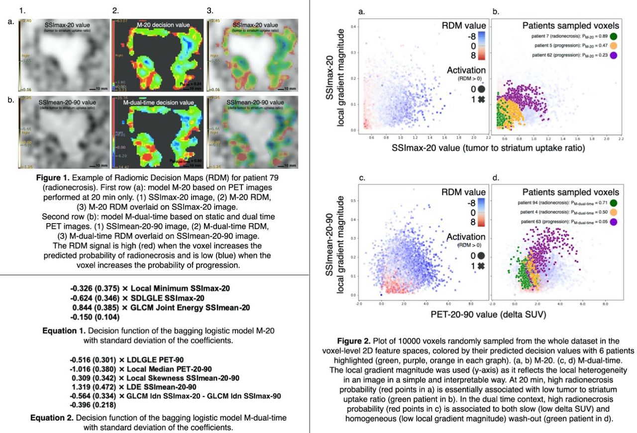
Methods: Ninety-six studies from 87 patients with suspicious uptake in [18F]-FDOPA PET were used. Each study included 2 static PET/CT scans: performed at 20 min (PET-20) and at 90 min (PET-90) post-injection. Final diagnosis of progression or radionecrosis was based on a 6-month follow-up and pathological data when available. PET-20 and PET-90 images were rigidly registered. The tumor and the contralateral striatum were segmented using a threshold set to 50% of the striatum SUVmax on PET-20 and propagated to PET-90. PET-20 and PET-90 images were scaled to the patient striatum uptake SUVmax or SUVmean by computing tumor to striatum uptake ratio to yield striatum-standardized images (SSI) [5, 6], denoted SSImax-20, SSImean-20, SSImax-90, and SSImean-90. Dual time point images were obtained by subtracting the 90 min images from the 20 min images, leading to PET-20-90, SSImax-20-90, and SSImean-20-90. A total of 924 voxel-wise radiomic feature maps were extracted by sliding a 5×5×5-voxel kernel over each image. The mean value over the tumor ROI was used for each feature to yield two feature vectors per patient, one for all images (924 features) and the other only for the standard 20 min images (231 features) to assess the added value of the dual time approach. After reducing redundancy using the variance inflation factor, we built two probabilistic models, M-20 and M-dual-time, to identify radionecrosis using forward feature selection and bagging regularized logistic regression. Radiomic decision maps (RDM) were computed by backprojecting the decision functions of the models at the voxel level. Based on the RDMs interpretation, we defined a simple and interpretable voxel-level 2D feature space for each model. We finally plotted 10,000 voxels randomly sampled from the whole dataset in these 2D spaces, colored by their predicted decision values and patient number to highlight the interpretations.

Results: There were 69 progressions and 27 radionecroses. Through cross-validation, 3 and 5 features were selected for M-20 and M-dual-time with a mean balanced accuracy of 71±9% (permutation test p-value = 0.04) and 75±9% (p-value = 0.03) respectively, with no significant difference between the 2 models. Sensitivity, specificity, and AUROC are reported in Table 1. Dual time features were preferentially selected by the model when available (4/5 features of M-dual-time). SSI features were also preferentially selected by both models (3/3 for M-20, 3/5 for M-dual-time). By interpreting the RDMs together with the models equations and then creating the 2D voxel-level feature spaces, we found that low local tumor to striatum uptake ratio was predictive of radionecrosis when using only the 20 min images, while radionecrosis identification was based on a locally homogeneous slow tracer wash-out when the 2 time points were used, consistently with other recent image-based studies [2, 7, 8].

Conclusions: Using RDMs for interpreting radiomic models built from a limited number of patients, our data-driven approach highlighted two patterns that discriminate radionecrosis from tumor progression without significant difference in terms of classification performances: low tumor to striatum uptake ratio at 20 min and locally homogeneous slow wash-out between 20 min and 90 min.

Figure
  • Download figure
  • Open in new tab
  • Download powerpoint
Previous
Back to top

In this issue

Journal of Nuclear Medicine
Vol. 63, Issue supplement 2
June 1, 2022
  • Table of Contents
  • Index by author
Article Alerts
Sign In to Email Alerts with your Email Address
Email Article

Thank you for your interest in spreading the word on Journal of Nuclear Medicine.

NOTE: We only request your email address so that the person you are recommending the page to knows that you wanted them to see it, and that it is not junk mail. We do not capture any email address.

Enter multiple addresses on separate lines or separate them with commas.
Radiomic decision maps reveal patterns discriminating between glioma progression and radiation-induced necrosis in static and dual time [18F]-FDOPA PET
(Your Name) has sent you a message from Journal of Nuclear Medicine
(Your Name) thought you would like to see the Journal of Nuclear Medicine web site.
Citation Tools
Radiomic decision maps reveal patterns discriminating between glioma progression and radiation-induced necrosis in static and dual time [18F]-FDOPA PET
Thibault Escobar, Fanny Orlhac, Anne-Capucine Rollet, Olivier Humbert, Sebastien Vauclin, Pascal Pineau, Jacques Darcourt, Irene Buvat
Journal of Nuclear Medicine Jun 2022, 63 (supplement 2) 2520;

Citation Manager Formats

  • BibTeX
  • Bookends
  • EasyBib
  • EndNote (tagged)
  • EndNote 8 (xml)
  • Medlars
  • Mendeley
  • Papers
  • RefWorks Tagged
  • Ref Manager
  • RIS
  • Zotero
Share
Radiomic decision maps reveal patterns discriminating between glioma progression and radiation-induced necrosis in static and dual time [18F]-FDOPA PET
Thibault Escobar, Fanny Orlhac, Anne-Capucine Rollet, Olivier Humbert, Sebastien Vauclin, Pascal Pineau, Jacques Darcourt, Irene Buvat
Journal of Nuclear Medicine Jun 2022, 63 (supplement 2) 2520;
Twitter logo Facebook logo LinkedIn logo Mendeley logo
  • Tweet Widget
  • Facebook Like
  • Google Plus One
Bookmark this article

Jump to section

  • Article
  • Figures & Data
  • Info & Metrics

Related Articles

  • No related articles found.
  • Google Scholar

Cited By...

  • No citing articles found.
  • Google Scholar

More in this TOC Section

  • Modular Model Architecture for Radiopharmaceutical Therapy Planning: Physiologically-Based RadioPharmacoKinetics (PBRPK) Implementation
  • Objective task-based evaluation of a deep-learning-based segmentation method for quantitative SPECT
  • Robustness and Reproducibility of Radiomics Features from Fusions of PET-CT Images
Show more Data Analysis & Management

Similar Articles

SNMMI

© 2023 Journal of Nuclear Medicine

Powered by HighWire