


Abstract
2399
Introduction: 227Th-labelled conjugates are an emerging class of α-particle radiopharmaceutical therapies (α-RPTs) [1-3]. 227Th decays to 223Ra, another α-particle emitting isotope that redistributes throughout the patient forming an independent biodistribution [4-5]. There is an important clinical need to quantify the dose with these therapies in lesions and organs at risk [6-7]. Since both 223Ra and 227Th emit gamma-ray photons, SPECT may provide a mechanism to quantify the uptake of these isotopes within the body. However, this quantification is challenged by the extremely low number of detected counts, multiple photopeaks in the emission spectra of these isotopes, and the significant overlap in the spectra of 223Ra and 227Th resulting in crosstalk [4-5, 8-10]. To address these challenges, we propose and evaluate a multiple-energy-window projection-domain quantitative SPECT (MEW-PDQ) method for joint regional uptake quantification of 227Th and 223Ra.
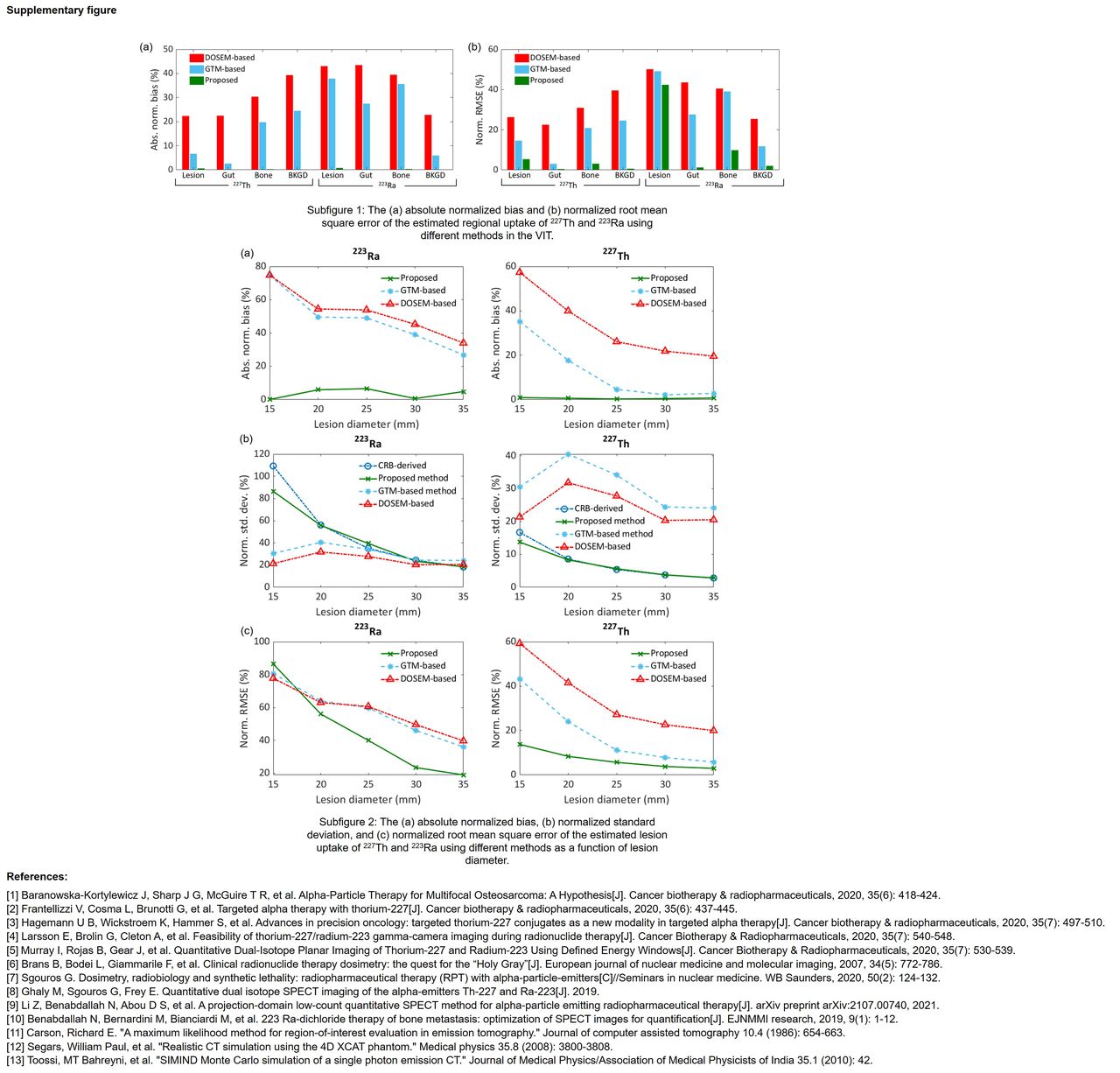
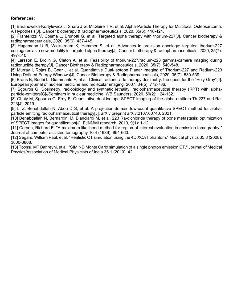
Methods: The proposed method directly estimates the regional activity uptake of 227Th and 223Ra from the SPECT projection data acquired over multiple energy windows, skipping the image-reconstruction step. A set of coupled equations are derived that relate the measured projections in the multiple energy windows to the uptake in different regions, while also modeling the crosstalk. Further, these equations model the 227Th/223Ra decay scheme. The coupled equations are solved iteratively to jointly estimate the regional uptake of the two isotopes. This proposed method has several advantages including reduced ill-posedness of the inverse problem, avoiding reconstruction-related information loss [9,11], and use of multiple energy window projections helping alleviate the low-count challenge. Evaluation of this method was performed using clinically realistic simulations. We conducted a virtual imaging trial (VIT) by simulating 50 digital 3D male patients with different anatomies using XCAT phantom [12]. The patients were simulated to have bone metastasis in the pelvic region and were administrated with 227Th-based α-RPT. We simulated the scenario that the patients were scanned on a GE Optima 640 SPECT/CT system with HEGP collimator after 120h of administering 227Th. Photons were acquired at 60 angular positions spaced uniformly over 360º and in four energy windows (75 – 100 keV, 135 – 165 keV, 215 – 260 keV, and 268 – 285 keV) corresponding to different photopeaks of 227Th and 223Ra. All relevant image-degrading processes and the isotope emission were simulated using SIMIND, a well-validated Monte Carlo simulation tool [13]. From the acquired projections, we estimated regional uptake of both isotopes in four regions, namely lesion, gut, bone, and the rest of the body (background) using the MEW-PDQ method. We also evaluated the sensitivity of the method to lesion sizes and compared the variance of the estimates to the lowest theoretical limit for any unbiased estimator, i.e., the Cramer-Rao bound (CRB). The method was compared to a dual-isotope ordered subset expectation maximization (DOSEM)-based method and a geometric transfer matrix (GTM)-based partial-volume compensation method.
Results: In the VIT, the MEW-PDQ method yielded the most accurate and reliable estimates (Subfigure 1). The normalized root mean square error (NRMSE) of the proposed method averaged over all regions was 2.4% and 13.9% for 227Th and 223Ra, respectively, in contrast to 29.8% and 39.9% for the DOSEM and 15.8% and 31.9% for the GTM-based methods. Further, the MEW-PDQ method yielded accurate (close to zero) and precise (close to CRB) estimates of mean regional uptakes of both 227Th and 223Ra and significantly outperformed the DOSEM and GTM-based methods for all lesion sizes (Subfigure 2).
Conclusions: The proposed MEW-PDQ method provided reliable joint absolute quantification of mean regional uptake of both 227Th and 223Ra as evaluated using realistic simulations. This method could thus provide a mechanism for dose quantifications in 227Th-based α-RPTs.


In this issue


Jump to section
Related Articles
Cited By...
- No citing articles found.