


Article Figures & Data
Figures
- FIGURE 1.
18F-FDG and cerebral blood flow (CBF) PET. Crossed cerebellar diaschisis (CCD) (A) seen in a right (Rt.) middle cerebral artery (MCA) stroke. Decreased glucose metabolism (18F-FDG) and perfusion (CBF) in contralateral left (Lt.) cerebellar hemisphere (white arrow) where no ischemic injury is present. Decreased 18F-FDG uptake and CBF are secondary to deactivation of cortico–ponto–cerebellar tract (“remote effect”). Such remote effects are known to occur in neurodegenerative disorders. 18F-FDG uptake not only reflects local pathology, but also could reflect remote pathology. Knowledge of cortical pathways is crucial for scan interpretation. (B) Coupling between glucose metabolism (18F-FDG) and CBF in AD measured by PET and 3D-SSP analysis. Statistical t maps (top 2 rows) represent regional hypometabolism and hypoperfusion seen in a group of AD patients. Both 18F-FDG and CBF PET show similar regional changes, though CBF PET appears slightly less sensitive. Ratio maps between CBF and 18F-FDG (bottom row) indicate that metabolic-flow coupling is relatively preserved in areas both affected and not affected by AD. This observation supports use of various flow-related measurements such as perfusion SPECT, early flow images obtained as a part of amyloid PET or tau PET, or MRI-based perfusion imaging for dementia evaluation.
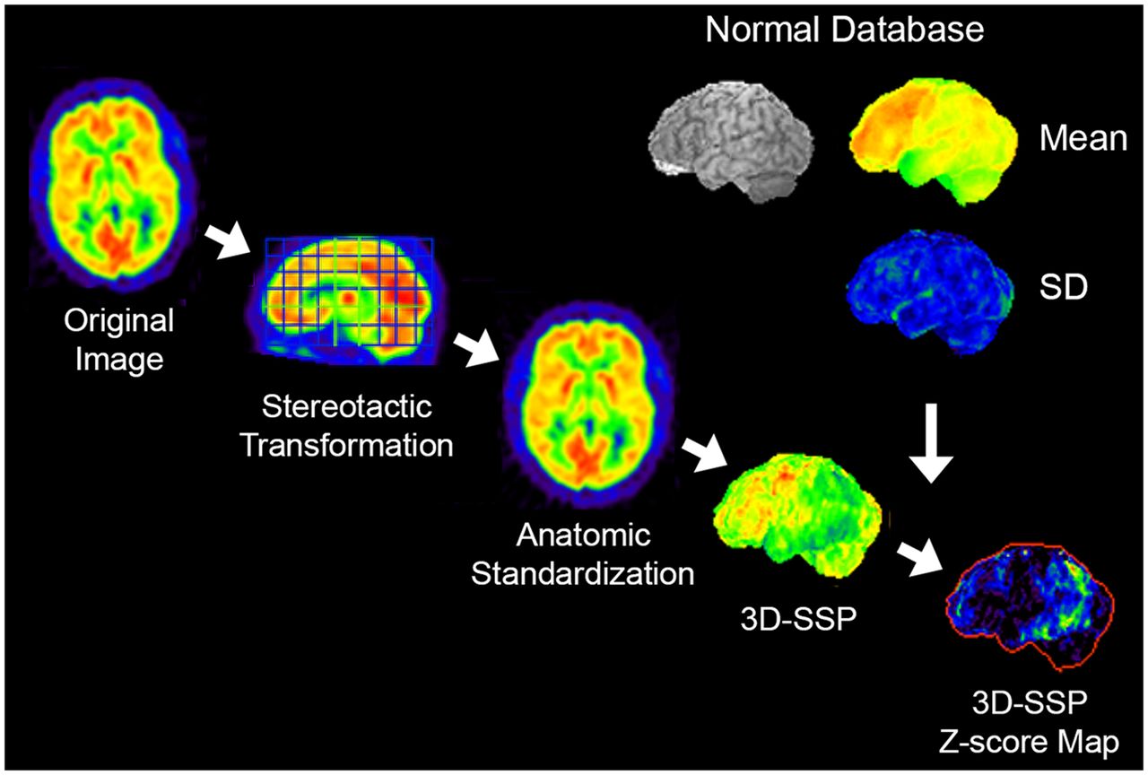
- FIGURE 2.
3D-SSP. Original transaxial images are anatomically standardized in the stereotactic coordinate system, and gray matter activity is extracted on a pixel-by-pixel basis by the 3D-SSP algorithm. Extracted data are then compared with a normal database (mean and SD), which is composed of similarly processed PET scans from multiple normal subjects. Differences between individual data and normal database are expressed as z score maps.
- FIGURE 3.
18F-FDG PET scan of patient with progressive mild cognitive decline. Original transaxial images (top 6 rows, black and white) demonstrate very mild metabolic reductions in parietal association cortex and posterior cingulate cortex/precuneus. However, such mild changes cannot be appreciated consistently. 3D-SSP z score maps (bottom row, from left to right: right lateral, left lateral, superior, inferior, anterior, posterior, right medial, and left medial views) from same patient demonstrate apparent metabolic reductions in posterior cingulate cortex/precuneus as well as parietal association cortex bilaterally seen on lateral, superior, and posterior views. Statistical mapping, if used appropriately, improves diagnostic accuracy and consistency.
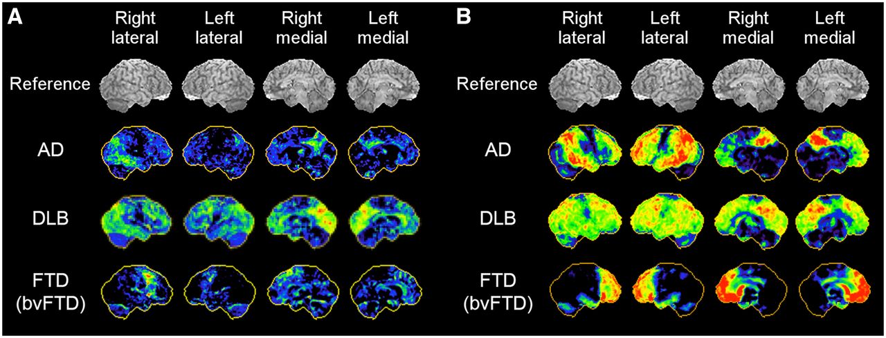
- FIGURE 4.
18F-FDG PET 3D-SSP maps from representative cases of AD, DLB, versus bvFTD. Red indicates more severe hypometabolism. Mild cases (A) and severe cases (B) are shown.
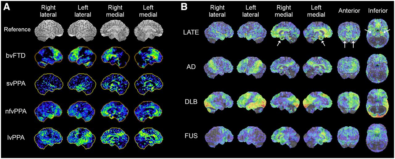
- FIGURE 5.
18F-FDG PET 3D-SSP z score maps of representative cases of FTLD/FTD variants, bvFTD, svPPA, nfvPPA, lvPPA, and PSP (A) and LATE, AD, DLB, and FUS (B, z score maps superimposed on reference MR image). The case of nfvPPA presented here shows bilateral temporofrontal involvement with right side slightly more prominent than left. LATE demonstrates prominent involvement of medial temporal lobe and hippocampus (white arrows) and medial and orbital frontal cortices. 18F-FDG uptake in medial temporal lobe is relatively mild in AD and DLB. FUS demonstrates pattern involving frontal and anterior temporal lobes, resembling FTLD/FLD spectrum.
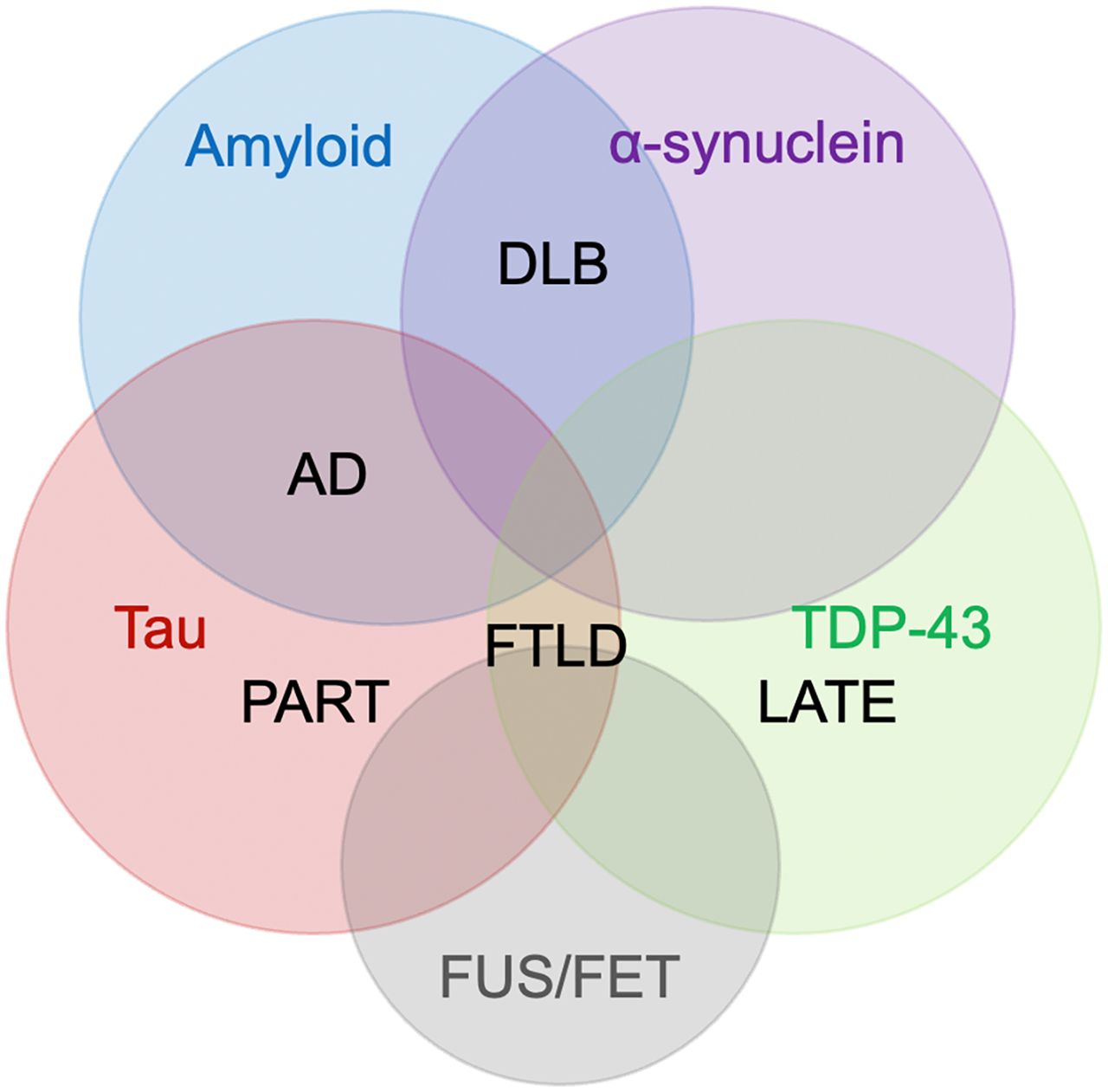
- FIGURE 6.
Cooccurrence of 4 major proteinopathies: amyloid, tau, α-synuclein, and TDP-43, and overlapping neurodegenerative disorders: AD, DLB, FTLD, PART, and LATE. Emerging proteinopathies such as fused in sarcoma (FUS), which belongs to the FET family of proteins (FUS, EWS, TAF15), have been identified in FTLD, comorbid with other proteinopathies, and awaiting further characterization. Pathologic diagnosis of neurodegenerative disorders involves new markers, and further investigations of clinicopathologic correlations including imaging will allow more precise antemortem diagnosis in the future.
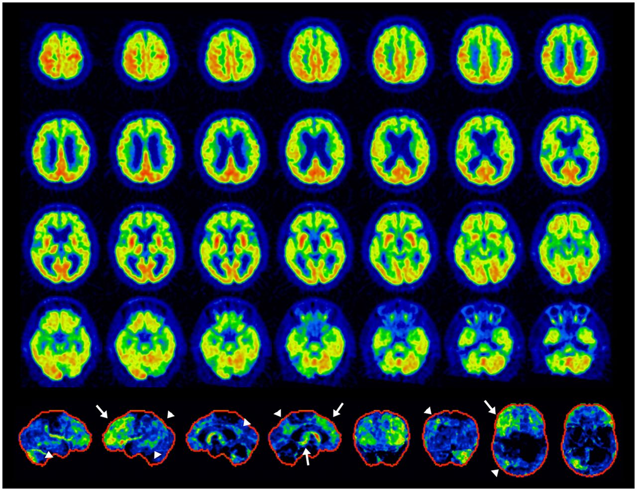
- FIGURE 7.
Example of mixed dementia, PSP+AD. Antemortem 18F-FDG PET: transaxial images (top 4 rows) and 3D-SSP z score maps (bottom row, from the left to right: right lateral, left lateral, right medial, and left medial, anterior, posterior, superior, inferior views). Findings are consistent with PSP (white arrows) and AD (arrowheads). Left-dominant pathologies associated with crossed-cerebellar diaschisis in right cerebellar hemisphere. It is important to note that these findings do not necessarily preclude other mixed pathologies. However, the frontal findings best explained the patient’s clinical symptoms.


Tables
Major differential diagnosis: standard of care Alzheimer disease (AD) Frontotemporal dementia (FTD) Dementia with Lewy bodies (DLB) Subtype Classification of FTLD/FTD Behavioral variant FTD (bvFTD) and Pick’s disease (PiD) Primary progressive aphasia (PPA) Semantic variant PPA (svPPA) or semantic dementia (SD) Nonfluent variant PPA (nfvPPA) or progressive nonfluent aphasia (PNFA) Logopenic variant PPA (lvPPA) or logopenic progressive aphasia (LPA) Movement disorders Progressive supranuclear palsy (PSP) Corticobasal degeneration (CBD) Recently recognized neurodegenerative disorders Limbic-predominant age-related TDP-43 encephalopathy (LATE) Hippocampus sclerosis (HS) Primary age-related tauopathy (PART) Argyrophilic grain disease (AGD) Fused in sarcoma (FUS) Mixed dementia with copathologies and overlapping disorders AD and vascular dementia (VaD) Dementia with multiple neurodegenerative copathologies +/− VaD Mixed/comorbid neurodegenerative dementing disorders Reference AD and TDP-43, cerebral amyloid angiopathy (91) AD and corticobasal syndrome, FTLD-TDP, Lewy body disease (92) AD and CBD (93) AD and tauopathy, TDP-43 (72) AD and DLB with TDP-43, tau, α-synuclein pathologies (94) DLB and AD pathology (95) FTLD-tau and AD and vascular copathologies (96) PSP and AD (97) PSP and AD and PD (98) PSP and AD, AGD, CBD, Lewy body disease (99) PiD and AD (100) PiD and AD, cerebral amyloid angiopathy, Lewy body disease (101) PiD and PSP (102) PiD = Pick’s disease.
In this issue


Jump to section
- Article
- Abstract
- 18F-FDG AS A NEUROCHEMICAL TRACER
- CELLULAR MECHANISMS OF 18F-FDG UPTAKE IN THE BRAIN
- REMOTE EFFECTS ON 18F-FDG UPTAKE (DIASCHISIS)
- IMAGE INTERPRETATION USING STATISTICAL MAPPING TECHNIQUES
- CLINICAL IMAGING ALTERNATIVES TO 18F-FDG PET FOR DEMENTIA EVALUATION
- DIFFERENTIAL DIAGNOSIS OF AD, FTD, AND DLB: STANDARD OF CARE
- PREDICTIVE VALUE OF 18F-FDG PET IN EVALUATION OF COGNITIVE DECLINE
- CLINICAL SUBTYPES OF AD
- CLINICAL SUBTYPES OF FRONTOTEMPORAL LOBAR DEGENERATION (FTLD)/FTD
- PPA: THREE SUBTYPES
- ATYPICAL PARKINSONIAN MOVEMENT DISORDERS (PSP AND CBD)
- RECENTLY RECOGNIZED NEURODEGENERATIVE DEMENTING DISORDERS
- LIMBIC-PREDOMINANT AGE-RELATED TDP-43 ENCEPHALOPATHY (LATE) AND HIPPOCAMPUS SCLEROSIS (HS)
- ARGYROPHILIC GRAIN DISEASE (AGD), PRIMARY AGE-RELATED TAUOPATHY (PART), AND FUSED IN SARCOMA (FUS)
- MIXED DEMENTIA AND COEXISTING PATHOLOGIES
- THE EVOLVING ROLE OF 18F-FDG PET IN DEMENTIA EVALUATION
- SUMMARY
- DISCLOSURE
- REFERENCES
- Figures & Data
- Info & Metrics
Related Articles
Cited By...
- 13C tracing in synaptosomes reveals that SGLT2 inhibition with dapagliflozin prevents metabolic deficits in the 5X-FAD model of Alzheimers Disease
- Macroscopic cerebral energy efficiency corresponds to neuron reorganization in awake and anesthetized mice
- BOLD Amplitude Correlates of Preclinical Alzheimers Disease
- SNMMI Procedure Standard/EANM Practice Guideline for Brain [18F]FDG PET Imaging, Version 2.0
- Evaluation of Neurodegenerative Disorders with Amyloid-{beta}, Tau, and Dopaminergic PET Imaging: Interpretation Pitfalls
- Glucose intolerance induces anxiety-like behaviors independent of obesity and insulin resistance in a novel model of nutritional metabolic stress
- Molecular Imaging of Neurodegeneration: The Way to New Horizons