
Main menu

  • Home
  • Content
    • Current
    • Ahead of print
    • Past Issues
    • JNM Supplement
    • SNMMI Annual Meeting Abstracts
    • Continuing Education
    • JNM Podcasts
  • Subscriptions
    • Subscribers
    • Institutional and Non-member
    • Rates
    • Journal Claims
    • Corporate & Special Sales
  • Authors
    • Submit to JNM
    • Information for Authors
    • Assignment of Copyright
    • AQARA requirements
  • Info
    • Reviewers
    • Permissions
    • Advertisers
  • About
    • About Us
    • Editorial Board
    • Contact Information
  • More
    • Alerts
    • Feedback
    • Help
    • SNMMI Journals
  • SNMMI
    • JNM
    • JNMT
    • SNMMI Journals
    • SNMMI

User menu

  • Subscribe
  • My alerts
  • Log in
  • My Cart

Search

  • Advanced search
Journal of Nuclear Medicine
  • SNMMI
    • JNM
    • JNMT
    • SNMMI Journals
    • SNMMI
  • Subscribe
  • My alerts
  • Log in
  • My Cart
Journal of Nuclear Medicine

Advanced Search

  • Home
  • Content
    • Current
    • Ahead of print
    • Past Issues
    • JNM Supplement
    • SNMMI Annual Meeting Abstracts
    • Continuing Education
    • JNM Podcasts
  • Subscriptions
    • Subscribers
    • Institutional and Non-member
    • Rates
    • Journal Claims
    • Corporate & Special Sales
  • Authors
    • Submit to JNM
    • Information for Authors
    • Assignment of Copyright
    • AQARA requirements
  • Info
    • Reviewers
    • Permissions
    • Advertisers
  • About
    • About Us
    • Editorial Board
    • Contact Information
  • More
    • Alerts
    • Feedback
    • Help
    • SNMMI Journals
  • View or Listen to JNM Podcast
  • Visit JNM on Facebook
  • Join JNM on LinkedIn
  • Follow JNM on Twitter
  • Subscribe to our RSS feeds
Research ArticleClinical Investigation

The Role of Amyloid PET in Imaging Neurodegenerative Disorders: A Review

Marianne Chapleau, Leonardo Iaccarino, David Soleimani-Meigooni and Gil D. Rabinovici
Journal of Nuclear Medicine June 2022, 63 (Supplement 1) 13S-19S; DOI: https://doi.org/10.2967/jnumed.121.263195
Marianne Chapleau
1Memory and Aging Center, Department of Neurology, University of California, San Francisco, San Francisco, California;
  • Find this author on Google Scholar
  • Find this author on PubMed
  • Search for this author on this site
Leonardo Iaccarino
1Memory and Aging Center, Department of Neurology, University of California, San Francisco, San Francisco, California;
  • Find this author on Google Scholar
  • Find this author on PubMed
  • Search for this author on this site
David Soleimani-Meigooni
1Memory and Aging Center, Department of Neurology, University of California, San Francisco, San Francisco, California;
  • Find this author on Google Scholar
  • Find this author on PubMed
  • Search for this author on this site
Gil D. Rabinovici
1Memory and Aging Center, Department of Neurology, University of California, San Francisco, San Francisco, California;
2Weill Institute for Neurosciences, University of California, San Francisco, San Francisco, California; and
3Department of Radiology and Biomedical Imaging, University of California, San Francisco, San Francisco, California
  • Find this author on Google Scholar
  • Find this author on PubMed
  • Search for this author on this site
  • Article
  • Figures & Data
  • Info & Metrics
  • PDF
Loading

Article Figures & Data

Figures

  • Tables
  • Figure
    • Download figure
    • Open in new tab
    • Download powerpoint
  • FIGURE 1.
    • Download figure
    • Open in new tab
    • Download powerpoint
    FIGURE 1.

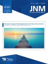
    Postmortem measures of amyloid pathology. (A) Types of amyloid deposits. (B) Amyloid angiopathy. (C) Distribution of diffuse and neuritic plaques. (D) Neuritic plaque density (highest density score observed in brain). (A, B, and D are from UCSF Neurodegenerative Disease Brain Bank; C is reprinted with permission of (53).) NP = neuritic plaques.

  • FIGURE 2.
    • Download figure
    • Open in new tab
    • Download powerpoint
    FIGURE 2.

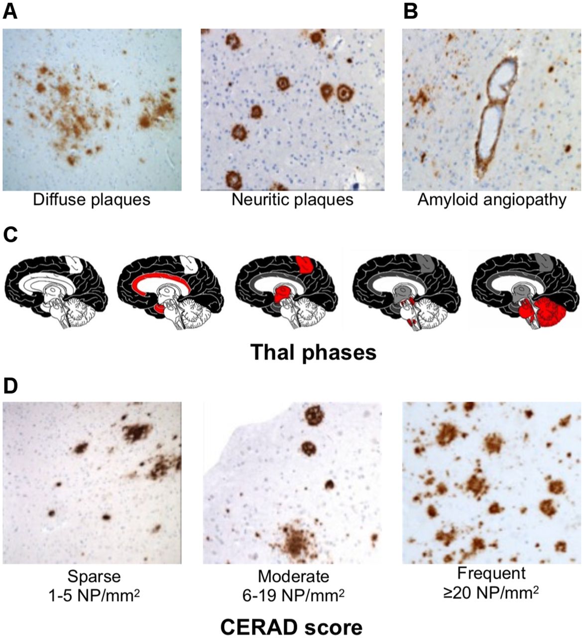
    Structures of thioflavin-T, 11C-PiB, 18F-flutafuranol, and Food and Drug Administration–approved Aβ PET tracers. (Reprinted from (54).)

  • FIGURE 3.
    • Download figure
    • Open in new tab
    • Download powerpoint
    FIGURE 3.

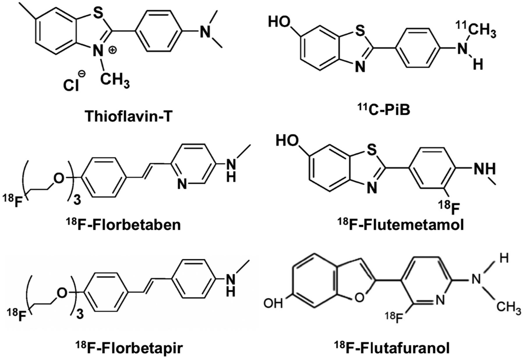
    Examples of negative and positive Aβ PET findings using different tracers. (18F-flutafuranol images are courtesy of Victor Villemagne and Christopher C. Rowe.)

  • FIGURE 4.
    • Download figure
    • Open in new tab
    • Download powerpoint
    FIGURE 4.

    Evolution of amyloid PET positivity across AD spectrum. (A) Positive 11C-PiB scan of cognitively normal (CN) participant, in which significant binding is observed in precuneus, posterior cingulate cortex, and medial prefrontal areas. (B) Positive 11C-PiB scan of MCI patient, in which significant and moderate binding is observed throughout cortex. (C) Positive 11C-PiB scan of AD patient, in which significant and severe binding is observed throughout cortex.

Tables

  • Figures
    • View popup
    TABLE 1.

    Summary Guidelines for Interpretation of Amyloid PET Scans Using Different Tracers

    Tracer categoryTracer nameDose and acquisition protocol (clinical)VisualizationInterpretation criteria for positive scan
    Food and Drug Administration– approved18F-florbetaben∼300 MBq; 15- to 20-min acquisition beginning at 45–130 min (research use, 20-min acquisition beginning at 90–110 min)Gray scale; window images to optimize GM/WM contrast in cerebellumIncreased GM uptake extending to cortical margin involving most slices in at least 1 of 4 target cortical regions: frontal, parietal, precuneus/posterior cingulate, lateral temporal; regional cortical tracer uptake/brain amyloid plaque load scores (20)
    18F-florbetapir∼370 MBq; 10- to 20-min acquisition beginning at 30–50 min (package insert guidelines) for clinical use or 50–70 min (optimized kinetics for quantification) for research useInverse gray scale; window images to optimize GM/WM contrast in cerebellumLoss of GM/WM contrast due to increased cortical binding in, first, 2 or more brain areas (each larger than single gyrus) with reduced or absent GM/WM contrast or, second, 1 or more areas with intense signal where GM > WM
    18F-flutemetamol∼185 MBq; 10- to 20-min acquisition at 60–120 min (research use, 20-min acquisition at 90–110 min)Color scale (NIH); normalize so that pons is at 90% of activityIncreased GM uptake (>50%–60% peak intensity) or loss of GM matter contrast in at least 1 of 4 cortical regions and 1 subcortical region: frontal, inferolateral parietal, precuneus/posterior cingulate, lateral temporal, striatum
    Research11C-PiB∼555 MBq; dynamic 60- to 90-min acquisition (distribution volume ratio) or 20-min acquisition at 50–70 min (SUV ratio)Color scale (NIH); window images to optimize GM/WM contrast in cerebellumNo formal guidelines for reading (research use only)
    18F-flutafuranol∼185 MBq; 20- to 30-min acquisition beginning at 40–50 minColor scale (NIH); window images to optimize GM/WM contrast in cerebellumNo formal guidelines for reading (research use only)
    • GM = gray matter; NIH = National Institutes of Health; WM = white matter.

PreviousNext
Back to top

In this issue

Journal of Nuclear Medicine: 63 (Supplement 1)
Journal of Nuclear Medicine
Vol. 63, Issue Supplement 1
June 1, 2022
  • Table of Contents
  • Table of Contents (PDF)
  • About the Cover
  • Index by author
  • Complete Issue (PDF)
Print
Download PDF
Article Alerts
Sign In to Email Alerts with your Email Address
Email Article

Thank you for your interest in spreading the word on Journal of Nuclear Medicine.

NOTE: We only request your email address so that the person you are recommending the page to knows that you wanted them to see it, and that it is not junk mail. We do not capture any email address.

Enter multiple addresses on separate lines or separate them with commas.
The Role of Amyloid PET in Imaging Neurodegenerative Disorders: A Review
(Your Name) has sent you a message from Journal of Nuclear Medicine
(Your Name) thought you would like to see the Journal of Nuclear Medicine web site.
Citation Tools
The Role of Amyloid PET in Imaging Neurodegenerative Disorders: A Review
Marianne Chapleau, Leonardo Iaccarino, David Soleimani-Meigooni, Gil D. Rabinovici
Journal of Nuclear Medicine Jun 2022, 63 (Supplement 1) 13S-19S; DOI: 10.2967/jnumed.121.263195

Citation Manager Formats

  • BibTeX
  • Bookends
  • EasyBib
  • EndNote (tagged)
  • EndNote 8 (xml)
  • Medlars
  • Mendeley
  • Papers
  • RefWorks Tagged
  • Ref Manager
  • RIS
  • Zotero
Share
The Role of Amyloid PET in Imaging Neurodegenerative Disorders: A Review
Marianne Chapleau, Leonardo Iaccarino, David Soleimani-Meigooni, Gil D. Rabinovici
Journal of Nuclear Medicine Jun 2022, 63 (Supplement 1) 13S-19S; DOI: 10.2967/jnumed.121.263195
Twitter logo Facebook logo LinkedIn logo Mendeley logo
  • Tweet Widget
  • Facebook Like
  • Google Plus One
Bookmark this article

Jump to section

  • Article
    • Visual Abstract
    • Abstract
    • VISUAL INTERPRETATION, QUANTIFICATION, AND THRESHOLDS
    • CLINICOIMAGING CORRELATES
    • APPROPRIATE-USE CRITERIA (AUCS)
    • CLINICAL UTILITY STUDIES
    • COMPARISON WITH FLUID BIOMARKERS
    • CLINICAL TRIALS
    • CONCLUSION
    • DISCLOSURE
    • REFERENCES
  • Figures & Data
  • Info & Metrics
  • PDF

Related Articles

  • No related articles found.
  • PubMed
  • Google Scholar

Cited By...

  • Development and clinical validation of blood-based multibiomarker models for the evaluation of brain amyloid pathology
  • PET Imaging in Alzheimer Disease: Pathology and Research Insights for Technologists
  • Concordance between amyloid-PET quantification and real-world visual reads: results from IDEAS
  • Evaluation of ComBat harmonization for reducing across-tracer differences in regional amyloid PET analyses
  • Sensitivity of unconstrained quantitative magnetization transfer MRI to Amyloid burden in preclinical Alzheimers disease
  • Opposing roles of physiological and pathological amyloid-{beta} on synapses in live human brain slice cultures
  • Molecular Imaging of Systemic and Cardiac Amyloidosis: Recent Advances and Focus on the Future
  • A novel ultrasensitive assay for plasma p-tau217: performance in individuals with subjective cognitive decline and early Alzheimers disease
  • Cell-type-specific Alzheimers disease polygenic risk scores are associated with distinct disease processes in Alzheimers disease
  • Tau PET Visual Reads: Research and Clinical Applications and Future Directions
  • A Visual Interpretation Algorithm for Assessing Brain Tauopathy with 18F-MK-6240 PET
  • Molecular Imaging of Neurodegeneration: The Way to New Horizons
  • Google Scholar

More in this TOC Section

  • SNMMI Procedure Standard/EANM Practice Guideline for Brain [18F]FDG PET Imaging, Version 2.0
  • Imaging Efficacy of [18F]CTT1057 PET for the Detection of PSMA-Positive Tumors Using Histopathology as Standard of Truth: Results from the GuideView Phase 2/3 Prospective Multicenter Study
  • Meeting Upcoming Clinical and Diagnostic Needs in Oncologic Imaging: A Structured Reporting System for Fibroblast-Activation-Protein–Targeted Imaging—FAP-RADS Version 1.0
Show more Clinical Investigation

Similar Articles

Keywords

  • neurology
  • PET
  • PET/MRI
  • Alzheimer disease
  • amyloid PET
  • neurodegenerative diseases
SNMMI

© 2025 SNMMI

Powered by HighWire