


Article Figures & Data
Figures
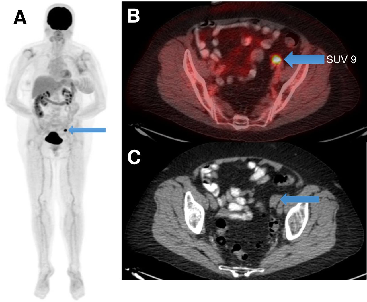
- FIGURE 1.
Asymptomatic 70-y-old woman with left arm MCC (s/p excision, left axillary lymphadenectomy, radiation to axilla) underwent surveillance 18F-FDG PET/CT scan 3.3 mo after treatment. 18F-FDG PET/CT (A; maximum-intensity projection, arrow) scan revealed solitary focal 18F-FDG uptake in left pelvis (B; fused PET/CT, SUV 9, arrow) in a nodular soft-tissue-density lesion in left adnexa (C; axial CT, arrow). USG pelvis showed solid mass in left ovary measuring 1.8 x 1.4 x 1.6 cm, corresponding to site of abnormality on 18F-FDG PET/CT. Patient underwent salpingo-oophorectomy and pathology was positive for MCC. s/p = status post; USG = ultrasound.
- FIGURE 2.
Receiver-operating-characteristic (ROC) curve for benign versus malignant lesion prediction based on SUVmax. FPR = false-positive rate; TPR = true-positive rate.
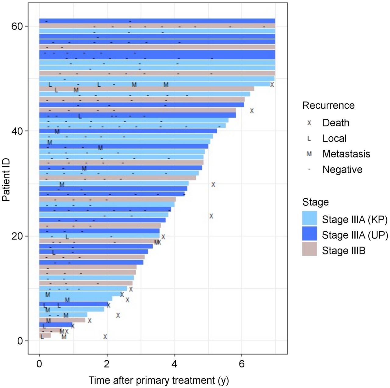
- FIGURE 3.
Swimmer plot illustrating information about local and distant recurrences, confirmed on pathology, during follow-up on surveillance 18F-FDG PET/CT scans since end of primary treatment for all included stage IIIA known primary (KP), IIIA unknown primary (UP), and IIIB MCC patients (n = 61).
- FIGURE 4.
OS of patients based on findings and timing of 18F-FDG PET/CT scan (A, at 3-mo; B, at 6 mo, and C, at 12 mo after definitive treatment, respectively).


Tables
Characteristic Data No. of patients 61 Sex (n) Male 44 (72%) Female 17 (28%) Mean age (±SD) at the time of diagnosis (y) 69 ± 13.0 (range, 25–93) Site of primary disease (n) Cheek/chin 5 (8.1%) Ear/eyelid/nose 4 (6.6%) Forehead/scalp 5 (8.1%) Neck node 2 (3.3%) Axilla 4 (6.6%) Back/chest 2 (3.3%) Groin 11 (18.0%) Buttocks 10 (16.3%) Finger/hand 2 (3.3%) Forearm/elbow/arm 8 (13.2%) Knee/leg/thigh 8 (13.2%) Stage of primary disease at diagnosis (n) Stage IIIA (known primary) 23 (37%) Stage IIIA (unknown primary) 20 (33%) Stage IIIB 18 (30%) Prior treatment received (n) Surgery 20 (33%) Surgery + chemotherapy/ radiation/chemoradiation 38 (62%) Chemoradiation 3 (5%) Number of 18F-FDG PET/CT scans 221 Scans performed per patient (n) 1–4 43 5–10 18 Univariate Multivariate Variable HR 95% CI P HR 95% CI P 18F-FDG PET/CT scan, positive 15.7 4.34, 56.5 <0.001 — — — SUVmax (for 1 unit) 1.27 1.17, 1.39 <0.001 1.17 1.05, 1.31 0.006 No. of positive lesions (for 1 lesion) 1.45 1.27, 1.64 <0.001 1.60 1.25, 2.04 <0.001 Stage 0.16 0.05 IIIA (KP) Ref. Ref. IIIA (UP) 0.27 0.06, 1.27 0.09 0.01, 0.99 IIIB 0.86 0.28, 2.64 0.38 0.08, 1.94 KP = known primary; Ref. = reference; UP = unknown primary.
Supplemental Data
Files in this Data Supplement:
In this issue


Jump to section
Related Articles
Cited By...
- No citing articles found.