


Article Figures & Data
Figures
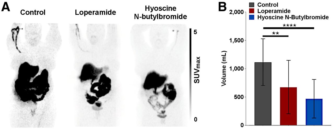
- FIGURE 1.
(A) Qualitative maximum-intensity-projection whole-body assessment of effect of pharmacologic interventions to slow progression of radioactive intestinal bolus. Without use of any intervention (left panel), 18F-4FMFES PET typically produces intense abdominal uptake caused by progression of radiometabolites excreted by gallbladder in intestines. Ingestion of 4 mg of loperamide 15 min before injection of radiotracer yielded mitigated results (center panel). Repeated intravenous injection of 20 mg of hyoscine N-butylbromide at 0, 20, and 40 min after 18F-4FMFES injection apparently reduced lower-abdomen background and slowed transit of radioactive intestinal bolus (right panel). (B) Measured volume extracted from application of SUV threshold of >4 on abdominal ROI. Both use of loperamide and use of hyoscine N-butylbromide significantly reduced intestinal background volume. **P < 0.01. ****P < 0.001.
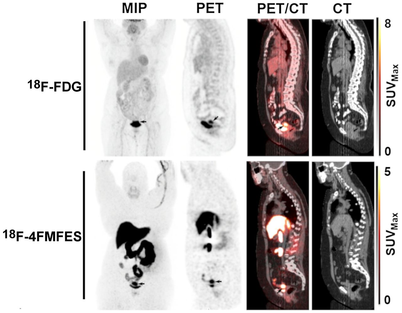
- FIGURE 2.
Representative case of endometrial carcinoma (arrows) imaged with 18F-FDG PET/CT (top row) and 18F-4FMFES PET/CT (bottom row), displayed in frontal maximum-intensity projection (MIP) and in sagittal views.
- FIGURE 3.
A 69-y-old endometroid adenocarcinoma patient with 18F-FDG–negative, 18F-4FMFES–positive primary tumor. 18F-FDG PET did not yield any abnormal uptake in uterus, whereas 18F-4FMFES PET revealed intense signal (SUVmax, 9.6; arrows) over 44 × 32 × 25 mm region. Postsurgery pathology report measured size of tumor to be 20 mm in its long axis, meaning 18F-4FMFES overestimated size of tumor in this case.
- FIGURE 4.
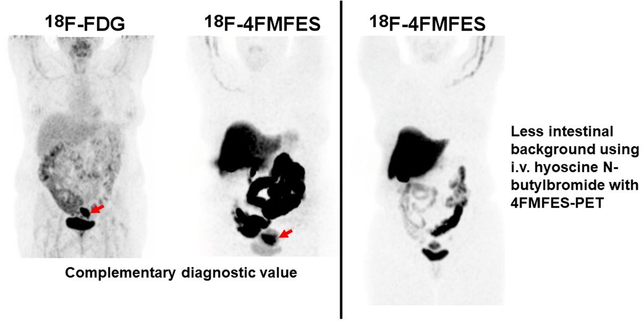
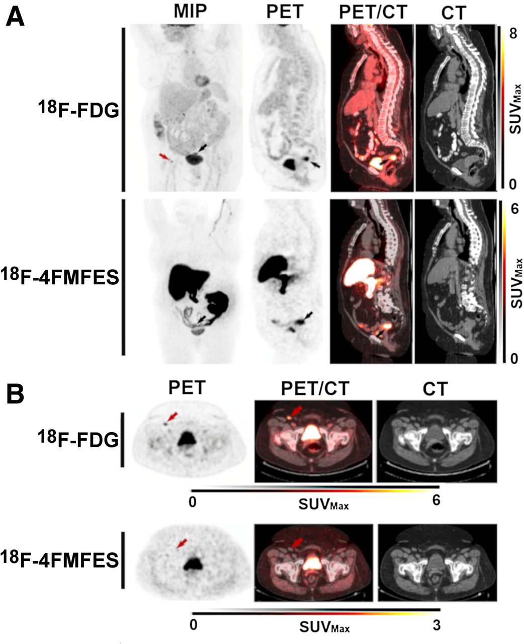
A 75-y-old endometroid adenocarcinoma patient with 18F-FDG false-positive inguinal node. (A) Endometroid adenocarcinoma primary tumor, with SUVmax uptake of 12.3 for 18F-FDG and 8.9 for 18F-4FMFES (black arrows). The 18F-FDG PET also revealed a suspected right inguinal node metastasis (red arrow), which yielded SUVmax of 5.2 (T/B, 7.2). (B) Transaxial slices of the suspected inguinal node metastasis (red arrows). The 18F-FDG–positive node was 18F-4FMFES–negative and of normal appearance in CT image. Pathology examination considered inguinal node as normal, meaning 18F-FDG signal was false-positive.
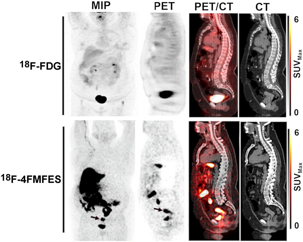
- FIGURE 5.
A 67-y-old endometrial carcinoma patient with 18F-4FMFES false-positive iliac node. (A) Endometrial carcinoma primary tumor, with SUVmax uptake of 12.9 for 18F-FDG and 12.7 for 18F-4FMFES (arrows). (B) Coronal (top) and transaxial (bottom) views centered on suspected left iliac sentinel node metastasis with 18F-4FMFES (arrows), which was of normal aspect in CT images. Pathology examination after surgery considered iliac node normal, confirming false-positive result for 18F-4FMFES.
- FIGURE 6.
Semiquantitative 18F-FDG and 18F-4FMFES uptake and T/Bs. (A) 18F-FDG and 18F-4FMFES uptake (SUVmax) for whole sample (left) and according to grade (right) (B) 18F-FDG and 18F-4FMFES T/Bs for whole studied sample (left) and according to grade (right). (C) 18F-FDG and 18F-4FMFES T/Bs according to grade. *P < 0.05. **P < 0.01. ****P < 0.001.


Tables
Parameter Data Patients (n) 25 Mean age ± SD (y) 63.4 ± 10.5 (median, 66; range, 41–79) Premenopausal (n) 4 Postmenopausal (n) 21 Histology (n) Endometrial carcinoma 23 Endometrial intraepithelial neoplasia 2 Grade (n) 1 5 2 12 3 8 Treatment (n) Loperamide (4 mg) 12 Hyoscine N-butylbromide (3 × 20 mg) 11 None 2 (plus 31 breast cancer patients (23))
In this issue


Jump to section
Related Articles
Cited By...
- No citing articles found.