


Article Figures & Data
Figures
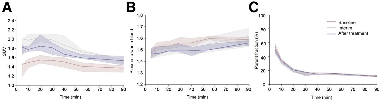
- FIGURE 1.
Venous blood sampling data of all patients obtained at the 3 different time points. (A) SUV whole blood data. (B) Plasma-to-whole blood ratios. (C) Parent fraction of 18F-FES in plasma. Data represent median of all values, with their corresponding IQRs.
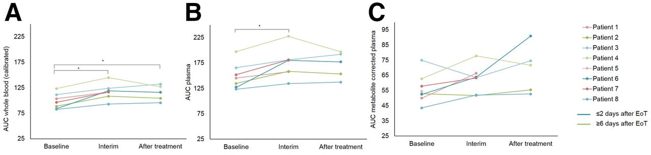
- FIGURE 2.
Tracer uptake in blood pool of each patient at 3 different time points. Whole blood calibrated with venous samples (A), plasma (B), and metabolite-corrected plasma (C) input curves. All curves have been corrected for administered dose and weight. At time of progression, patients were scanned ≤2 d (blue curves) or ≥6 d (green curves) after EoT. * P < 0.05
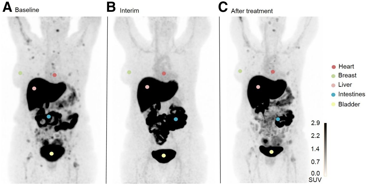
- FIGURE 3.
Visual assessment of 18F-FES uptake in various healthy tissues in 1 patient at baseline (A), interim (B), and after treatment (C). This patient underwent interim scanning 10 d after EoT. Uterus, an ER-expressing organ, is not visible in these images as it is located behind bladder. Images are maximal-intensity projections.
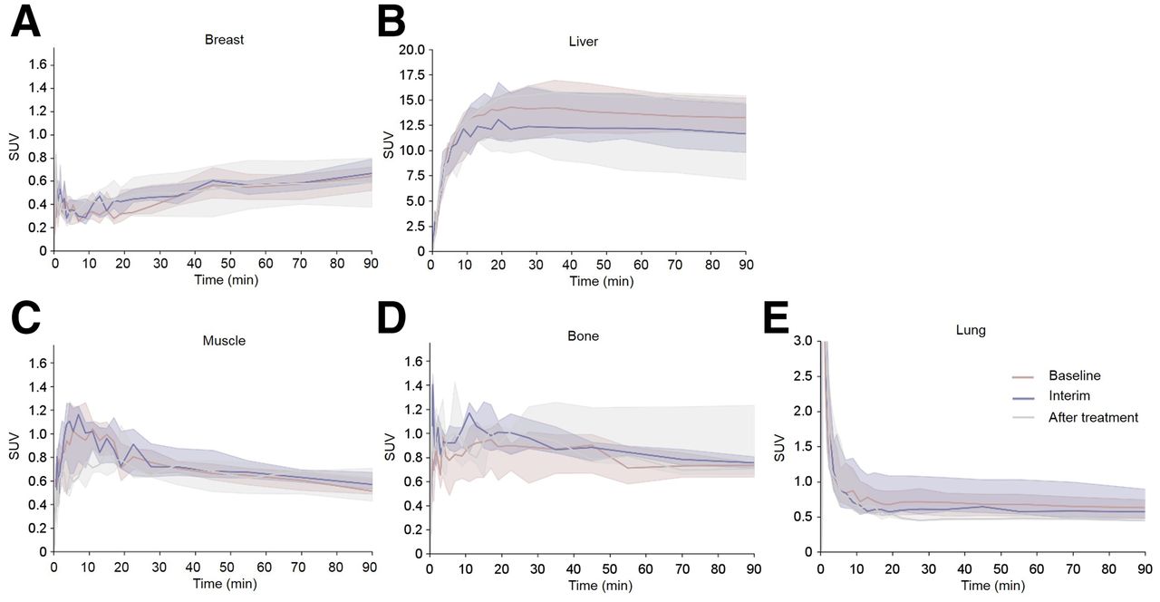
- FIGURE 4.
SUV time–activity curves (derived from dynamic scans) for various healthy tissue regions ([A] breast, [B] liver, [C] muscle, [D] bone. and [E] lung) of all patients at 3 different time points. Curves represent median of all values, with their corresponding IQRs.


Additional Files
Supplemental Data
Files in this Data Supplement:
In this issue


Jump to section
Related Articles
Cited By...
- No citing articles found.