
Main menu

  • Home
  • Content
    • Current
    • Ahead of print
    • Past Issues
    • JNM Supplement
    • SNMMI Annual Meeting Abstracts
    • Continuing Education
    • JNM Podcasts
  • Subscriptions
    • Subscribers
    • Institutional and Non-member
    • Rates
    • Journal Claims
    • Corporate & Special Sales
  • Authors
    • Submit to JNM
    • Information for Authors
    • Assignment of Copyright
    • AQARA requirements
  • Info
    • Reviewers
    • Permissions
    • Advertisers
  • About
    • About Us
    • Editorial Board
    • Contact Information
  • More
    • Alerts
    • Feedback
    • Help
    • SNMMI Journals
  • SNMMI
    • JNM
    • JNMT
    • SNMMI Journals
    • SNMMI

User menu

  • Subscribe
  • My alerts
  • Log in
  • My Cart

Search

  • Advanced search
Journal of Nuclear Medicine
  • SNMMI
    • JNM
    • JNMT
    • SNMMI Journals
    • SNMMI
  • Subscribe
  • My alerts
  • Log in
  • My Cart
Journal of Nuclear Medicine

Advanced Search

  • Home
  • Content
    • Current
    • Ahead of print
    • Past Issues
    • JNM Supplement
    • SNMMI Annual Meeting Abstracts
    • Continuing Education
    • JNM Podcasts
  • Subscriptions
    • Subscribers
    • Institutional and Non-member
    • Rates
    • Journal Claims
    • Corporate & Special Sales
  • Authors
    • Submit to JNM
    • Information for Authors
    • Assignment of Copyright
    • AQARA requirements
  • Info
    • Reviewers
    • Permissions
    • Advertisers
  • About
    • About Us
    • Editorial Board
    • Contact Information
  • More
    • Alerts
    • Feedback
    • Help
    • SNMMI Journals
  • View or Listen to JNM Podcast
  • Visit JNM on Facebook
  • Join JNM on LinkedIn
  • Follow JNM on Twitter
  • Subscribe to our RSS feeds
Research ArticleBasic Science Investigation

Dynamic Tumor-Specific MHC-II Immuno-PET Predicts the Efficacy of Checkpoint Inhibitor Immunotherapy in Melanoma

Zhen Yang, Feng Li, Yuqian Huang, Na Yin, Junjun Chu, Ying Ma, Roderic I. Pettigrew, Dale J. Hamilton, Diego R. Martin and Zheng Li
Journal of Nuclear Medicine November 2022, 63 (11) 1708-1714; DOI: https://doi.org/10.2967/jnumed.121.263151
Zhen Yang
1Department of Radiology, Houston Methodist Academic Institute, Houston, Texas;
2Center for Bioenergetics, Houston Methodist Academic Institute, Houston, Texas;
  • Find this author on Google Scholar
  • Find this author on PubMed
  • Search for this author on this site
Feng Li
1Department of Radiology, Houston Methodist Academic Institute, Houston, Texas;
2Center for Bioenergetics, Houston Methodist Academic Institute, Houston, Texas;
  • Find this author on Google Scholar
  • Find this author on PubMed
  • Search for this author on this site
Yuqian Huang
2Center for Bioenergetics, Houston Methodist Academic Institute, Houston, Texas;
3Xiangya Hospital, Central South University, Hunan, China;
  • Find this author on Google Scholar
  • Find this author on PubMed
  • Search for this author on this site
Na Yin
4Department of Pediatrics, Baylor College of Medicine, Houston, Texas;
  • Find this author on Google Scholar
  • Find this author on PubMed
  • Search for this author on this site
Junjun Chu
5Department of Medicine, Kech School of Medicine, University of Southern California, Los Angeles, California;
  • Find this author on Google Scholar
  • Find this author on PubMed
  • Search for this author on this site
Ying Ma
6NIBIB, NIH, Bethesda, Maryland;
  • Find this author on Google Scholar
  • Find this author on PubMed
  • Search for this author on this site
Roderic I. Pettigrew
7Engineering Medicine, Texas A&M University, College Station, Texas;
  • Find this author on Google Scholar
  • Find this author on PubMed
  • Search for this author on this site
Dale J. Hamilton
2Center for Bioenergetics, Houston Methodist Academic Institute, Houston, Texas;
8Department of Medicine, Weil Cornell Medicine, New York, New York; and
  • Find this author on Google Scholar
  • Find this author on PubMed
  • Search for this author on this site
Diego R. Martin
1Department of Radiology, Houston Methodist Academic Institute, Houston, Texas;
9Department of Radiology, Weil Cornell Medicine, New York, New York
  • Find this author on Google Scholar
  • Find this author on PubMed
  • Search for this author on this site
Zheng Li
1Department of Radiology, Houston Methodist Academic Institute, Houston, Texas;
2Center for Bioenergetics, Houston Methodist Academic Institute, Houston, Texas;
9Department of Radiology, Weil Cornell Medicine, New York, New York
  • Find this author on Google Scholar
  • Find this author on PubMed
  • Search for this author on this site
  • Article
  • Figures & Data
  • Supplemental
  • Info & Metrics
  • PDF
Loading

Article Figures & Data

Figures

  • Additional Files
  • Figure
    • Download figure
    • Open in new tab
    • Download powerpoint
  • FIGURE 1.
    • Download figure
    • Open in new tab
    • Download powerpoint
    FIGURE 1.

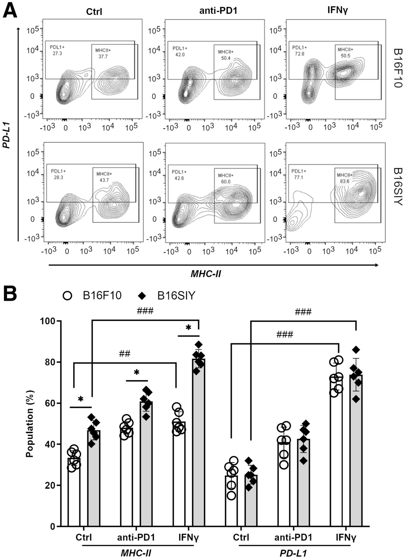
    (A) Fluorescence-activated cell sorting analysis of tsMHC-II and PD-L1 population in B16F10 and B16SIY tumors and population changes when treated with anti-PD1 and IFNγ immunity stimulants. (B) Summary of fluorescence-activated cell sorting assay (n = 6 for each cohort). Statistical analysis for MHC-II: B16F10 control vs. B16SIY control (P = 0.0382), B16F10 PD1 vs. B16SIY PD1 (P = 0.0158), B16F10 IFNγ vs. B16SIY IFNγ (P = 0.0112), B16F10 control vs. B16F10 IFNγ (P = 0.0026), and B16SIY control vs. B16SIY IFNγ (P = 0.0001). Statistical analysis for PD-L1: B16F10 control vs. B16F10 IFNγ (P = 0.0001) and B16SIY control vs. B16SIY IFNγ (P = 0.0001). *P < 0.05. ##P < 0.01. ###P < 0.001. Ctrl = control.

  • FIGURE 2.
    • Download figure
    • Open in new tab
    • Download powerpoint
    FIGURE 2.

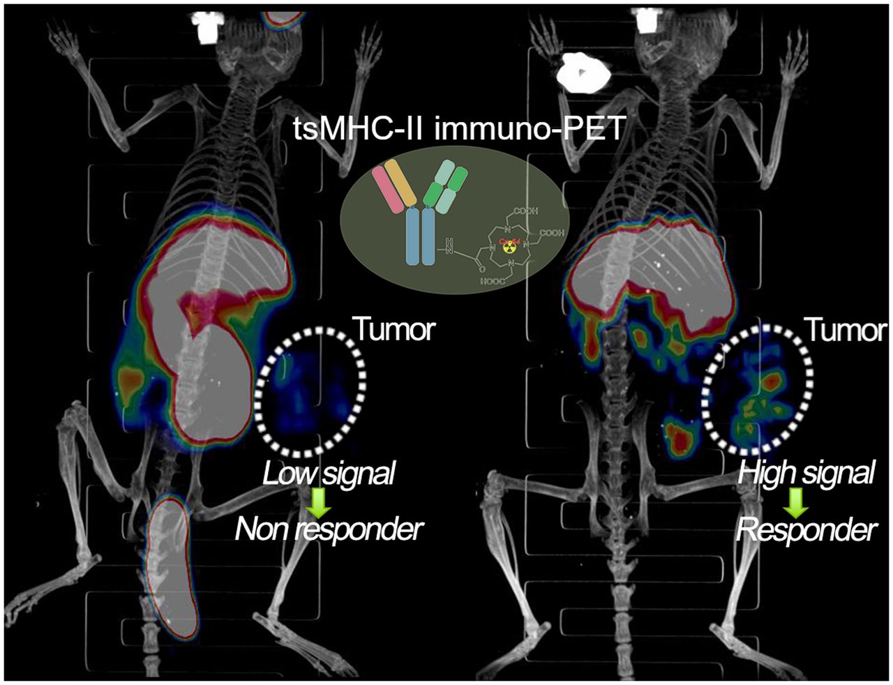
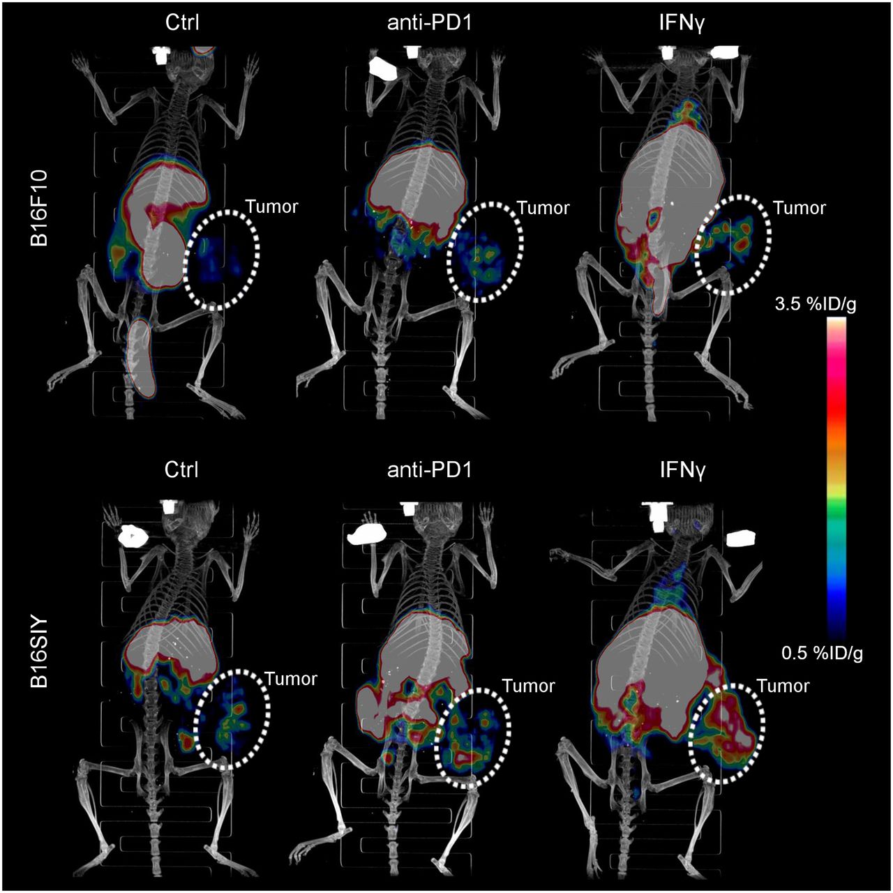
    Three-dimensionally rendered maximum-intensity projection of PET/CT images of control, anti-PD1–treated, and IFNγ-stimulated B16F10 and B16SIY tumor-bearing mice 48 h after injection of 64Cu-DOTA-MHCII. Circled areas are tumor site. Ctrl = control.

  • FIGURE 3.
    • Download figure
    • Open in new tab
    • Download powerpoint
    FIGURE 3.

    (A) Region-of-interest quantification of tumor-accumulated 64Cu-DOTA-MHCII in PET images (n = 6 per group). Unpaired Student t test was performed to compare B16F10 control vs. B16SIY control (P = 0.0082), B16F10 PD1 vs. B16SIY PD1 (P = 0.0035), B16F10 IFNγ vs. B16SIY IFNγ (P = 0.0002), B16F10 control vs. B16F10 IFNγ (P = 0.034), and B16SIY control vs. B16SIY IFNγ (P = 0.0003). (B) Tumor uptake of 64Cu-DOTA-MHCII in biodistribution study at 48 h after injection. Unpaired Student t test was performed to compare B16F10 control vs. B16SIY control (P = 0.0002), B16F10 PD1 vs. B16SIY PD1 (P = 0.0003), B16F10 IFNγ vs. B16SIY IFNγ (P = 0.0002), B16F10 control vs. B16F10 IFNγ (P = 0.0044), and B16SIY control vs. B16SIY IFNγ (P = 0.0001). (C) Biodistribution study of 64Cu-DOTA-MHCII in B16F10 tumor model. (D) Biodistribution study of 64Cu-DOTA-MHCII in B16SIY tumor model (n = 6). *P < 0.05. **P < 0.01. ***P < 0.001. #P < 0.05. ##P < 0.01. ###P < 0.001. GI = gastrointestinal.

  • FIGURE 4.
    • Download figure
    • Open in new tab
    • Download powerpoint
    FIGURE 4.

    Western blot of tsMHC-II expression (A) and dot density quantified by ImageJ (B) in control, anti-PD1–treated, and IFNγ-treated B16F10 and B16SIY tumors. Expression level was normalized by GAPDH across various groups. (C) Immunohistochemistry staining of MHC-II in tumors. Ctrl = control.

  • FIGURE 5.
    • Download figure
    • Open in new tab
    • Download powerpoint
    FIGURE 5.

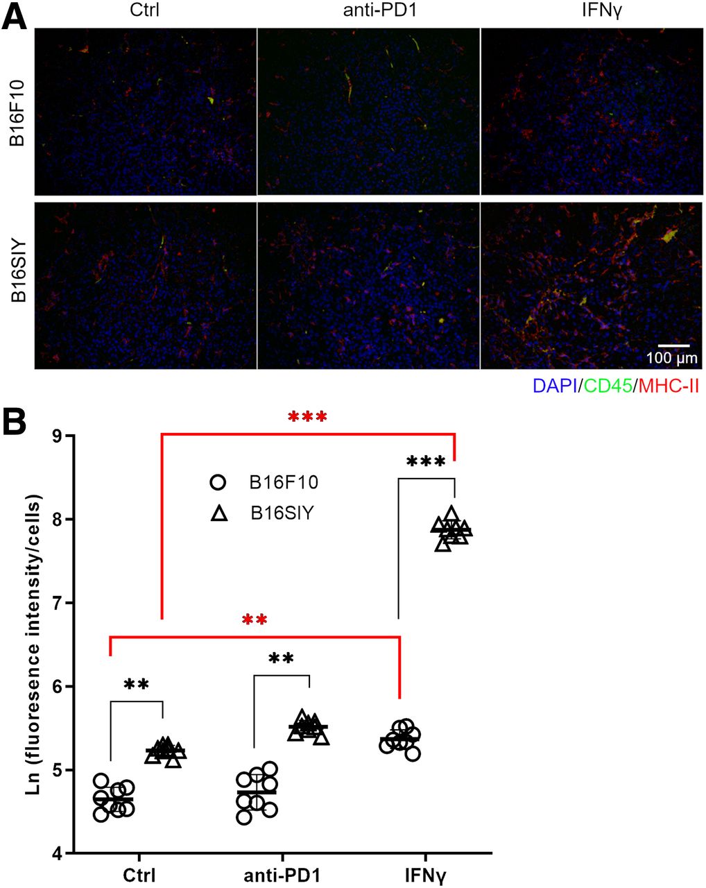
    (A) Immunofluorescent staining of CD45 and MHC-II in control, anti-PD1–treated, and IFNγ-treated B16F10 and B16SIY tumors. (B) Fluorescence intensity quantified by ImageJ. Fluorescence intensity was normalized by nucleus numbers that represent cell numbers, and ratio was plotted as natural logarithm to compare differences across groups. Eight different views were analyzed per sample to obtain statistics. Unpaired Student t test was performed to compare B16F10 control vs. B16SIY control (P = 0.0046), B16F10 PD1 vs. B16SIY PD1 (P = 0.0031), and B16F10 IFNγ vs. B16SIY IFNγ (P < 0.0001). Meanwhile, dynamic change was also compared: B16F10 control vs. B16F10 IFNγ (P = 0.0024) and B16SIY control vs. B16SIY IFNγ (P < 0.0001). **P < 0.01. ***P < 0.001. Ctrl = control.

Additional Files

  • Figures
  • Supplemental Data

    Files in this Data Supplement:

    • Supplemental Data
PreviousNext
Back to top

In this issue

Journal of Nuclear Medicine: 63 (11)
Journal of Nuclear Medicine
Vol. 63, Issue 11
November 1, 2022
  • Table of Contents
  • Table of Contents (PDF)
  • About the Cover
  • Index by author
  • Complete Issue (PDF)
Print
Download PDF
Article Alerts
Sign In to Email Alerts with your Email Address
Email Article

Thank you for your interest in spreading the word on Journal of Nuclear Medicine.

NOTE: We only request your email address so that the person you are recommending the page to knows that you wanted them to see it, and that it is not junk mail. We do not capture any email address.

Enter multiple addresses on separate lines or separate them with commas.
Dynamic Tumor-Specific MHC-II Immuno-PET Predicts the Efficacy of Checkpoint Inhibitor Immunotherapy in Melanoma
(Your Name) has sent you a message from Journal of Nuclear Medicine
(Your Name) thought you would like to see the Journal of Nuclear Medicine web site.
Citation Tools
Dynamic Tumor-Specific MHC-II Immuno-PET Predicts the Efficacy of Checkpoint Inhibitor Immunotherapy in Melanoma
Zhen Yang, Feng Li, Yuqian Huang, Na Yin, Junjun Chu, Ying Ma, Roderic I. Pettigrew, Dale J. Hamilton, Diego R. Martin, Zheng Li
Journal of Nuclear Medicine Nov 2022, 63 (11) 1708-1714; DOI: 10.2967/jnumed.121.263151

Citation Manager Formats

  • BibTeX
  • Bookends
  • EasyBib
  • EndNote (tagged)
  • EndNote 8 (xml)
  • Medlars
  • Mendeley
  • Papers
  • RefWorks Tagged
  • Ref Manager
  • RIS
  • Zotero
Share
Dynamic Tumor-Specific MHC-II Immuno-PET Predicts the Efficacy of Checkpoint Inhibitor Immunotherapy in Melanoma
Zhen Yang, Feng Li, Yuqian Huang, Na Yin, Junjun Chu, Ying Ma, Roderic I. Pettigrew, Dale J. Hamilton, Diego R. Martin, Zheng Li
Journal of Nuclear Medicine Nov 2022, 63 (11) 1708-1714; DOI: 10.2967/jnumed.121.263151
Twitter logo Facebook logo LinkedIn logo Mendeley logo
  • Tweet Widget
  • Facebook Like
  • Google Plus One
Bookmark this article

Jump to section

  • Article
    • Visual Abstract
    • Abstract
    • MATERIALS AND METHODS
    • RESULTS
    • DISCUSSION
    • CONCLUSION
    • DISCLOSURE
    • ACKNOWLEDGMENTS
    • Footnotes
    • REFERENCES
  • Figures & Data
  • Supplemental
  • Info & Metrics
  • PDF

Related Articles

  • PubMed
  • Google Scholar

Cited By...

  • No citing articles found.
  • Google Scholar

More in this TOC Section

  • [11C]ZTP-1: An Effective Short-Lived Radioligand for PET of Rat and Monkey Brain Phosphodiesterase Type 4 Subtype B
  • Reduced Renal Uptake of Various Radiopharmaceuticals with Sodium Paraaminohippurate Coadministration in a Rat Model
  • In Vivo Visualization and Quantification of Brain Heat Shock Protein 90 with [11C]HSP990 in Healthy Aging and Neurodegeneration
Show more Basic Science Investigation

Similar Articles

Keywords

  • immuno-PET
  • MHC-II
  • anti-PD1 immunotherapy
  • melanoma
  • Molecular imaging
SNMMI

© 2025 SNMMI

Powered by HighWire