
Main menu

  • Home
  • Content
    • Current
    • Ahead of print
    • Past Issues
    • JNM Supplement
    • SNMMI Annual Meeting Abstracts
    • Continuing Education
    • JNM Podcasts
  • Subscriptions
    • Subscribers
    • Institutional and Non-member
    • Rates
    • Journal Claims
    • Corporate & Special Sales
  • Authors
    • Submit to JNM
    • Information for Authors
    • Assignment of Copyright
    • AQARA requirements
  • Info
    • Reviewers
    • Permissions
    • Advertisers
  • About
    • About Us
    • Editorial Board
    • Contact Information
  • More
    • Alerts
    • Feedback
    • Help
    • SNMMI Journals
  • SNMMI
    • JNM
    • JNMT
    • SNMMI Journals
    • SNMMI

User menu

  • Subscribe
  • My alerts
  • Log in
  • My Cart

Search

  • Advanced search
Journal of Nuclear Medicine
  • SNMMI
    • JNM
    • JNMT
    • SNMMI Journals
    • SNMMI
  • Subscribe
  • My alerts
  • Log in
  • My Cart
Journal of Nuclear Medicine

Advanced Search

  • Home
  • Content
    • Current
    • Ahead of print
    • Past Issues
    • JNM Supplement
    • SNMMI Annual Meeting Abstracts
    • Continuing Education
    • JNM Podcasts
  • Subscriptions
    • Subscribers
    • Institutional and Non-member
    • Rates
    • Journal Claims
    • Corporate & Special Sales
  • Authors
    • Submit to JNM
    • Information for Authors
    • Assignment of Copyright
    • AQARA requirements
  • Info
    • Reviewers
    • Permissions
    • Advertisers
  • About
    • About Us
    • Editorial Board
    • Contact Information
  • More
    • Alerts
    • Feedback
    • Help
    • SNMMI Journals
  • View or Listen to JNM Podcast
  • Visit JNM on Facebook
  • Join JNM on LinkedIn
  • Follow JNM on Twitter
  • Subscribe to our RSS feeds
Meeting ReportOral - PhysicianPharm

Clinical Evaluation of Data-Driven Motion Corrected Reconstruction for PET Brain Imaging

Matthew Spangler-Bickell, Samuel Hurley, Ali Pirasteh, Scott Perlman, Timothy Deller and Alan McMillan
Journal of Nuclear Medicine May 2021, 62 (supplement 1) 59;
Matthew Spangler-Bickell
1Radiology University of Wisconsin - Madison Madison WI United States
  • Find this author on Google Scholar
  • Find this author on PubMed
  • Search for this author on this site
Samuel Hurley
1Radiology University of Wisconsin - Madison Madison WI United States
  • Find this author on Google Scholar
  • Find this author on PubMed
  • Search for this author on this site
Ali Pirasteh
1Radiology University of Wisconsin - Madison Madison WI United States
  • Find this author on Google Scholar
  • Find this author on PubMed
  • Search for this author on this site
Scott Perlman
1Radiology University of Wisconsin - Madison Madison WI United States
  • Find this author on Google Scholar
  • Find this author on PubMed
  • Search for this author on this site
Timothy Deller
2PET/MR Engineering GE Healthcare Waukesha WI United States
  • Find this author on Google Scholar
  • Find this author on PubMed
  • Search for this author on this site
Alan McMillan
1Radiology University of Wisconsin - Madison Madison WI United States
  • Find this author on Google Scholar
  • Find this author on PubMed
  • Search for this author on this site
  • Article
  • Figures & Data
  • Info & Metrics
Loading

Abstract

59

Introduction: Head motion during a brain PET scan can cause motion blurring and loss of image quality. In this work we present a completely data-driven motion estimation approach with full event-by-event motion corrected list-mode (LM) reconstruction. A reader study and atlas-based quantitative analysis comparing the motion corrected (MoCo) and uncorrected (nMoCo) reconstructions were performed on 50 clinical FDG brain scans.

Methods: Ultra-fast LM reconstructions of very short frames (0.6 - 1.8 sec) were performed. Head motion was estimated using image registration [1]. Event-by-event motion corrected LM reconstruction was then performed on the entire acquisition. Normalization, deadtime, attenuation, scatter, and randoms corrections were performed during reconstruction. A cohort of 50 retrospective clinical metabolic FDG brain scans was obtained from 3 scanners: SIGNA PET/MR (n=21), DMI PET/CT (n=11), and D710 PET/CT (n=18) (GE Healthcare, Chicago, IL). Experienced readers evaluated the MoCo and nMoCo images in a blinded read using a Likert scale (1-5) for image sharpness and diagnostic quality. Atlas-based quantitation analysis was performed on the SUVmax values of 9 regions-of-interest (ROIs) [2]. Paired Wilcoxon tests were used to assess differences in reader scores and ROI-wise SUVmax. A Levene test was used to assess if the variance of the relative difference of MoCo and nMoCo was different between motion groups. Statistical tests were corrected for multiple comparisons.

Results: The range of estimated motion in the cohort, as quantified by a point within the brain displaced by the motion, is shown in Figure 1. Figure 1 also shows how the cohort was divided into 4 motion groups. Figure 2 and 3 show examples of MoCo applied to 2 data sets. Figure 4 shows the results of the reader study. Figure 5 shows the results of the ROI analysis.

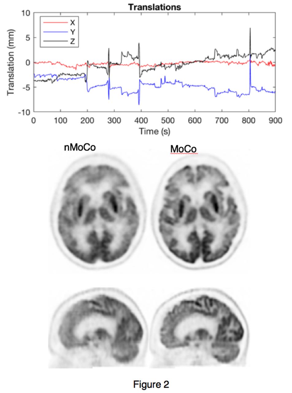
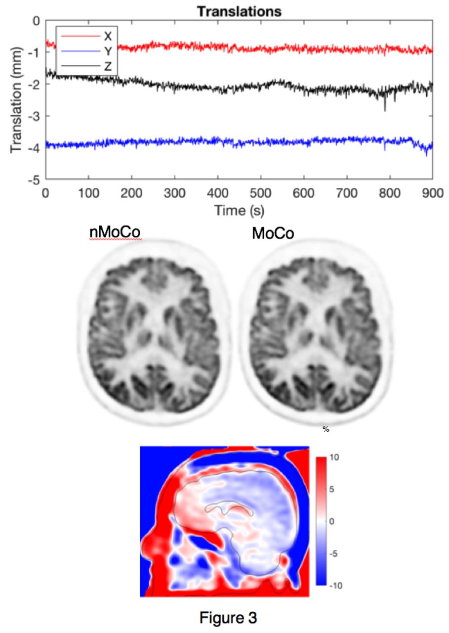
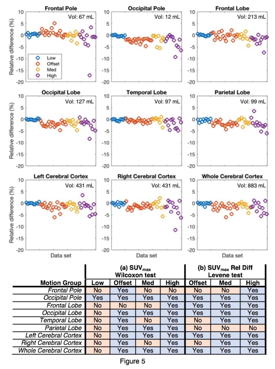
Conclusions: MoCo improved both quantitative and qualitive assessment of the images. Using an atlas-based approach we demonstrated that, when motion occurs, MoCo makes a significant difference in the quantitation of the reconstruction. The reader study demonstrated that MoCo improved diagnostic quality in 10% of the data sets, with 8% changing from diagnostically “unacceptable” to “acceptable” with MoCo. When there is little motion, MoCo reconstructions are not significantly different to the nMoCo reconstructions, as expected. The developed approach was completely data-driven, yielding motion estimates with ~1 Hz frame rate utilizing an event-by-event LM reconstruction, and is compatible with routine imaging workflows suggesting that motion-robust PET is clinically attainable. Fig. 1: (Left) Box plots of estimated motion. Shaded regions indicate motion groups: “Offset” includes low motion during the scan with an offset between PET and attenuation map of >2 mm. (Right) Initial offset between PET and attenuation map. Fig. 2: Example data set in the “High” motion group. (Top) Translations of the motion (rotations not shown). (Bottom) The nMoCo and MoCo images. The nMoCo image is visibly blurred. Fig. 3: Example data set in the “Offset” motion group. (Top) Translations of the estimated motion. (Bottom) The nMoCo and MoCo images, and a sagittal difference image showing a relative gradient. Fig. 4: Difference of the averaged reader scores between the MoCo and nMoCo images, grouped according to motion group for (Top) image sharpness and (Middle) diagnostic quality. (Bottom) Significant differences in sharpness and quality for MoCo and nMoCo reconstructions were found for the “High” motion group and the pooled cohort data (“All”). Fig. 5: (Top) Relative differences between the SUVmax values of the MoCo and nMoCo images in 9 ROIs. (Bottom) Significant differences in SUVmax were present in many ROIs between the MoCo and nMoCo images. Similarly, the variance of the relative difference between MoCo and nMoCo was significantly different for many ROIs in the “Offset”, “Med”, and “High” motion groups compared to the “Low” motion group.

Figure
  • Download figure
  • Open in new tab
  • Download powerpoint
Figure
  • Download figure
  • Open in new tab
  • Download powerpoint
Figure
  • Download figure
  • Open in new tab
  • Download powerpoint
Figure
  • Download figure
  • Open in new tab
  • Download powerpoint
Figure
  • Download figure
  • Open in new tab
  • Download powerpoint
Previous
Back to top

In this issue

Journal of Nuclear Medicine
Vol. 62, Issue supplement 1
May 1, 2021
  • Table of Contents
  • Index by author
Article Alerts
Sign In to Email Alerts with your Email Address
Email Article

Thank you for your interest in spreading the word on Journal of Nuclear Medicine.

NOTE: We only request your email address so that the person you are recommending the page to knows that you wanted them to see it, and that it is not junk mail. We do not capture any email address.

Enter multiple addresses on separate lines or separate them with commas.
Clinical Evaluation of Data-Driven Motion Corrected Reconstruction for PET Brain Imaging
(Your Name) has sent you a message from Journal of Nuclear Medicine
(Your Name) thought you would like to see the Journal of Nuclear Medicine web site.
Citation Tools
Clinical Evaluation of Data-Driven Motion Corrected Reconstruction for PET Brain Imaging
Matthew Spangler-Bickell, Samuel Hurley, Ali Pirasteh, Scott Perlman, Timothy Deller, Alan McMillan
Journal of Nuclear Medicine May 2021, 62 (supplement 1) 59;

Citation Manager Formats

  • BibTeX
  • Bookends
  • EasyBib
  • EndNote (tagged)
  • EndNote 8 (xml)
  • Medlars
  • Mendeley
  • Papers
  • RefWorks Tagged
  • Ref Manager
  • RIS
  • Zotero
Share
Clinical Evaluation of Data-Driven Motion Corrected Reconstruction for PET Brain Imaging
Matthew Spangler-Bickell, Samuel Hurley, Ali Pirasteh, Scott Perlman, Timothy Deller, Alan McMillan
Journal of Nuclear Medicine May 2021, 62 (supplement 1) 59;
Twitter logo Facebook logo LinkedIn logo Mendeley logo
  • Tweet Widget
  • Facebook Like
  • Google Plus One
Bookmark this article

Jump to section

  • Article
  • Figures & Data
  • Info & Metrics

Related Articles

  • No related articles found.
  • Google Scholar

Cited By...

  • Evaluation of Data-Driven Rigid Motion Correction in Clinical Brain PET Imaging
  • Google Scholar

More in this TOC Section

Oral - PhysicianPharm

  • Dynamic 68Ga-DOTA PET/CT and Compartmental Modelling to Non-invasively Estimate the Glomerular Filtration Rate
  • Myocardial glucose suppression interferes with the detection of inflammatory cells with FDG-PET in a canine model of myocardial infarction
  • Global cardiac atherosclerotic burden assessed by fast automated artificial intelligence-based heart segmentation in 18F-sodium fluoride PET/CT scans: head-to-head comparison with manual segmentation
Show more Oral - PhysicianPharm

Advances in Imaging Technologies

  • Human Research Studies on the PennPET Explorer
  • Total-Body Parametric Reconstruction with Deep Learning-based Data-driven Motion Compensation
  • A mouse brain PET prototype with a staggered 3-layer DOI detector
Show more Advances in Imaging Technologies

Similar Articles

SNMMI

© 2025 SNMMI

Powered by HighWire