
Main menu

  • Home
  • Content
    • Current
    • Ahead of print
    • Past Issues
    • JNM Supplement
    • SNMMI Annual Meeting Abstracts
    • Continuing Education
    • JNM Podcasts
  • Subscriptions
    • Subscribers
    • Institutional and Non-member
    • Rates
    • Journal Claims
    • Corporate & Special Sales
  • Authors
    • Submit to JNM
    • Information for Authors
    • Assignment of Copyright
    • AQARA requirements
  • Info
    • Reviewers
    • Permissions
    • Advertisers
  • About
    • About Us
    • Editorial Board
    • Contact Information
  • More
    • Alerts
    • Feedback
    • Help
    • SNMMI Journals
  • SNMMI
    • JNM
    • JNMT
    • SNMMI Journals
    • SNMMI

User menu

  • Subscribe
  • My alerts
  • Log in
  • My Cart

Search

  • Advanced search
Journal of Nuclear Medicine
  • SNMMI
    • JNM
    • JNMT
    • SNMMI Journals
    • SNMMI
  • Subscribe
  • My alerts
  • Log in
  • My Cart
Journal of Nuclear Medicine

Advanced Search

  • Home
  • Content
    • Current
    • Ahead of print
    • Past Issues
    • JNM Supplement
    • SNMMI Annual Meeting Abstracts
    • Continuing Education
    • JNM Podcasts
  • Subscriptions
    • Subscribers
    • Institutional and Non-member
    • Rates
    • Journal Claims
    • Corporate & Special Sales
  • Authors
    • Submit to JNM
    • Information for Authors
    • Assignment of Copyright
    • AQARA requirements
  • Info
    • Reviewers
    • Permissions
    • Advertisers
  • About
    • About Us
    • Editorial Board
    • Contact Information
  • More
    • Alerts
    • Feedback
    • Help
    • SNMMI Journals
  • View or Listen to JNM Podcast
  • Visit JNM on Facebook
  • Join JNM on LinkedIn
  • Follow JNM on Twitter
  • Subscribe to our RSS feeds
Meeting ReportOral - PhysicianPharm

Super-resolution in brain PET Using a Real Time Motion Capture System

Yanis Chemli, Marc-Andre Tetrault, Marc Normandin, Thibault Marin, Isabelle Bloch, Georges El Fakhri, Jinsong Ouyang and Yoann Petibon
Journal of Nuclear Medicine May 2021, 62 (supplement 1) 34;
Yanis Chemli
2LTCI, Telecom Paris, Institut Polytechnique de Paris Paris France
1Gordon Center for Medical Imaging Boston MA United States
  • Find this author on Google Scholar
  • Find this author on PubMed
  • Search for this author on this site
Marc-Andre Tetrault
3Computer Engineering Universite de Sherbrooke Sherbrooke QC Canada
  • Find this author on Google Scholar
  • Find this author on PubMed
  • Search for this author on this site
Marc Normandin
4Department of Radiology Massachusetts General Hospital, Harvard Medical Sc Boston MA United States
  • Find this author on Google Scholar
  • Find this author on PubMed
  • Search for this author on this site
Thibault Marin
1Gordon Center for Medical Imaging Boston MA United States
  • Find this author on Google Scholar
  • Find this author on PubMed
  • Search for this author on this site
Isabelle Bloch
5Sorbonne Universite, CNRS, LIP6 Paris France
2LTCI, Telecom Paris, Institut Polytechnique de Paris Paris France
  • Find this author on Google Scholar
  • Find this author on PubMed
  • Search for this author on this site
Georges El Fakhri
4Department of Radiology Massachusetts General Hospital, Harvard Medical Sc Boston MA United States
  • Find this author on Google Scholar
  • Find this author on PubMed
  • Search for this author on this site
Jinsong Ouyang
4Department of Radiology Massachusetts General Hospital, Harvard Medical Sc Boston MA United States
  • Find this author on Google Scholar
  • Find this author on PubMed
  • Search for this author on this site
Yoann Petibon
4Department of Radiology Massachusetts General Hospital, Harvard Medical Sc Boston MA United States
  • Find this author on Google Scholar
  • Find this author on PubMed
  • Search for this author on this site
  • Article
  • Figures & Data
  • Info & Metrics
Loading

Abstract

34

Objectives: Super-resolution (SR) is a powerful methodology seeking to improve image resolution by exploiting the increased spatial sampling information obtained from multiple acquisitions of the same object performed with accurately known sub-resolution shifts [1]. The goal of this work is to develop and evaluate a SR estimation framework for brain positron emission tomography (PET), taking advantage of a high-resolution infra-red tracking camera to measure object shifts precisely and continuously.

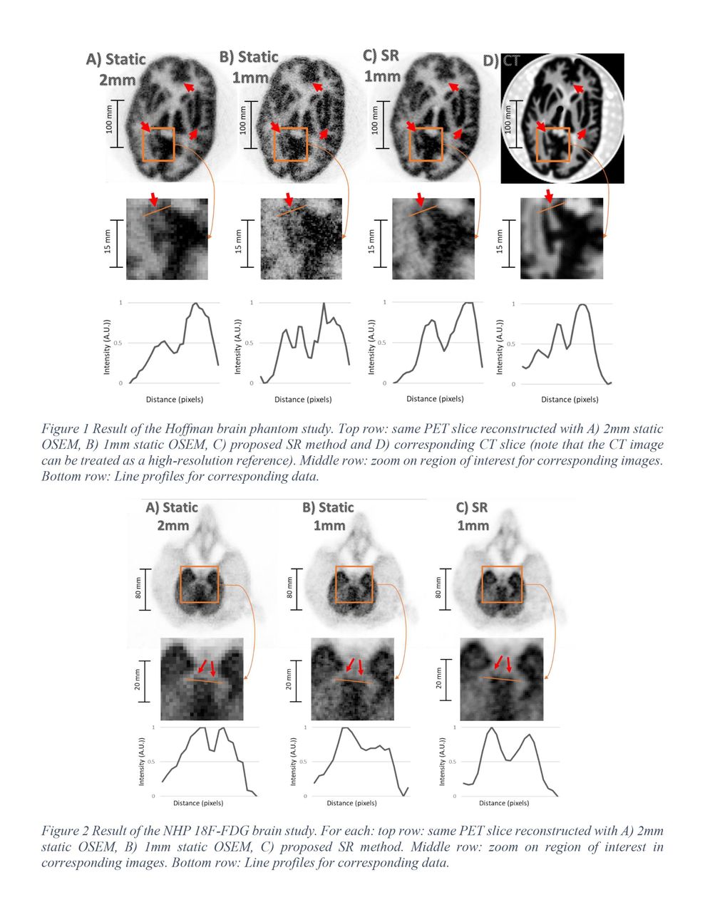
Methods: Moving phantom and non-human primate (NHP) experiments were performed on a PET/CT scanner (Discovery MI, GE Healthcare) using an external motion tracking device (Polaris Vega, NDI). The Polaris Vega, which yields a tracking accuracy up to 0.12 mm isotropic and a 60 Hz frame rate, was integrated with the scanner through a custom-built interface sending trigger events synchronized with the motion capture frames to the list mode. A transformation linking the Polaris and scanner coordinates space was determined by six paired measurements of high-resolution CT scans (0.7x0.7x0.6 mm) and Polaris tracking of the same reference object by a least squares minimization to spatially align the two instruments. To enable SR, a list-mode Ordered Subset Expectation Maximization (OSEM) PET reconstruction algorithm was developed, incorporating the high-resolution tracking data from the Polaris Vega to correct motion for measured line of responses (LORs) on an event-by-event basis. A Hoffman phantom, filled with 3mCi 18F, was scanned for 15 min in list mode while undergoing continuous rotation/translation movements introduced by a QUASAR system (Modus QA). Similarly, a rhesus monkey administered with 11 mCi 18F-FDG was scanned for 15-min in list mode with continuous head motion induced manually. For both scans, motion was tracked at all time using the Vega and reflective markers rigidly attached to the targets. Reference static PET acquisitions were also performed without inducing movement. PET images were reconstructed with 3 different methods: (A) OSEM algorithm applied to the static reference scan data (2 mm voxel size), (B) OSEM algorithm applied to the static scan on smaller voxel size (1 mm), (C) proposed SR algorithm applied to the moving scan with 1 mm voxel size. Attenuation and sensitivity effects were accounted for during reconstruction. Iteration numbers were chosen to obtain matched image noise levels for method (A) and (C). For method (B), we used the same number of iterations as for (C).

Results: For both phantom and NHP studies, the developed SR reconstruction method yielded PET images with visibly increased spatial resolution as compared to static acquisitions reconstructed using 1 or 2 mm voxels, allowing for improved visualization of small cortical and subcortical brain phantom structures (Fig. 1 and 2). Overall, the SR method achieved better noise control than the static reconstruction with the same voxel size, owing to interpolation effects introduced by the continuous application of motion transformations during image estimation. Line profiles confirmed improved spatial resolution for the SR images in both studies. For the Hoffman phantom, the SR images showed improved correspondence with the high-resolution CT compared to the conventional methods.

Conclusions: The results demonstrate that super resolution can be achieved in brain PET by measuring object movement in real time using a high-resolution infrared tracking camera. Acknowledgments: This work was supported in part by U01EB027003 and P41EB022544. References: [1] Kennedy et. al, IEEE Trans. Med. Imag.,25(2), pp. 137-147, 2006.

Figure
  • Download figure
  • Open in new tab
  • Download powerpoint
Previous
Back to top

In this issue

Journal of Nuclear Medicine
Vol. 62, Issue supplement 1
May 1, 2021
  • Table of Contents
  • Index by author
Article Alerts
Sign In to Email Alerts with your Email Address
Email Article

Thank you for your interest in spreading the word on Journal of Nuclear Medicine.

NOTE: We only request your email address so that the person you are recommending the page to knows that you wanted them to see it, and that it is not junk mail. We do not capture any email address.

Enter multiple addresses on separate lines or separate them with commas.
Super-resolution in brain PET Using a Real Time Motion Capture System
(Your Name) has sent you a message from Journal of Nuclear Medicine
(Your Name) thought you would like to see the Journal of Nuclear Medicine web site.
Citation Tools
Super-resolution in brain PET Using a Real Time Motion Capture System
Yanis Chemli, Marc-Andre Tetrault, Marc Normandin, Thibault Marin, Isabelle Bloch, Georges El Fakhri, Jinsong Ouyang, Yoann Petibon
Journal of Nuclear Medicine May 2021, 62 (supplement 1) 34;

Citation Manager Formats

  • BibTeX
  • Bookends
  • EasyBib
  • EndNote (tagged)
  • EndNote 8 (xml)
  • Medlars
  • Mendeley
  • Papers
  • RefWorks Tagged
  • Ref Manager
  • RIS
  • Zotero
Share
Super-resolution in brain PET Using a Real Time Motion Capture System
Yanis Chemli, Marc-Andre Tetrault, Marc Normandin, Thibault Marin, Isabelle Bloch, Georges El Fakhri, Jinsong Ouyang, Yoann Petibon
Journal of Nuclear Medicine May 2021, 62 (supplement 1) 34;
Twitter logo Facebook logo LinkedIn logo Mendeley logo
  • Tweet Widget
  • Facebook Like
  • Google Plus One
Bookmark this article

Jump to section

  • Article
  • Figures & Data
  • Info & Metrics

Related Articles

  • No related articles found.
  • Google Scholar

Cited By...

  • No citing articles found.
  • Google Scholar

More in this TOC Section

Oral - PhysicianPharm

  • Optimal [18]F-Misonidazole PET threshold to locate SCC7 tumor hypoxia using EPR pO2 as ground truth
  • The value of 18F-FDG PET in the diagnostic workup of pediatric fever of unknown origin: a systematic review and meta-analysis
  • Prediction of absorbed dose by tumors and critical organs after Lu-177-DOTATATE therapy using pretherapeutic Ga-68-DOTATOC PET/CT
Show more Oral - PhysicianPharm

Physics, Instrumentation and Data Science Young Investigator Award Session

  • Design study of a high-resolution and ultrahigh-sensitivity brain SPECT system for imaging medically intractable epilepsy
  • Pre-selecting radiomic features based on their robustness to changes in imaging properties of multicentre data: impact on predictive modelling performance compared to ComBat harmonization of all available features
  • Data-driven motion compensation using cGAN for total-body [18F]FDG-PET imaging
Show more Physics, Instrumentation and Data Science Young Investigator Award Session

Similar Articles

SNMMI

© 2025 SNMMI

Powered by HighWire