
Main menu

  • Home
  • Content
    • Current
    • Ahead of print
    • Past Issues
    • JNM Supplement
    • SNMMI Annual Meeting Abstracts
  • Subscriptions
    • Subscribers
    • Institutional and Non-member
    • Rates
    • Corporate & Special Sales
    • Journal Claims
  • Authors
    • Submit to JNM
    • Information for Authors
    • Assignment of Copyright
    • AQARA requirements
  • Info
    • Permissions
    • Advertisers
    • Continuing Education
  • About
    • About Us
    • Editorial Board
    • Contact Information
  • More
    • Alerts
    • Feedback
    • Help
    • SNMMI Journals
  • SNMMI
    • JNM
    • JNMT
    • SNMMI Journals
    • SNMMI

User menu

  • Subscribe
  • My alerts
  • Log in
  • My Cart

Search

  • Advanced search
Journal of Nuclear Medicine
  • SNMMI
    • JNM
    • JNMT
    • SNMMI Journals
    • SNMMI
  • Subscribe
  • My alerts
  • Log in
  • My Cart
Journal of Nuclear Medicine

Advanced Search

  • Home
  • Content
    • Current
    • Ahead of print
    • Past Issues
    • JNM Supplement
    • SNMMI Annual Meeting Abstracts
  • Subscriptions
    • Subscribers
    • Institutional and Non-member
    • Rates
    • Corporate & Special Sales
    • Journal Claims
  • Authors
    • Submit to JNM
    • Information for Authors
    • Assignment of Copyright
    • AQARA requirements
  • Info
    • Permissions
    • Advertisers
    • Continuing Education
  • About
    • About Us
    • Editorial Board
    • Contact Information
  • More
    • Alerts
    • Feedback
    • Help
    • SNMMI Journals
  • Follow JNM on Twitter
  • Visit JNM on Facebook
  • Join JNM on LinkedIn
  • Subscribe to our RSS feeds
Meeting ReportPoster - PhysicianPharm

An original voxel-wise supervised analysis of tumors with multimodal radiomics to highlight predictive biological patterns

Thibault Escobar, Sebastien Vauclin, Fanny Orlhac, Christophe Nioche, Pascal Pineau and Irene Buvat
Journal of Nuclear Medicine May 2021, 62 (supplement 1) 1404;
Thibault Escobar
2Laboratoire d’Imagerie Translationnelle en Oncologie Universite Paris Saclay/Inserm/Institut Curie Orsay France
1DOSIsoft SA Cachan France
  • Find this author on Google Scholar
  • Find this author on PubMed
  • Search for this author on this site
Sebastien Vauclin
1DOSIsoft SA Cachan France
  • Find this author on Google Scholar
  • Find this author on PubMed
  • Search for this author on this site
Fanny Orlhac
2Laboratoire d’Imagerie Translationnelle en Oncologie Universite Paris Saclay/Inserm/Institut Curie Orsay France
  • Find this author on Google Scholar
  • Find this author on PubMed
  • Search for this author on this site
Christophe Nioche
2Laboratoire d’Imagerie Translationnelle en Oncologie Universite Paris Saclay/Inserm/Institut Curie Orsay France
  • Find this author on Google Scholar
  • Find this author on PubMed
  • Search for this author on this site
Pascal Pineau
1DOSIsoft SA Cachan France
  • Find this author on Google Scholar
  • Find this author on PubMed
  • Search for this author on this site
Irene Buvat
2Laboratoire d’Imagerie Translationnelle en Oncologie Universite Paris Saclay/Inserm/Institut Curie Orsay France
  • Find this author on Google Scholar
  • Find this author on PubMed
  • Search for this author on this site
  • Article
  • Figures & Data
  • Info & Metrics
Loading

Abstract

1404

Objectives: Translational applications of predictive and prognostic image-based learning models are challenging due to their lack of interpretability. When using deep learning, Class Activation Maps (CAM) give information about the regions driving the models. Yet, due to the high-level abstraction of deep features, deep CAM are difficult to interpret. We propose a method that combines the interpretability of handcrafted radiomics with a voxel-wise analysis and facilitates the biological interpretation of models.

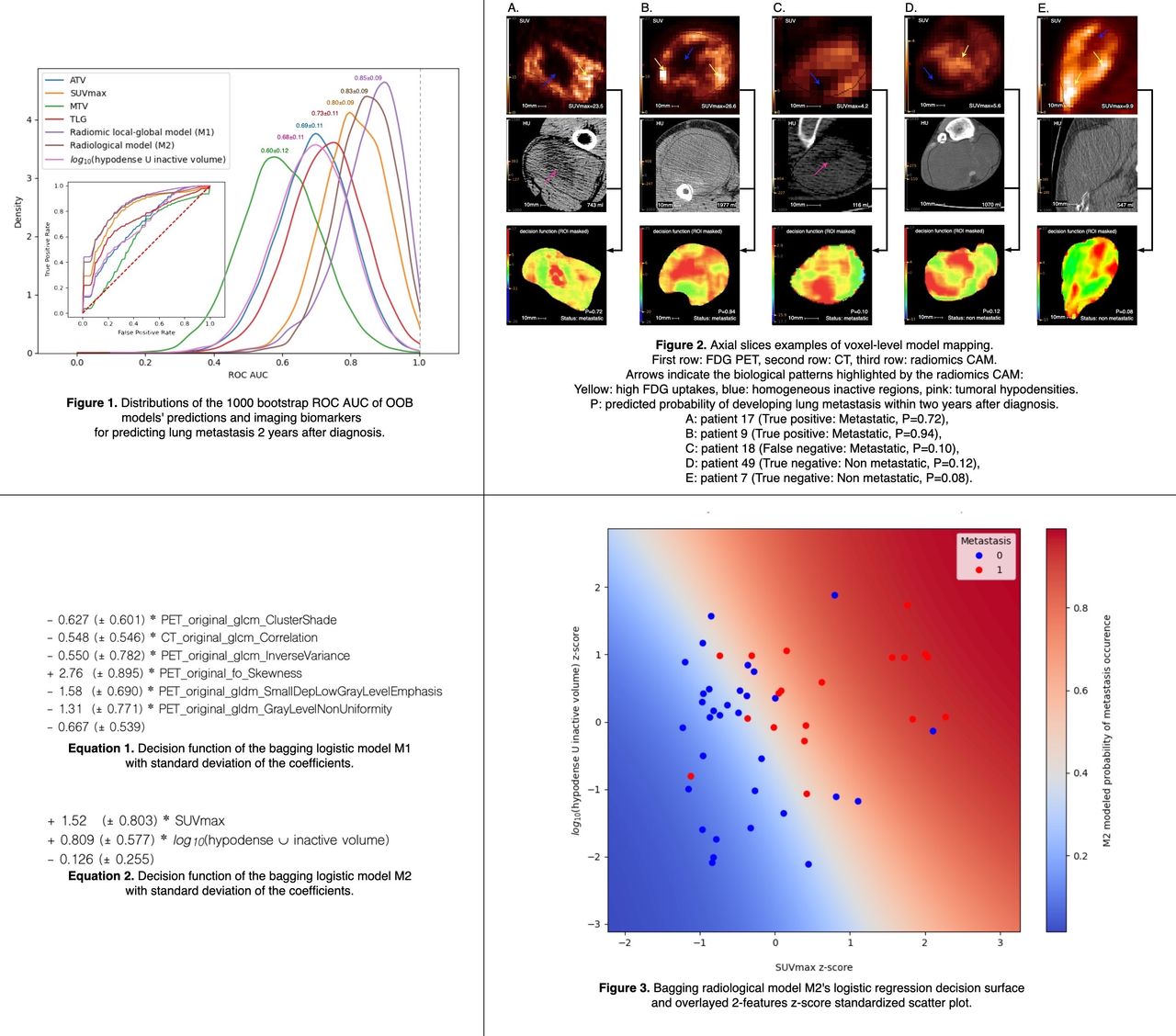
Methods: A public dataset of 51 soft tissue sarcoma patients including FDG PET and CT scans, and clinical and follow-up information was used [1]. The task was to predict the occurrence of lung metastases within two years after the diagnosis (19 patients with metastases and 32 without). PET and CT images were pre-processed according to IBSI guidelines [2]. A total of 154 radiomic features per voxel were computed with Pyradiomics [3] by sliding a 93-voxels kernel to mimic the mean local receptive field of a weakly-supervised U-net convolutional neural network (CNN) with 2 max-pooling layers [4]. An “ROI-Global-Average-Pooling” was performed within each tumor region to yield one global 154-features vector per patient. To address the multicollinearity problem in supervised machine learning, global features were then selected by iteratively removing the one with the highest variance inflation factor (VIF) [5] until the maximum VIF in the feature set was <10. Regularized logistic regression with wrapped forward feature selection and weighted loss was used to model the probability of lung metastasis occurrence. Hyper-parameters tuning was performed using 200*5 folds repeated cross-validation. The performance of the model was characterized based on out of bag (OOB) samples using 1000 bootstrap drawings and was compared to those obtained with anatomical tumor volume (ATV), SUVmax, metabolic tumor volume (MTV) and total lesion glycolysis (TLG). For interpretability, bagging of all 1000 models was used to obtain the final radiomic model M1. To avoid data leakage, the data from each patient were analyzed using a sub-bagging model obtained from the aggregation of all models in which the patient data were not used for training. Radiomics CAM were computed by backpropagating the global decision function of the model M1 at the voxel level. Based on the interpretation of the radiomics CAM, we created simple and radiologically interpretable new features. We finally built a “radiological” model M2 following the same machine learning process but using only these new features combined with ATV, SUVmax, MTV and TLG.

Results: Six features were selected through the cross-validation procedure to design the radiomic model M1. The mean OOB ROC AUC of model M1 was 0.85±0.09, to be compared to 0.80±0.09 for SUVmax, 0.73±0.11 for TLG, 0.69±0.11 for ATV, and 0.60±0.12 for MTV. The radiomics CAM highlighted 3 biological patterns associated with the risk of metastasis: substantial and homogeneous non-metabolic tumor region, localized high FDG uptake sub-regions, and some hypodense sub-regions on CT. These findings are concordant with the soft tissue sarcoma grading system based on the biopsy [6]. The radiomics CAM associated with M1 made it possible to define a new feature reflecting the volume of the tumor sub-region devoid of metabolic activity (SUV<40%SUVmax) or with reduced Hounsfield units (<20HU). A 2-features model M2 combining that feature with SUVmax yielded a mean OOB ROC AUC of 0.83±0.09.

Conclusions: We describe a method based on locally-calculated handcrafted radiomic features that highlights the sub-regions and biological signal driving the model prediction. In a situation where the number of data is limited, we demonstrate how that method makes it possible to spatially and quantitatively interpret radiomic models and design simple and robust biomarkers amenable to a biological interpretation for patient stratification.

Figure
  • Download figure
  • Open in new tab
  • Download powerpoint
Previous
Back to top

In this issue

Journal of Nuclear Medicine
Vol. 62, Issue supplement 1
May 1, 2021
  • Table of Contents
  • Index by author
Article Alerts
Sign In to Email Alerts with your Email Address
Email Article

Thank you for your interest in spreading the word on Journal of Nuclear Medicine.

NOTE: We only request your email address so that the person you are recommending the page to knows that you wanted them to see it, and that it is not junk mail. We do not capture any email address.

Enter multiple addresses on separate lines or separate them with commas.
An original voxel-wise supervised analysis of tumors with multimodal radiomics to highlight predictive biological patterns
(Your Name) has sent you a message from Journal of Nuclear Medicine
(Your Name) thought you would like to see the Journal of Nuclear Medicine web site.
Citation Tools
An original voxel-wise supervised analysis of tumors with multimodal radiomics to highlight predictive biological patterns
Thibault Escobar, Sebastien Vauclin, Fanny Orlhac, Christophe Nioche, Pascal Pineau, Irene Buvat
Journal of Nuclear Medicine May 2021, 62 (supplement 1) 1404;

Citation Manager Formats

  • BibTeX
  • Bookends
  • EasyBib
  • EndNote (tagged)
  • EndNote 8 (xml)
  • Medlars
  • Mendeley
  • Papers
  • RefWorks Tagged
  • Ref Manager
  • RIS
  • Zotero
Share
An original voxel-wise supervised analysis of tumors with multimodal radiomics to highlight predictive biological patterns
Thibault Escobar, Sebastien Vauclin, Fanny Orlhac, Christophe Nioche, Pascal Pineau, Irene Buvat
Journal of Nuclear Medicine May 2021, 62 (supplement 1) 1404;
Twitter logo Facebook logo LinkedIn logo Mendeley logo
  • Tweet Widget
  • Facebook Like
  • Google Plus One
Bookmark this article

Jump to section

  • Article
  • Figures & Data
  • Info & Metrics

Related Articles

  • No related articles found.
  • Google Scholar

Cited By...

  • No citing articles found.
  • Google Scholar

More in this TOC Section

Poster - PhysicianPharm

  • Preliminary result of Texture Analysis on prediction of overall outcome of neuroendocrine tumors based on pre-therapy heterogeneity of somatostatin receptors on 68Ga Dotatate PET/CT scans.
  • 177Lu-PSMA-617 versus Cabazitaxel in Metastatic Castration-Resistant Prostate Cancer: a randomised, open-label, phase 2 trial (TheraP)
  • Artificial Intelligence based segmental quantification of pulmonary perfusion for pre-transplant workup.
Show more Poster - PhysicianPharm

PIDS - Data Analysis and Management

  • An empirical update of left ventricular 3D segmentation algorithm in myocardial perfusion SPECT imaging
  • No-gold-standard evaluation of partial volume compensation methods for brain PET
Show more PIDS - Data Analysis and Management

Similar Articles

SNMMI

© 2023 Journal of Nuclear Medicine

Powered by HighWire