
Main menu

  • Home
  • Content
    • Current
    • Ahead of print
    • Past Issues
    • JNM Supplement
    • SNMMI Annual Meeting Abstracts
    • Continuing Education
    • JNM Podcasts
  • Subscriptions
    • Subscribers
    • Institutional and Non-member
    • Rates
    • Journal Claims
    • Corporate & Special Sales
  • Authors
    • Submit to JNM
    • Information for Authors
    • Assignment of Copyright
    • AQARA requirements
  • Info
    • Reviewers
    • Permissions
    • Advertisers
  • About
    • About Us
    • Editorial Board
    • Contact Information
  • More
    • Alerts
    • Feedback
    • Help
    • SNMMI Journals
  • SNMMI
    • JNM
    • JNMT
    • SNMMI Journals
    • SNMMI

User menu

  • Subscribe
  • My alerts
  • Log in
  • Log out
  • My Cart

Search

  • Advanced search
Journal of Nuclear Medicine
  • SNMMI
    • JNM
    • JNMT
    • SNMMI Journals
    • SNMMI
  • Subscribe
  • My alerts
  • Log in
  • Log out
  • My Cart
Journal of Nuclear Medicine

Advanced Search

  • Home
  • Content
    • Current
    • Ahead of print
    • Past Issues
    • JNM Supplement
    • SNMMI Annual Meeting Abstracts
    • Continuing Education
    • JNM Podcasts
  • Subscriptions
    • Subscribers
    • Institutional and Non-member
    • Rates
    • Journal Claims
    • Corporate & Special Sales
  • Authors
    • Submit to JNM
    • Information for Authors
    • Assignment of Copyright
    • AQARA requirements
  • Info
    • Reviewers
    • Permissions
    • Advertisers
  • About
    • About Us
    • Editorial Board
    • Contact Information
  • More
    • Alerts
    • Feedback
    • Help
    • SNMMI Journals
  • View or Listen to JNM Podcast
  • Visit JNM on Facebook
  • Join JNM on LinkedIn
  • Follow JNM on Twitter
  • Subscribe to our RSS feeds
Meeting ReportOral - PhysicianPharm

Multi-Parametric Total-body Dynamic PET/CT imaging in Differential Diagnosis of Benign and Malignant Lymph Nodes

Fangfang Fu, Nan Meng, Yaping Wu, Tao Feng, Tianyi Xu, Zhun Huang, Yan Bai, Xiaochen Li and Meiyun Wang
Journal of Nuclear Medicine May 2021, 62 (supplement 1) 128;
Fangfang Fu
1Henan Provincial People’s Hospital & the People’s Hospital of Zhengzhou University Zhengzhou China
  • Find this author on Google Scholar
  • Find this author on PubMed
  • Search for this author on this site
Nan Meng
2Henan Provincial People’s Hospital & the People’s Hospital of Zhengzhou University, Zhengzhou China
  • Find this author on Google Scholar
  • Find this author on PubMed
  • Search for this author on this site
Yaping Wu
3HenanProvincialPeople’sHospital&thePeople’sHospitalofZhengzhouUniversity, Zhengzhou China
  • Find this author on Google Scholar
  • Find this author on PubMed
  • Search for this author on this site
Tao Feng
4United Imaging Healthcare Houston TX United States
  • Find this author on Google Scholar
  • Find this author on PubMed
  • Search for this author on this site
Tianyi Xu
5CT & MI BU, UIH Group, Shanghai, China Shanghai China
  • Find this author on Google Scholar
  • Find this author on PubMed
  • Search for this author on this site
Zhun Huang
1Henan Provincial People’s Hospital & the People’s Hospital of Zhengzhou University Zhengzhou China
  • Find this author on Google Scholar
  • Find this author on PubMed
  • Search for this author on this site
Yan Bai
6Henan Provincial People’s Hospital & the People’s Hospital of Zhengzhou University Henan China
  • Find this author on Google Scholar
  • Find this author on PubMed
  • Search for this author on this site
Xiaochen Li
1Henan Provincial People’s Hospital & the People’s Hospital of Zhengzhou University Zhengzhou China
  • Find this author on Google Scholar
  • Find this author on PubMed
  • Search for this author on this site
Meiyun Wang
1Henan Provincial People’s Hospital & the People’s Hospital of Zhengzhou University Zhengzhou China
  • Find this author on Google Scholar
  • Find this author on PubMed
  • Search for this author on this site
  • Article
  • Figures & Data
  • Info & Metrics
Loading

Abstract

128

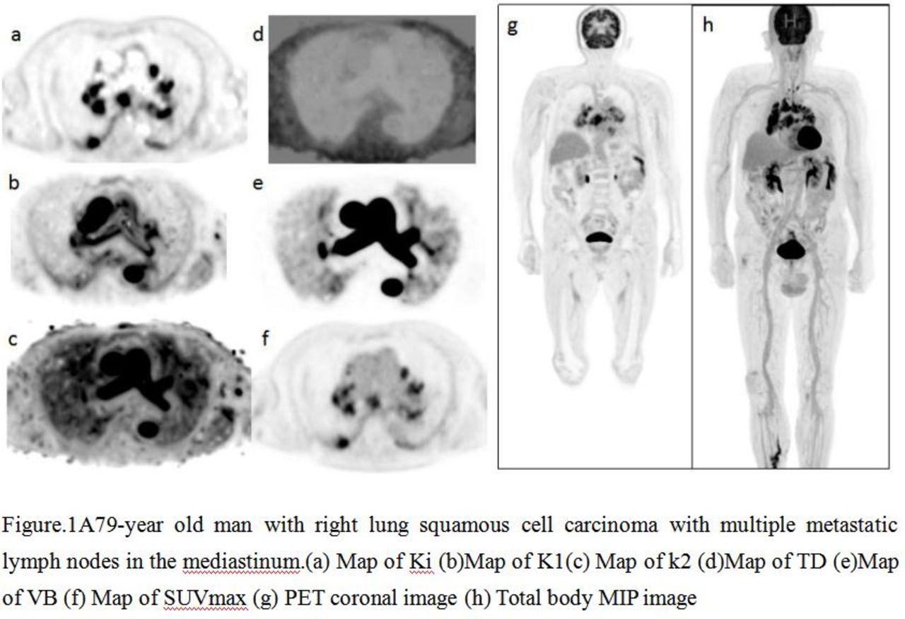
Introduction: The world’s first 2-meter-long total-body PET scanner (uEXPLORER) provides a versatile platform for biomedical research and opens a new venue for lesion detection throughout the body. Our study aims to explore the value of multi-parametric total-body dynamic PET/CT imaging in distinguishing benign and malignant lymph nodes of the whole body. Materials and Methods: A total number of 40 patients with lymph node lesions were recruited in this study, including 25 males and 15 females, with a median age of 55.13 (range 31-79), a median height of 1.68m (range 1.55-1.85m), and a median weight of 69.61kg (range43-110kg). We conducted total body kinetic analysis on each patient using the uEXPLORER. The patients were told to fast for at least 6 hours prior to the study. The leg was chosen as the injection site due to easier access. A low dose CT with a slice thickness of 5mm and slice increment of 5mm was applied to provide the attenuation information for the PET image reconstruction. A dynamic PET scan of 60 minutes was acquired subsequently. The FDG is given on a dosage of 3.7MBq/kg (0.1mCi/kg) and is to be injected right after the PET scan starts. An image-derived input function is acquired from the dynamic images using a region-of-interest placed on the descending aorta. A set of parametric images were produced using two methods. The net flux rate Ki images were generated by the uKinetics software (United Imaging Healthcare) using the linear Patlak model. The K1, k2, plasma fraction (VB), and the time delay (TD) were estimated jointly via a non-linear 1-tissue compartment model using early phase data (5 minutes post-injection). SUVmax was measured from PET image.The Mann-Whitney U test and receiver operating characteristic (ROC) analysis were performed for all dynamic PET/CT parameters to compare their diagnosis performance in distinguishing benign and malignant lymph nodes.

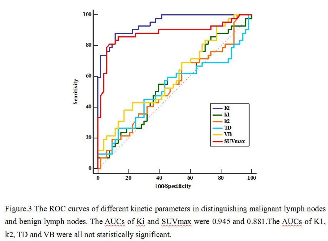
Results: Ninety-five lymph nodes (including 42 benign lymph nodes in 16 cases and 53 malignant lymph nodes in 24 cases) confirmed by pathological diagnosis or clinical follow-up were included in the study.Both Ki and SUVmax values of benign lymph nodes were lower than those of malignant lymph nodes (0.66×10⁻² vs 2.64×10⁻² ml/g/min and 2.43 vs 5.46), and there was a statistically significant difference between them (all P < 0.001). But there was no statistically significant difference between K1values of benign lymph nodes and malignant lymph nodes (3.83×10⁻³ vs 3.99×10⁻³ml/g/s), between k2 values of benign lymph nodes and malignant lymph nodes (1.54×10⁻² vs 1.7×10⁻²s-1), between TD values of benign lymph nodes and malignant lymph nodes (0.482vs 0.128 s), between VB values of benign lymph nodes and malignant lymph nodes (4.66×10⁻² vs 8.43×10⁻²) (all P > 0.05). The area under the ROC curve (AUC) of Ki (AUC=0.945) was significantly higher than that of SUVmax (AUC=0.881) in discriminating malignant and benign lymph nodes (P < 0.05).

Conclusions: Among all different kinetic parameters and traditional SUVmax, Ki was shown to provide the highest diagnostic efficiency and was an promising indicator in the differential diagnosis of benign and malignant lymph nodes.

Figure
  • Download figure
  • Open in new tab
  • Download powerpoint
Figure
  • Download figure
  • Open in new tab
  • Download powerpoint
Figure
  • Download figure
  • Open in new tab
  • Download powerpoint
Previous
Back to top

In this issue

Journal of Nuclear Medicine
Vol. 62, Issue supplement 1
May 1, 2021
  • Table of Contents
  • Index by author
Article Alerts
Sign In to Email Alerts with your Email Address
Email Article

Thank you for your interest in spreading the word on Journal of Nuclear Medicine.

NOTE: We only request your email address so that the person you are recommending the page to knows that you wanted them to see it, and that it is not junk mail. We do not capture any email address.

Enter multiple addresses on separate lines or separate them with commas.
Multi-Parametric Total-body Dynamic PET/CT imaging in Differential Diagnosis of Benign and Malignant Lymph Nodes
(Your Name) has sent you a message from Journal of Nuclear Medicine
(Your Name) thought you would like to see the Journal of Nuclear Medicine web site.
Citation Tools
Multi-Parametric Total-body Dynamic PET/CT imaging in Differential Diagnosis of Benign and Malignant Lymph Nodes
Fangfang Fu, Nan Meng, Yaping Wu, Tao Feng, Tianyi Xu, Zhun Huang, Yan Bai, Xiaochen Li, Meiyun Wang
Journal of Nuclear Medicine May 2021, 62 (supplement 1) 128;

Citation Manager Formats

  • BibTeX
  • Bookends
  • EasyBib
  • EndNote (tagged)
  • EndNote 8 (xml)
  • Medlars
  • Mendeley
  • Papers
  • RefWorks Tagged
  • Ref Manager
  • RIS
  • Zotero
Share
Multi-Parametric Total-body Dynamic PET/CT imaging in Differential Diagnosis of Benign and Malignant Lymph Nodes
Fangfang Fu, Nan Meng, Yaping Wu, Tao Feng, Tianyi Xu, Zhun Huang, Yan Bai, Xiaochen Li, Meiyun Wang
Journal of Nuclear Medicine May 2021, 62 (supplement 1) 128;
Twitter logo Facebook logo LinkedIn logo Mendeley logo
  • Tweet Widget
  • Facebook Like
  • Google Plus One
Bookmark this article

Jump to section

  • Article
  • Figures & Data
  • Info & Metrics

Related Articles

  • No related articles found.
  • Google Scholar

Cited By...

  • No citing articles found.
  • Google Scholar

More in this TOC Section

Oral - PhysicianPharm

  • Safety and efficacy of radioligand therapy with 177lutetium-PSMA-617 within 3 months after 223Radium-dichloride
  • Usefulness of99mTc SESTAMIBI Scintigraphy in Persistent Hyperparathyroidism after Kidney Transplant
  • Toll-like receptor 5 in triple-negative breast cancer: a novel reporter for tumor progression
Show more Oral - PhysicianPharm

Advances in PET Imaging for Cancer: Highlights of Clinical Diagnostics in Oncology

  • A Phase 3 study of 18F-DCFPyL-PET/CT in Patients with Biochemically Recurrent Prostate Cancer (CONDOR): An Analysis of Disease Detection Rate and Positive Predictive Value (PPV) by Anatomic Region
  • Ga-68-FAPI for sarcoma imaging: Data from the FAPI-PET prospective observational trial
  • Response monitoring in metastatic breast cancer: a comparison of survival times between FDG-PET/CT and CE-CT
Show more Advances in PET Imaging for Cancer: Highlights of Clinical Diagnostics in Oncology

Similar Articles

SNMMI

© 2025 SNMMI

Powered by HighWire