
Main menu

  • Home
  • Content
    • Current
    • Ahead of print
    • Past Issues
    • JNM Supplement
    • SNMMI Annual Meeting Abstracts
    • Continuing Education
    • JNM Podcasts
  • Subscriptions
    • Subscribers
    • Institutional and Non-member
    • Rates
    • Journal Claims
    • Corporate & Special Sales
  • Authors
    • Submit to JNM
    • Information for Authors
    • Assignment of Copyright
    • AQARA requirements
  • Info
    • Reviewers
    • Permissions
    • Advertisers
  • About
    • About Us
    • Editorial Board
    • Contact Information
  • More
    • Alerts
    • Feedback
    • Help
    • SNMMI Journals
  • SNMMI
    • JNM
    • JNMT
    • SNMMI Journals
    • SNMMI

User menu

  • Subscribe
  • My alerts
  • Log in
  • My Cart

Search

  • Advanced search
Journal of Nuclear Medicine
  • SNMMI
    • JNM
    • JNMT
    • SNMMI Journals
    • SNMMI
  • Subscribe
  • My alerts
  • Log in
  • My Cart
Journal of Nuclear Medicine

Advanced Search

  • Home
  • Content
    • Current
    • Ahead of print
    • Past Issues
    • JNM Supplement
    • SNMMI Annual Meeting Abstracts
    • Continuing Education
    • JNM Podcasts
  • Subscriptions
    • Subscribers
    • Institutional and Non-member
    • Rates
    • Journal Claims
    • Corporate & Special Sales
  • Authors
    • Submit to JNM
    • Information for Authors
    • Assignment of Copyright
    • AQARA requirements
  • Info
    • Reviewers
    • Permissions
    • Advertisers
  • About
    • About Us
    • Editorial Board
    • Contact Information
  • More
    • Alerts
    • Feedback
    • Help
    • SNMMI Journals
  • View or Listen to JNM Podcast
  • Visit JNM on Facebook
  • Join JNM on LinkedIn
  • Follow JNM on Twitter
  • Subscribe to our RSS feeds
Research ArticleChallenging Case Studies

Distinguishing Primary Lateral Sclerosis from Parkinsonian Syndromes with the Help of Advanced Imaging

Pegah Masrori, Donatienne Van Weehaeghe, Koen Van Laere and Philip Van Damme
Journal of Nuclear Medicine September 2021, 62 (9) 1318-1319; DOI: https://doi.org/10.2967/jnumed.121.261942
Pegah Masrori
1Neuromuscular Reference Centre, Department of Neurology, University Hospitals Leuven, Leuven, Belgium;
2Department of Neurosciences, Experimental Neurology, and Leuven Brain Institute, KU Leuven, University of Leuven, Leuven, Belgium;
3Laboratory of Neurobiology, Center for Brain and Disease Research, VIB, Leuven, Belgium;
  • Find this author on Google Scholar
  • Find this author on PubMed
  • Search for this author on this site
Donatienne Van Weehaeghe
4Division of Nuclear Medicine, University Hospitals Leuven, Leuven, Belgium; and
5Nuclear Medicine and Molecular Imaging, Department of Imaging and Pathology, University Hospitals Leuven and KU Leuven, Leuven, Belgium
  • Find this author on Google Scholar
  • Find this author on PubMed
  • Search for this author on this site
Koen Van Laere
4Division of Nuclear Medicine, University Hospitals Leuven, Leuven, Belgium; and
5Nuclear Medicine and Molecular Imaging, Department of Imaging and Pathology, University Hospitals Leuven and KU Leuven, Leuven, Belgium
  • Find this author on Google Scholar
  • Find this author on PubMed
  • Search for this author on this site
Philip Van Damme
1Neuromuscular Reference Centre, Department of Neurology, University Hospitals Leuven, Leuven, Belgium;
2Department of Neurosciences, Experimental Neurology, and Leuven Brain Institute, KU Leuven, University of Leuven, Leuven, Belgium;
3Laboratory of Neurobiology, Center for Brain and Disease Research, VIB, Leuven, Belgium;
  • Find this author on Google Scholar
  • Find this author on PubMed
  • Search for this author on this site
  • Article
  • Figures & Data
  • Info & Metrics
  • PDF
Loading

Abstract

We describe a unique case of a patient presenting with unilateral mild paresis, slowing of the upper limb, and parkinsonism, who underwent a full imaging work-up including MRI, 123I-FP-CIT PET, 18F-FE-PE2I PET, and 18F-FDG PET. This case demonstrates that imaging may aid substantially in the diagnostic work-up of complex neurologic disorders.

  • neurology
  • PET/CT
  • PET/MRI
  • motor neuron disease
  • neurodegenerative disorders
  • functional imaging

PART 1

Case Report

We report the case of a 60-y-old man who initially presented after 6 mo of subacute and slowly progressive symptoms including muscle stiffness, decreased fine motor skills, slowing of movements, and mild muscle weakness in his right hand. The patient was right-handed and experienced writing difficulties—without cramps or dystonia—that affected legibility. He had no complaints of postural symptoms associated with orthostatic hypotension or other autonomic failure. No sleep disturbance, periodic limb movement during sleep, or restless legs syndrome was mentioned.

The patient had a history of a myocardial infarction, diabetes mellitus, and hypercholesterolemia; the latter two were relatively well controlled with medication. His family history was negative for neurodegenerative diseases.

The neurologic examination showed slowness in whole-hand grasping and on tapping tasks, as well as mild paresis of the dorsal interosseous muscles of the right hand. Furthermore, we found an asymmetric mild hypertonia in the lower right limb. A unilateral positive Hoffman sign (right) was noted, but we found no Babinski sign and tendon reflexes were symmetrically brisk. When the patient walked, we observed a reduced arm swing on the right side without postural imbalance or gait disorder. There was no evidence of myoclonus. All sensory modalities were normal. Eye movements, both saccades and vertical gaze, were normal. Furthermore, detailed language testing and a behavioral assessment showed no abnormalities. Obtaining a diagnosis remained challenging, as it was difficult to distinguish between parkinsonism and a motor neuron disorder.

Hematologic and serum biochemistry tests had normal findings. The electromyography results were strictly normal.

MRI of the brain demonstrated mild white matter lesions normal for his age, without any hyperintensity in the corticospinal tracts. The cervical MR images showed no evidence of myelopathy or radiculopathy. 18F-FDG brain PET showed asymmetric mild hypometabolism in the left sensorimotor cortex compatible with the right arm symptoms, in favor of a motor neuron disease. To exclude neurodegenerative parkinsonism 123I-N-ω-fluoropropyl-2β-carbomethoxy-3β-(4-iodophenyl)nortropane (123I-FP-CIT) SPECT was performed 9 mo later and showed normal presynaptic dopaminergic transmission (Fig. 1).

FIGURE 1.
  • Download figure
  • Open in new tab
  • Download powerpoint
FIGURE 1.

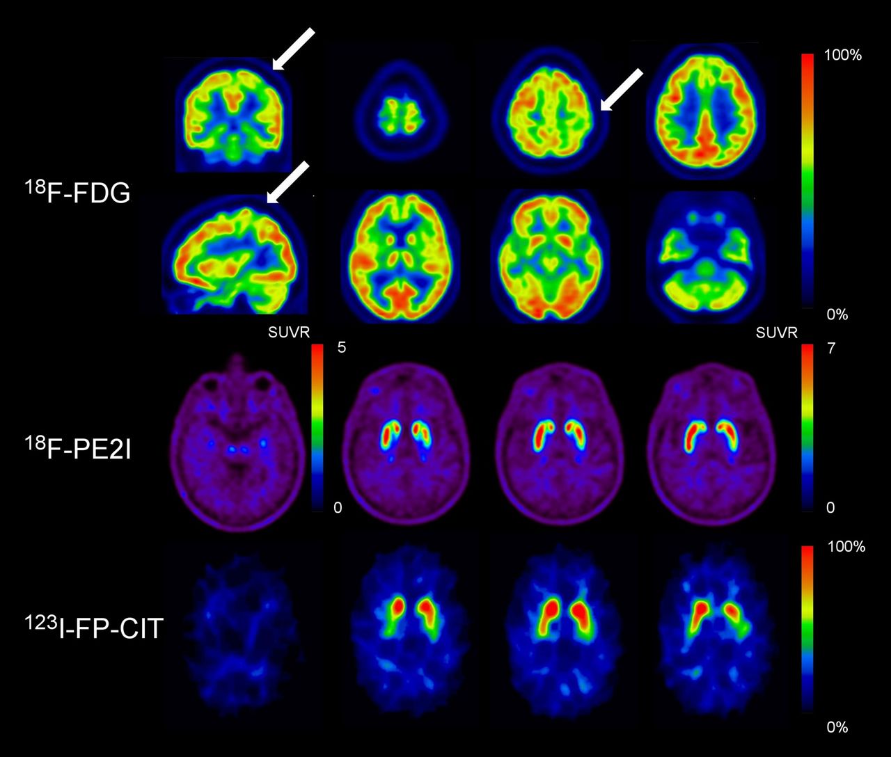
(Top) Orthogonal 18F-FDG PET slices showing mild hypometabolism (arrows) in left sensorimotor cortex corresponding to right arm location of cortical homunculus. (Middle and bottom) Transverse slices of dopamine transporter imaging. Both classic 123I-FP-CIT SPECT and high-resolution, selective 18F-PE2I PET showed normal presynaptic dopaminergic uptake. Images are in radiologic orientation. SUVR = SUV ratio.

Notably, there was no response to dopaminergic treatment with levodopa and benserazide.

Discussion

The differential diagnosis was broad. After excluding structural, metabolic, inflammatory, and infectious disorders, we were leaning toward diagnosis of a motor neuron disorder. Finally, we found no mutations in the SOD1, TARDBP, FUS, or C9orf72 genes. Whole-exome sequencing was done but showed no mutations related to neurodegenerative disorders.

Besides a slow progression of isolated paresis of the right hand, the patient’s condition remained unchanged during a 1-y follow-up period. Repetition of the electromyography showed no signs of lower motor neuron degeneration. We requested a more sensitive 18F-(E)-N-(3-iodoprop-2-enyl)-2β-carbofluoroethoxy-3β-(4′-methyl-phenyl) nortropane (18F-FE-PE2I) PET scan to exclude subtle presynaptic dopaminergic transmission deficits. This imaging showed no abnormalities in the striatum or in the substantia nigra.

PART 2

Final Diagnosis

Together, the clinical picture and ancillary tests were compatible with an asymmetric form of pure upper motor neuron disorder. The imaging allowed diagnosis of primary lateral sclerosis (PLS) by showing on the one hand a hypometabolism of the sensory-motor cortex contralateral to the clinical involvement and on the other hand a presynaptic integrity of the dopaminergic pathways.

Conclusion

PLS is a rare neurodegenerative disorder characterized by a slowly progressive upper motor neuron syndrome. Compared with classic variants of amyotrophic lateral sclerosis, PLS has a significantly slower rate of disease progression and longer survival. Diagnosis of PLS is challenging and is based on excluding structural disorders (e.g., cervical spondylotic myelopathy, Arnold–Chiari malformation, spinal arteriovenous fistula, and tumor), hereditary spastic paraparesis, leukodystrophies, metabolic and toxic disorders (e.g., vitamin E deficiency, cerebrotendinous xanthomatosis, and hexosaminidase deficiency), neurodegenerative disorders (e.g., Parkinson disease, multiple systems atrophy, and corticobasal syndrome), inflammatory disorders (e.g., primary progressive multiple sclerosis), and infections (e.g., neurosyphilis, tropical spastic paraparesis, neuroborreliosis, spinal sarcoidosis, and AIDS). Thalamic, hippocampal, and basal ganglia atrophy, as well as subcortical gray matter degeneration, have been demonstrated in imaging studies of PLS (1). Postmortem studies have reported atrophy of the thalamus and striatum, as well as TDP-43 inclusions in the striatum, amygdala, hippocampus, and basal ganglia.

In this case, molecular imaging had a substantial added value to aid in the differential diagnosis between neurodegenerative parkinsonian syndromes and amyotrophic lateral sclerosis. Neurodegenerative parkinsonian syndromes are characterized by a presynaptic dopaminergic deficit. 123I-FP-CIT SPECT is widely used in clinical practice to visualize dopaminergic system integrity at the level of the presynaptic terminals in the striatum (2). 18F-FE-PE2I PET is a novel highly selective and high-resolution DAT imaging technique that allows visualization and quantification of both the striatum and the substantia nigra (3). As both techniques, when repeated after 12 mo of follow-up, had normal results, a presynaptic dopaminergic deficit could be excluded. Although dopaminergic deficits have been described in amyotrophic lateral sclerosis patients, a recent report on PLS patients with atypical signs of parkinsonism did not find any dopaminergic deficits (4), in line with our results.

Moreover, 18F-FDG PET also showed typical signs of a motor neuron disease, as a mild hypometabolism in the left sensorimotor cortex was found corresponding to the right arm location of the homunculus (5,6). Therefore, advanced brain imaging guided the diagnosis.

DISCLOSURE

No potential conflict of interest relevant to this article was reported.

Footnotes

  • Published online May 20, 2021.

  • © 2021 by the Society of Nuclear Medicine and Molecular Imaging.

REFERENCES

  1. 1.↵
    1. Finegan E,
    2. Li Hi Shing S,
    3. Chipika RH,
    4. et al
    . Widespread subcortical grey matter degeneration in primary lateral sclerosis: a multimodal imaging study with genetic profiling. Neuroimage Clin. 2019;24:102089.
    OpenUrl
  2. 2.↵
    1. Calvo A,
    2. Chio A,
    3. Pagani M,
    4. et al
    . Parkinsonian traits in amyotrophic lateral sclerosis (ALS): a prospective population-based study. J Neurol. 2019;266:1633–1642.
    OpenUrl
  3. 3.↵
    1. Delva A,
    2. Van Weehaeghe D,
    3. van Aalst J,
    4. et al
    . Quantification and discriminative power of 18F-FE-PE2I PET in patients with Parkinson’s disease. Eur J Nucl Med Mol Imaging. 2020;47:1913–1926.
    OpenUrl
  4. 4.↵
    1. Norlinah IM,
    2. Bhatia KP,
    3. Ostergaard K,
    4. Howard R,
    5. Arabia G,
    6. Quinn NP.
    Primary lateral sclerosis mimicking atypical parkinsonism. Mov Disord. 2007;22:2057–2062.
    OpenUrlCrossRefPubMed
  5. 5.↵
    1. Van Laere K,
    2. Wilms G,
    3. Van Damme P.
    Neurological picture. FDG-PET findings in three cases of Mills’ syndrome. J Neurol Neurosurg Psychiatry. 2016;87:222–223.
    OpenUrlFREE Full Text
  6. 6.↵
    1. Claassen DO,
    2. Josephs KA,
    3. Peller PJ.
    The stripe of primary lateral sclerosis: focal primary motor cortex hypometabolism seen on fluorodeoxyglucose F18 positron emission tomography. Arch Neurol. 2010;67:122–125.
    OpenUrlCrossRefPubMed
  • Received for publication January 22, 2021.
  • Accepted for publication April 13, 2021.
PreviousNext
Back to top

In this issue

Journal of Nuclear Medicine: 62 (9)
Journal of Nuclear Medicine
Vol. 62, Issue 9
September 1, 2021
  • Table of Contents
  • Table of Contents (PDF)
  • About the Cover
  • Index by author
  • Complete Issue (PDF)
Print
Download PDF
Article Alerts
Sign In to Email Alerts with your Email Address
Email Article

Thank you for your interest in spreading the word on Journal of Nuclear Medicine.

NOTE: We only request your email address so that the person you are recommending the page to knows that you wanted them to see it, and that it is not junk mail. We do not capture any email address.

Enter multiple addresses on separate lines or separate them with commas.
Distinguishing Primary Lateral Sclerosis from Parkinsonian Syndromes with the Help of Advanced Imaging
(Your Name) has sent you a message from Journal of Nuclear Medicine
(Your Name) thought you would like to see the Journal of Nuclear Medicine web site.
Citation Tools
Distinguishing Primary Lateral Sclerosis from Parkinsonian Syndromes with the Help of Advanced Imaging
Pegah Masrori, Donatienne Van Weehaeghe, Koen Van Laere, Philip Van Damme
Journal of Nuclear Medicine Sep 2021, 62 (9) 1318-1319; DOI: 10.2967/jnumed.121.261942

Citation Manager Formats

  • BibTeX
  • Bookends
  • EasyBib
  • EndNote (tagged)
  • EndNote 8 (xml)
  • Medlars
  • Mendeley
  • Papers
  • RefWorks Tagged
  • Ref Manager
  • RIS
  • Zotero
Share
Distinguishing Primary Lateral Sclerosis from Parkinsonian Syndromes with the Help of Advanced Imaging
Pegah Masrori, Donatienne Van Weehaeghe, Koen Van Laere, Philip Van Damme
Journal of Nuclear Medicine Sep 2021, 62 (9) 1318-1319; DOI: 10.2967/jnumed.121.261942
Twitter logo Facebook logo LinkedIn logo Mendeley logo
  • Tweet Widget
  • Facebook Like
  • Google Plus One
Bookmark this article

Jump to section

  • Article
    • Abstract
    • PART 1
    • PART 2
    • DISCLOSURE
    • Footnotes
    • REFERENCES
  • Figures & Data
  • Info & Metrics
  • PDF

Related Articles

  • This Month in JNM
  • PubMed
  • Google Scholar

Cited By...

  • No citing articles found.
  • Google Scholar

More in this TOC Section

  • Liver Enzyme Elevation After 177Lu-PSMA Radioligand Therapy for Metastasized Castration-Resistant Prostate Cancer
Show more Challenging Case Studies

Similar Articles

Keywords

  • Neurology
  • PET/CT
  • PET/MRI
  • motor neuron disease
  • Neurodegenerative disorders
  • functional imaging
SNMMI

© 2025 SNMMI

Powered by HighWire