


Article Figures & Data
Figures
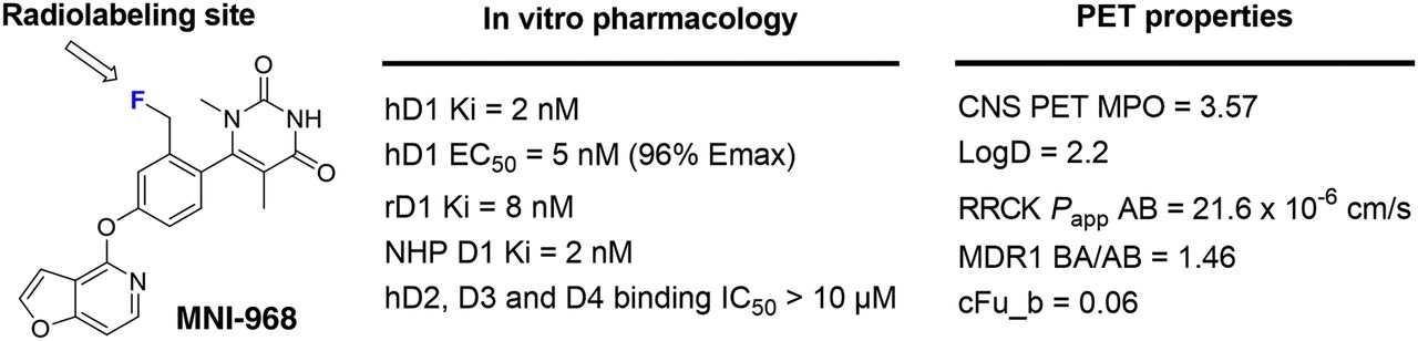
- FIGURE 1.
Profile of D1R agonist PET ligand lead MNI-968. AB = apical to basolateral; BA = basolateral to apical; CNS = central nervous system; cFub = fraction unbound in brain; EC50 = half-maximal effective concentration; Emax = maximal effect; hD1 = human D1; hD2 = human D2; IC50 = half-maximal inhibitory concentration; Ki = inhibition constant; MDR1 = multi-drug resistance 1; MPO = multiparameter optimization; rD1 = rat D1; Papp = apparent permeability; RRCK = Ralph Russ canine kidney assay.
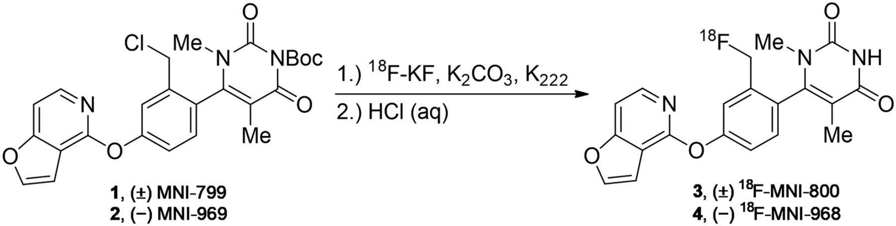
- FIGURE 2.
Radiosynthesis of 18F-MNI-800 and 18F-MNI-968.
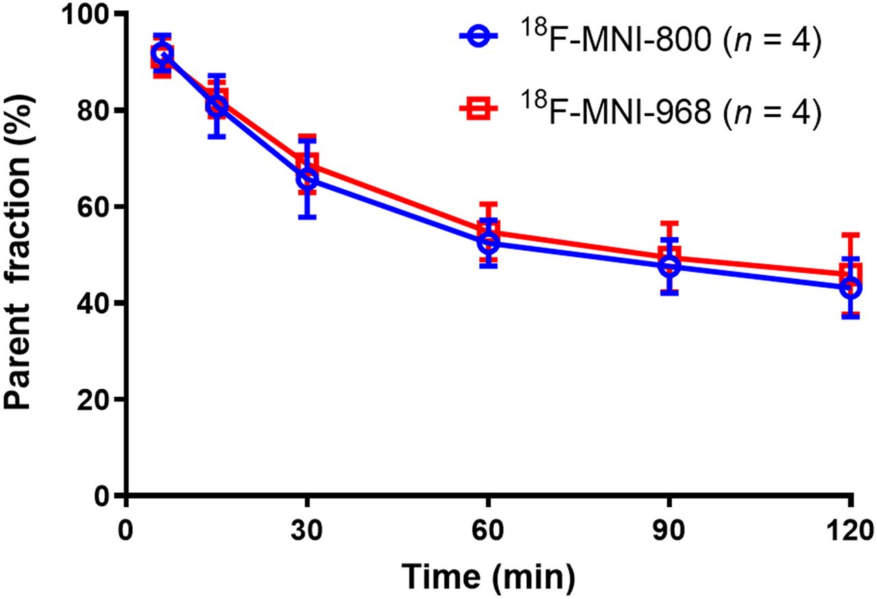
- FIGURE 3.
Parent fraction profile in arterial plasma after intravenous administration of 18F-MNI-800 (mean ± SD, n = 4) or 18F-MNI-968 (mean ± SD, n = 4).
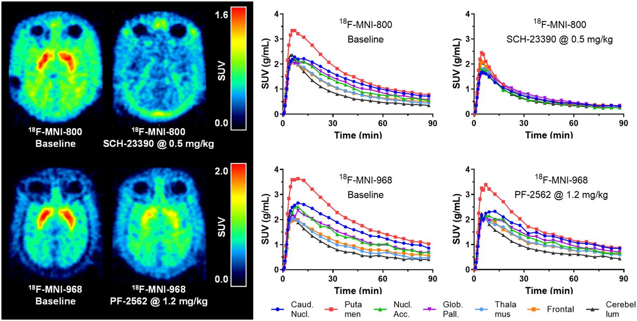
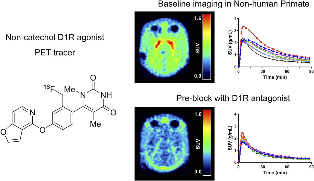
- FIGURE 4.
(Left) Average PET images from 30 to 90 min after injection for rhesus macaque (NHP A) in transverse plane of 18F-MNI-800 at baseline and after dosing with SCH-23390 at 0.5 mg/kg (occupancy of ∼85%) and of 18F-MNI-968 at baseline and after dosing with PF-2562 at 1.2 mg/kg (occupancy of ∼40%). (Right) Time–activity curves in some brain regions for same rhesus macaque for studies with 18F-MNI-800 and 18F-MNI-968. Caud. Nucl. = caudate nucleus; Glob. Pall. = globus pallidus; Nucl. Acc. = nucleus accumbens.
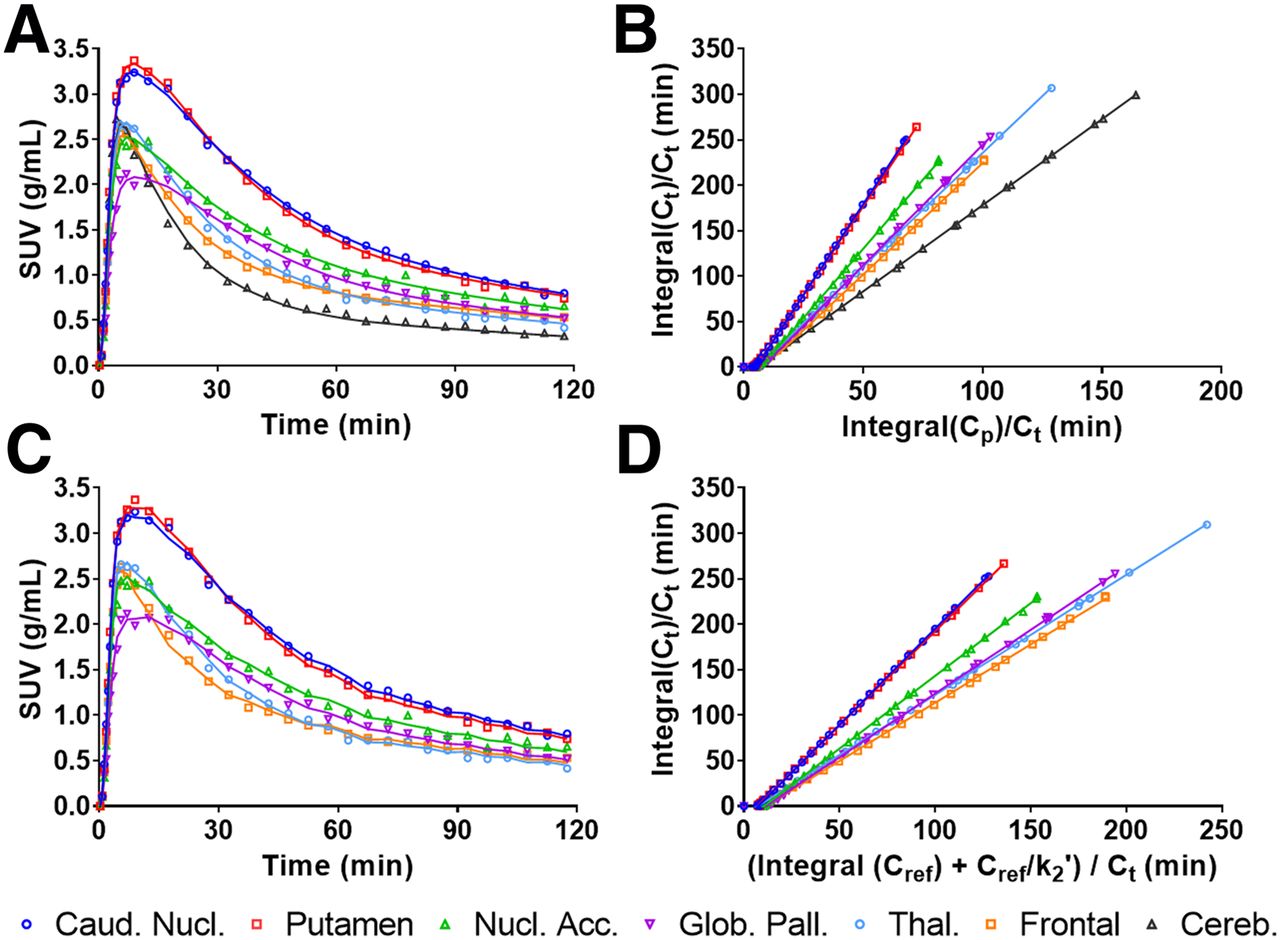
- FIGURE 5.
(A and C) Representative time–activity curves at baseline for rhesus macaque (NHP B) in some brain regions after bolus injection of 18F-MNI-968, showing 2T compartment model fits (A) and SRTM fits (C). (B and D) Graphical analysis with LGA with plasma input function (t* = 15 min) (B) and NI-LGA with reference region input function (t* = 10 min) (D). Caud. Nucl. = caudate nucleus; Cp = activity concentration in plasma; Cref = activity concentration in reference region; Ct = activity concentration in region of interest; Glob. Pall. = globus pallidus; Nucl. Acc. = nucleus accumbens; Thal. = thalamus.
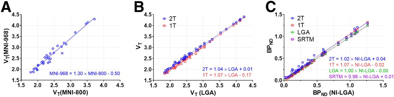
- FIGURE 6.
(A) Within-animal comparison (n = 2) of 18F-MNI-800 and 18F-MNI-968 2T VT estimates. (B) Comparison of 18F-MNI-968 VT estimates across models (n = 3). (C) Comparison of 18F-MNI-968 BPND estimates across models (n = 3). 1T = 1 tissue compartment.
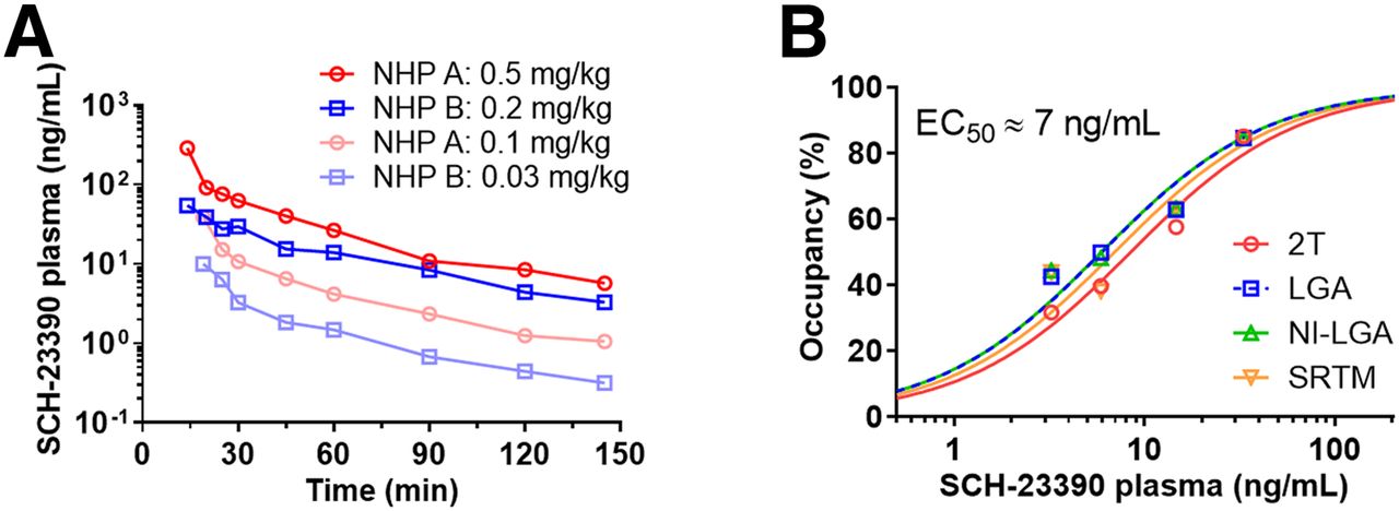
- FIGURE 7.
(A) SCH-23390 plasma levels for 4 doses, with 18F-MNI-800 injection at 25 min after drug administration. (B) Striatal D1R occupancy against average plasma levels between 25 and 145 min after administration of SCH-23390. EC50 = half-maximal effective concentration.


Tables
Species NHP no. 18F-MNI-800 18F-MNI-968 Rhesus A Test, retest, SCH23390 (0.5 and 0.1 mg/kg) Test, retest,* PF-2562 (1.2 mg/kg)* B Test, retest, SCH23390 (0.2 and 0.03 mg/kg), dosimetry Baseline C Dosimetry Baseline,* PF-2562 (1.2 mg/kg)* Cynomolgus D Baseline Baseline E Baseline F Baseline ↵* 90-min scan.
Scans are 120 min unless otherwise indicated.
VT BPND Region 2T LGA 2T LGA SRTM NI-LGA Striatum 3.6 ± 0.3 (8%) 3.5 ± 0.3 (8%) 0.86 ± 0.10 (11%) 0.83 ± 0.08 (9%) 0.83 ± 0.07 (8%) 0.83 ± 0.07 (8%) Caudate 3.5 ± 0.4 (11%) 3.4 ± 0.4 (11%) 0.81 ± 0.11 (14%) 0.78 ± 0.10 (12%) 0.78 ± 0.09 (11%) 0.78 ± 0.09 (12%) Putamen 3.7 ± 0.3 (7%) 3.6 ± 0.3 (7%) 0.91 ± 0.14 (15%) 0.88 ± 0.12 (13%) 0.89 ± 0.11 (12%) 0.89 ± 0.11 (13%) Nucleus accumbens 2.9 ± 0.2 (7%) 2.8 ± 0.2 (7%) 0.48 ± 0.03 (6%) 0.46 ± 0.02 (5%) 0.45 ± 0.02 (5%) 0.46 ± 0.02 (5%) Globus pallidus 2.9 ± 0.3 (9%) 2.8 ± 0.3 (9%) 0.50 ± 0.06 (13%) 0.48 ± 0.06 (12%) 0.48 ± 0.06 (12%) 0.48 ± 0.05 (11%) Thalamus 2.4 ± 0.2 (9%) 2.4 ± 0.2 (8%) 0.25 ± 0.06 (22%) 0.25 ± 0.04 (17%) 0.26 ± 0.04 (16%) 0.26 ± 0.04 (16%) Frontal cortex 2.3 ± 0.0 (0%) 2.2 ± 0.0 (1%) 0.24 ± 0.04 (18%) 0.20 ± 0.05 (24%) 0.20 ± 0.05 (23%) 0.20 ± 0.05 (25%) Cerebellum 1.9 ± 0.1 (7%) 1.9 ± 0.1 (6%) Data are mean ± SD, followed by coefficient of variation in parentheses (n = 4).
VT BPND Region 2T LGA 2T LGA SRTM NI-LGA Striatum 4.1 ± 0.2 (4%) 4.0 ± 0.2 (4%) 1.14 ± 0.05 (5%) 1.07 ± 0.02 (2%) 1.07 ± 0.03 (2%) 1.08 ± 0.03 (3%) Caudate 4.0 ± 0.3 (8%) 3.9 ± 0.3 (8%) 1.08 ± 0.12 (11%) 1.02 ± 0.09 (9%) 1.06 ± 0.09 (8%) 1.06 ± 0.08 (8%) Putamen 4.3 ± 0.2 (4%) 4.1 ± 0.2 (4%) 1.20 ± 0.09 (8%) 1.13 ± 0.10 (9%) 1.11 ± 0.08 (7%) 1.12 ± 0.08 (8%) Nucleus accumbens 3.2 ± 0.2 (5%) 3.1 ± 0.2 (6%) 0.65 ± 0.02 (4%) 0.61 ± 0.00 (0%) 0.59 ± 0.07 (12%) 0.60 ± 0.07 (12%) Globus pallidus 3.2 ± 0.6 (18%) 3.1 ± 0.5 (18%) 0.65 ± 0.20 (31%) 0.60 ± 0.21 (35%) 0.58 ± 0.18 (31%) 0.58 ± 0.18 (31%) Thalamus 2.6 ± 0.2 (9%) 2.6 ± 0.2 (9%) 0.35 ± 0.04 (13%) 0.33 ± 0.04 (13%) 0.29 ± 0.05 (19%) 0.29 ± 0.06 (20%) Frontal cortex 2.6 ± 0.1 (2%) 2.4 ± 0.1 (2%) 0.33 ± 0.08 (25%) 0.27 ± 0.04 (15%) 0.28 ± 0.05 (17%) 0.28 ± 0.05 (18%) Cerebellum 1.9 ± 0.1 (6%) 1.9 ± 0.1 (5%) Data are mean ± SD, followed by coefficient of variation in parentheses (n = 3 for 2T and LGA, and n = 5 for SRTM and NI-LGA).
Supplemental Data
Files in this Data Supplement:
In this issue


Jump to section
Related Articles
Cited By...
- No citing articles found.