
Main menu

  • Home
  • Content
    • Current
    • Ahead of print
    • Past Issues
    • JNM Supplement
    • SNMMI Annual Meeting Abstracts
    • Continuing Education
    • JNM Podcasts
  • Subscriptions
    • Subscribers
    • Institutional and Non-member
    • Rates
    • Journal Claims
    • Corporate & Special Sales
  • Authors
    • Submit to JNM
    • Information for Authors
    • Assignment of Copyright
    • AQARA requirements
  • Info
    • Reviewers
    • Permissions
    • Advertisers
  • About
    • About Us
    • Editorial Board
    • Contact Information
  • More
    • Alerts
    • Feedback
    • Help
    • SNMMI Journals
  • SNMMI
    • JNM
    • JNMT
    • SNMMI Journals
    • SNMMI

User menu

  • Subscribe
  • My alerts
  • Log in
  • My Cart

Search

  • Advanced search
Journal of Nuclear Medicine
  • SNMMI
    • JNM
    • JNMT
    • SNMMI Journals
    • SNMMI
  • Subscribe
  • My alerts
  • Log in
  • My Cart
Journal of Nuclear Medicine

Advanced Search

  • Home
  • Content
    • Current
    • Ahead of print
    • Past Issues
    • JNM Supplement
    • SNMMI Annual Meeting Abstracts
    • Continuing Education
    • JNM Podcasts
  • Subscriptions
    • Subscribers
    • Institutional and Non-member
    • Rates
    • Journal Claims
    • Corporate & Special Sales
  • Authors
    • Submit to JNM
    • Information for Authors
    • Assignment of Copyright
    • AQARA requirements
  • Info
    • Reviewers
    • Permissions
    • Advertisers
  • About
    • About Us
    • Editorial Board
    • Contact Information
  • More
    • Alerts
    • Feedback
    • Help
    • SNMMI Journals
  • View or Listen to JNM Podcast
  • Visit JNM on Facebook
  • Join JNM on LinkedIn
  • Follow JNM on Twitter
  • Subscribe to our RSS feeds
Research ArticleTheranostics
Open Access

Head-to-Head Comparison of 68Ga-Prostate-Specific Membrane Antigen PET/CT and Ferumoxtran-10–Enhanced MRI for the Diagnosis of Lymph Node Metastases in Prostate Cancer Patients

Melline G.M. Schilham, Patrik Zamecnik, Bastiaan M. Privé, Bas Israël, Mark Rijpkema, Tom Scheenen, Jelle O. Barentsz, James Nagarajah and Martin Gotthardt
Journal of Nuclear Medicine September 2021, 62 (9) 1258-1263; DOI: https://doi.org/10.2967/jnumed.120.258541
Melline G.M. Schilham
1Department of Medical Imaging, Nuclear Medicine, Radboud University Medical Centre, Nijmegen, The Netherlands; and
  • Find this author on Google Scholar
  • Find this author on PubMed
  • Search for this author on this site
Patrik Zamecnik
1Department of Medical Imaging, Nuclear Medicine, Radboud University Medical Centre, Nijmegen, The Netherlands; and
  • Find this author on Google Scholar
  • Find this author on PubMed
  • Search for this author on this site
Bastiaan M. Privé
1Department of Medical Imaging, Nuclear Medicine, Radboud University Medical Centre, Nijmegen, The Netherlands; and
  • Find this author on Google Scholar
  • Find this author on PubMed
  • Search for this author on this site
Bas Israël
1Department of Medical Imaging, Nuclear Medicine, Radboud University Medical Centre, Nijmegen, The Netherlands; and
  • Find this author on Google Scholar
  • Find this author on PubMed
  • Search for this author on this site
Mark Rijpkema
1Department of Medical Imaging, Nuclear Medicine, Radboud University Medical Centre, Nijmegen, The Netherlands; and
  • Find this author on Google Scholar
  • Find this author on PubMed
  • Search for this author on this site
Tom Scheenen
1Department of Medical Imaging, Nuclear Medicine, Radboud University Medical Centre, Nijmegen, The Netherlands; and
  • Find this author on Google Scholar
  • Find this author on PubMed
  • Search for this author on this site
Jelle O. Barentsz
1Department of Medical Imaging, Nuclear Medicine, Radboud University Medical Centre, Nijmegen, The Netherlands; and
  • Find this author on Google Scholar
  • Find this author on PubMed
  • Search for this author on this site
James Nagarajah
1Department of Medical Imaging, Nuclear Medicine, Radboud University Medical Centre, Nijmegen, The Netherlands; and
2Department of Nuclear Medicine, Technical University Munich, Klinikum rechts der Isar, Munich, Germany
  • Find this author on Google Scholar
  • Find this author on PubMed
  • Search for this author on this site
Martin Gotthardt
1Department of Medical Imaging, Nuclear Medicine, Radboud University Medical Centre, Nijmegen, The Netherlands; and
  • Find this author on Google Scholar
  • Find this author on PubMed
  • Search for this author on this site
  • Article
  • Figures & Data
  • Info & Metrics
  • PDF
Loading

Article Figures & Data

Figures

  • Tables
  • Figure
    • Download figure
    • Open in new tab
    • Download powerpoint
  • FIGURE 1.
    • Download figure
    • Open in new tab
    • Download powerpoint
    FIGURE 1.

    Examples of iron-sensitive T2*-weighted fat-saturated nano-MRI images (left) and PSMA PET/CT images (right). (A) Seven-millimeter-diameter LN in left pararectal region that is positive on both nano-MRI and PSMA PET/CT. (B) Four-millimeter-diameter LN in region of left external iliac artery that is positive on nano-MRI but negative on PSMA PET/CT. (C) Suspicious LN dorsal to left external iliac artery that is negative on nano-MRI (nano-MRI also shows no left ureter in this area) but positive on PSMA PET/CT. LNs are encircled; arrows indicate right ureter.
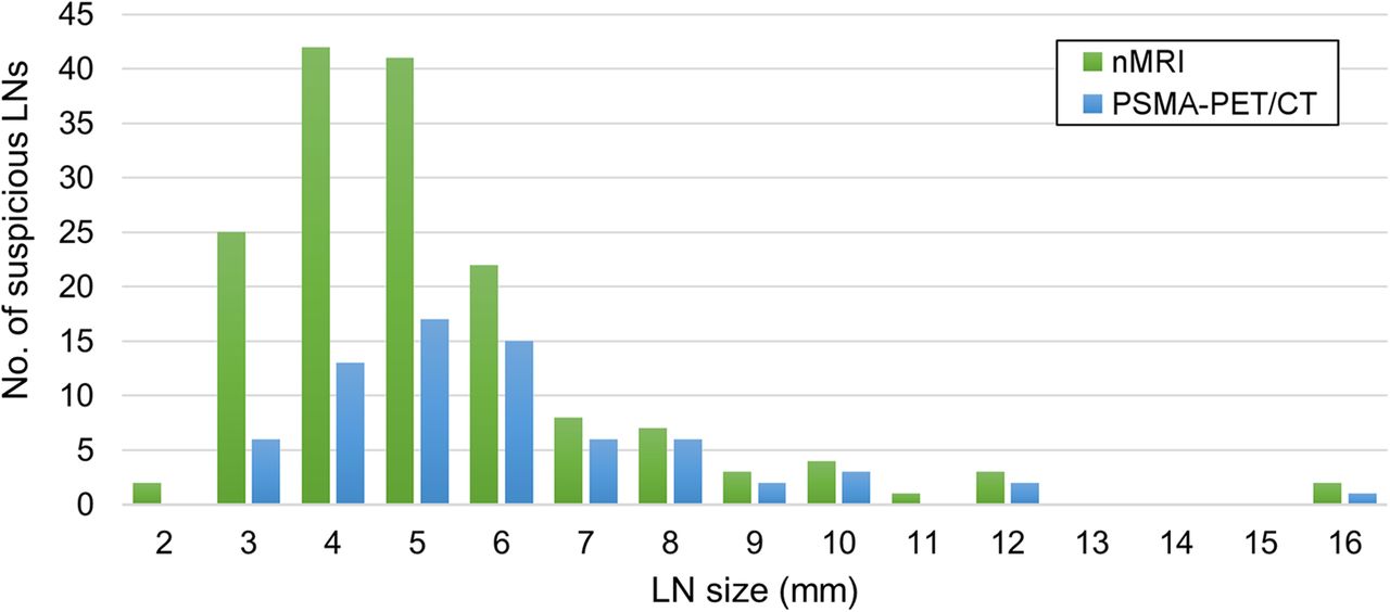
  • FIGURE 2.
    • Download figure
    • Open in new tab
    • Download powerpoint
    FIGURE 2.

    Size distribution of suspicious LNs as detected by nano-MRI (nMRI) and PSMA PET/CT.

  • FIGURE 3.
    • Download figure
    • Open in new tab
    • Download powerpoint
    FIGURE 3.

    Anatomic distribution of identified suspicious LNs as detected by nano-MRI (nMRI) and PSMA PET/CT.

Tables

  • Figures
    • View popup
    TABLE 1

    Patient Characteristics

    CharacteristicData
    Patients45 (100%)
    Age (y)64 (48–82)
    Serum PSA level (ng/mL)*
     Overall, n = 429.9 (0.0–150)
     Primary setting, n = 828.9 (5.6–150)
     Recurrence setting, n = 335.0 (0.0–46)
    Time between diagnosis and scans (mo)†50 (1–202)
    Time between scans (d)3.0 (1–18)
    Before imaging
     Any PCa treatment
      Yes36 (80%)
      No8 (18%)
      Unknown1 (2%)
     PLND
      Yes22 (49%)
      No19 (42%)
      Unknown4 (9%)
     Clinical ISUP grade
      15 (11%)
      26 (13%)
      37 (16%)
      413 (29%)
      58 (18%)
      Unknown6 (13%)
    • ↵* No data available for 3 patients.

    • ↵† No data available for 1 patient.

    • ISUP = International Society of Urological Pathology.

    • Qualitative data are number and percentage; continuous data are mean and range.

    • View popup
    TABLE 2

    Node Detection and Characteristics for Nano-MRI and PSMA PET/CT

    CharacteristicTotalNano-MRIPSMA PET/CTP
    Total scans90 (100%)45 (100%)45 (100%)
    Total positive scans58 (64%)33 (73%)25 (56%)
    Total suspicious LNs179 (100%)160 (89%)71 (40%)
    Suspicious LNs per patient4.0 (range, 0–6)3.6 (range, 0–15)1.6 (range, 0–12)<0.001*
    Suspicious LN size (mm)5.2 (range, 2–16)5.3 (range, 2–16)6.0 (range, 3–16)0.006†
    LoS4 (IQR, 4–5)4 (IQR, 4–5)5 (IQR, 4–5)
    • ↵* Positive scan defined as at least 1 LN with LoS ≥ 3.

    • ↵† Wilcoxon signed-rank test.

    • ‡Mann–Whitney U test.

    • IQR = interquartile range.

    • Qualitative data are number and percentage; continuous data are mean and range or IQR.

    • View popup
    TABLE 3

    Conformity of Nano-MRI and PSMA PET/CT

    Suspicious LNs as detected by…
    CharacteristicBoth nano-MRI and PSMA PET/CTNano-MRI onlyPSMA PET/CT only
    No. of patients203014
    Total suspicious LNs52 (29%)108 (60%)19 (11%)
    Suspicious LNs per patient1.2 (range, 0–10)2.4 (range, 0–8)0.4 (range, 0–3)
    LN size (mm)6.5 (range, 4–16)4.7 (range, 2–16)4.4 (range, 3–8)
    LoS5 (IQR, 4–5)4 (IQR, 4–5)3 (IQR, 3–4)
    • IQR = interquartile range.

    • Qualitative data are number and percentage; continuous data are mean and range or IQR.

PreviousNext
Back to top

In this issue

Journal of Nuclear Medicine: 62 (9)
Journal of Nuclear Medicine
Vol. 62, Issue 9
September 1, 2021
  • Table of Contents
  • Table of Contents (PDF)
  • About the Cover
  • Index by author
  • Complete Issue (PDF)
Print
Download PDF
Article Alerts
Sign In to Email Alerts with your Email Address
Email Article

Thank you for your interest in spreading the word on Journal of Nuclear Medicine.

NOTE: We only request your email address so that the person you are recommending the page to knows that you wanted them to see it, and that it is not junk mail. We do not capture any email address.

Enter multiple addresses on separate lines or separate them with commas.
Head-to-Head Comparison of 68Ga-Prostate-Specific Membrane Antigen PET/CT and Ferumoxtran-10–Enhanced MRI for the Diagnosis of Lymph Node Metastases in Prostate Cancer Patients
(Your Name) has sent you a message from Journal of Nuclear Medicine
(Your Name) thought you would like to see the Journal of Nuclear Medicine web site.
Citation Tools
Head-to-Head Comparison of 68Ga-Prostate-Specific Membrane Antigen PET/CT and Ferumoxtran-10–Enhanced MRI for the Diagnosis of Lymph Node Metastases in Prostate Cancer Patients
Melline G.M. Schilham, Patrik Zamecnik, Bastiaan M. Privé, Bas Israël, Mark Rijpkema, Tom Scheenen, Jelle O. Barentsz, James Nagarajah, Martin Gotthardt
Journal of Nuclear Medicine Sep 2021, 62 (9) 1258-1263; DOI: 10.2967/jnumed.120.258541

Citation Manager Formats

  • BibTeX
  • Bookends
  • EasyBib
  • EndNote (tagged)
  • EndNote 8 (xml)
  • Medlars
  • Mendeley
  • Papers
  • RefWorks Tagged
  • Ref Manager
  • RIS
  • Zotero
Share
Head-to-Head Comparison of 68Ga-Prostate-Specific Membrane Antigen PET/CT and Ferumoxtran-10–Enhanced MRI for the Diagnosis of Lymph Node Metastases in Prostate Cancer Patients
Melline G.M. Schilham, Patrik Zamecnik, Bastiaan M. Privé, Bas Israël, Mark Rijpkema, Tom Scheenen, Jelle O. Barentsz, James Nagarajah, Martin Gotthardt
Journal of Nuclear Medicine Sep 2021, 62 (9) 1258-1263; DOI: 10.2967/jnumed.120.258541
Twitter logo Facebook logo LinkedIn logo Mendeley logo
  • Tweet Widget
  • Facebook Like
  • Google Plus One
Bookmark this article

Jump to section

  • Article
    • Visual Abstract
    • Abstract
    • MATERIALS AND METHODS
    • RESULTS
    • DISCUSSION
    • CONCLUSION
    • DISCLOSURE
    • Acknowledgments
    • Footnotes
    • REFERENCES
  • Figures & Data
  • Info & Metrics
  • PDF

Related Articles

  • This Month in JNM
  • PubMed
  • Google Scholar

Cited By...

  • No citing articles found.
  • Google Scholar

More in this TOC Section

Theranostics

  • Determination of the Intralesional Distribution of Theranostic 124I-Omburtamab Convection-Enhanced Delivery in Treatment of Diffuse Intrinsic Pontine Glioma
  • Evidence-Based Clinical Protocols to Monitor Efficacy of [177Lu]Lu-PSMA Radiopharmaceutical Therapy in Metastatic Castration-Resistant Prostate Cancer Using Real-World Data
  • 177Lu-Labeled Anticlaudin 6 Monoclonal Antibody for Targeted Therapy in Esophageal Cancer
Show more Theranostics

Clinical

  • Determination of the Intralesional Distribution of Theranostic 124I-Omburtamab Convection-Enhanced Delivery in Treatment of Diffuse Intrinsic Pontine Glioma
  • Evidence-Based Clinical Protocols to Monitor Efficacy of [177Lu]Lu-PSMA Radiopharmaceutical Therapy in Metastatic Castration-Resistant Prostate Cancer Using Real-World Data
  • 177Lu-Labeled Anticlaudin 6 Monoclonal Antibody for Targeted Therapy in Esophageal Cancer
Show more Clinical

Similar Articles

Keywords

  • prostate cancer
  • lymph node
  • prostate specific membrane antigen
  • 68Ga-PSMA PET/CT
  • ferumoxtran-10
SNMMI

© 2025 SNMMI

Powered by HighWire