
Main menu

  • Home
  • Content
    • Current
    • Ahead of print
    • Past Issues
    • JNM Supplement
    • SNMMI Annual Meeting Abstracts
    • Continuing Education
    • JNM Podcasts
  • Subscriptions
    • Subscribers
    • Institutional and Non-member
    • Rates
    • Journal Claims
    • Corporate & Special Sales
  • Authors
    • Submit to JNM
    • Information for Authors
    • Assignment of Copyright
    • AQARA requirements
  • Info
    • Reviewers
    • Permissions
    • Advertisers
  • About
    • About Us
    • Editorial Board
    • Contact Information
  • More
    • Alerts
    • Feedback
    • Help
    • SNMMI Journals
  • SNMMI
    • JNM
    • JNMT
    • SNMMI Journals
    • SNMMI

User menu

  • Subscribe
  • My alerts
  • Log in
  • My Cart

Search

  • Advanced search
Journal of Nuclear Medicine
  • SNMMI
    • JNM
    • JNMT
    • SNMMI Journals
    • SNMMI
  • Subscribe
  • My alerts
  • Log in
  • My Cart
Journal of Nuclear Medicine

Advanced Search

  • Home
  • Content
    • Current
    • Ahead of print
    • Past Issues
    • JNM Supplement
    • SNMMI Annual Meeting Abstracts
    • Continuing Education
    • JNM Podcasts
  • Subscriptions
    • Subscribers
    • Institutional and Non-member
    • Rates
    • Journal Claims
    • Corporate & Special Sales
  • Authors
    • Submit to JNM
    • Information for Authors
    • Assignment of Copyright
    • AQARA requirements
  • Info
    • Reviewers
    • Permissions
    • Advertisers
  • About
    • About Us
    • Editorial Board
    • Contact Information
  • More
    • Alerts
    • Feedback
    • Help
    • SNMMI Journals
  • View or Listen to JNM Podcast
  • Visit JNM on Facebook
  • Join JNM on LinkedIn
  • Follow JNM on Twitter
  • Subscribe to our RSS feeds
Research ArticleClinical Investigation

Metabolic Scar Assessment with18F-FDG PET: Correlation to Ischemic Ventricular Tachycardia Substrate and Successful Ablation Sites

Yousra Ghzally, Hasan Imanli, Mark Smith, Jagat Mahat, Wengen Chen, Alejandro Jimenez, Mariem A. Sawan, Mohamed Aboel-Kassem F. Abdelmegid, Hatem Abd el Rahman Helmy, Salwa Demitry, Vincent See, Stephen Shorofsky, Vasken Dilsizian and Timm Dickfeld
Journal of Nuclear Medicine November 2021, 62 (11) 1591-1598; DOI: https://doi.org/10.2967/jnumed.120.246413
Yousra Ghzally
  • Find this author on Google Scholar
  • Find this author on PubMed
  • Search for this author on this site
Hasan Imanli
1Maryland Arrhythmia and Cardiology Imaging Group, Baltimore, Maryland;
2Department of Cardiovascular Medicine, Assiut University, Assiut, Egypt;
  • Find this author on Google Scholar
  • Find this author on PubMed
  • Search for this author on this site
Mark Smith
4Department of Cardiovascular Medicine and Department of Diagnostic Radiology and Nuclear Medicine, University of Maryland School of Medicine, Baltimore, Maryland
  • Find this author on Google Scholar
  • Find this author on PubMed
  • Search for this author on this site
Jagat Mahat
1Maryland Arrhythmia and Cardiology Imaging Group, Baltimore, Maryland;
  • Find this author on Google Scholar
  • Find this author on PubMed
  • Search for this author on this site
Wengen Chen
1Maryland Arrhythmia and Cardiology Imaging Group, Baltimore, Maryland;
3Assiut University, Assiut, Egypt; and
  • Find this author on Google Scholar
  • Find this author on PubMed
  • Search for this author on this site
Alejandro Jimenez
1Maryland Arrhythmia and Cardiology Imaging Group, Baltimore, Maryland;
2Department of Cardiovascular Medicine, Assiut University, Assiut, Egypt;
  • Find this author on Google Scholar
  • Find this author on PubMed
  • Search for this author on this site
Mariem A. Sawan
1Maryland Arrhythmia and Cardiology Imaging Group, Baltimore, Maryland;
2Department of Cardiovascular Medicine, Assiut University, Assiut, Egypt;
  • Find this author on Google Scholar
  • Find this author on PubMed
  • Search for this author on this site
Mohamed Aboel-Kassem F. Abdelmegid
3Assiut University, Assiut, Egypt; and
  • Find this author on Google Scholar
  • Find this author on PubMed
  • Search for this author on this site
Hatem Abd el Rahman Helmy
3Assiut University, Assiut, Egypt; and
  • Find this author on Google Scholar
  • Find this author on PubMed
  • Search for this author on this site
Salwa Demitry
3Assiut University, Assiut, Egypt; and
  • Find this author on Google Scholar
  • Find this author on PubMed
  • Search for this author on this site
Vincent See
1Maryland Arrhythmia and Cardiology Imaging Group, Baltimore, Maryland;
2Department of Cardiovascular Medicine, Assiut University, Assiut, Egypt;
  • Find this author on Google Scholar
  • Find this author on PubMed
  • Search for this author on this site
Stephen Shorofsky
1Maryland Arrhythmia and Cardiology Imaging Group, Baltimore, Maryland;
2Department of Cardiovascular Medicine, Assiut University, Assiut, Egypt;
  • Find this author on Google Scholar
  • Find this author on PubMed
  • Search for this author on this site
Vasken Dilsizian
4Department of Cardiovascular Medicine and Department of Diagnostic Radiology and Nuclear Medicine, University of Maryland School of Medicine, Baltimore, Maryland
  • Find this author on Google Scholar
  • Find this author on PubMed
  • Search for this author on this site
Timm Dickfeld
1Maryland Arrhythmia and Cardiology Imaging Group, Baltimore, Maryland;
2Department of Cardiovascular Medicine, Assiut University, Assiut, Egypt;
  • Find this author on Google Scholar
  • Find this author on PubMed
  • Search for this author on this site
  • Article
  • Figures & Data
  • Supplemental
  • Info & Metrics
  • PDF
Loading

Article Figures & Data

Figures

  • Tables
  • Additional Files
  • Figure
    • Download figure
    • Open in new tab
    • Download powerpoint
  • FIGURE 1.
    • Download figure
    • Open in new tab
    • Download powerpoint
    FIGURE 1.

    Workflow of 18F-FDG reconstructions. (A) Raw 18F-FDG PET DICOM showing left ventricle with central region of interest (purple). (B) Resulting 3D LV (purple) and right ventricle (blue) reconstruction. (C) Same slice as shown in A, with color map according to value of 18F-FDG uptake in each segment, showing decreased uptake in inferolateral scar area. (D) Generated 3D color PET reconstruction from short-axis slices with inferolateral scar based on 18F-FDG uptake percentage.
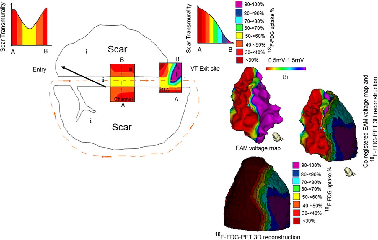
  • FIGURE 2.
    • Download figure
    • Open in new tab
    • Download powerpoint
    FIGURE 2.

    Metabolic channel. (A) EAM showing inferior scar with 0.5- to 1.5-mV setting and VT channel or exit site (white point and arrow). (B) Corresponding PET 3D reconstruction showing metabolic channel (dashed lines). (C) Coregistration of EAM and PET 3D reconstruction showing VT channel or exit (arrow) within metabolic channel (dashed lines). Additional example is shown in supplemental materials.
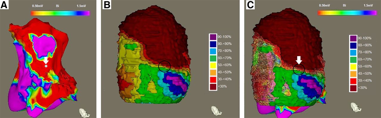
  • FIGURE 3.
    • Download figure
    • Open in new tab
    • Download powerpoint
    FIGURE 3.

    RTA. (A) EAM showing apical and inferior scars with 0.5- to 1.5-mV setting and VT channel or exit site (white point and arrow). (B) PET 3D reconstruction demonstrating RTA (circle, change of ≥50% uptake/15 mm [red to blue color shift]). (C) Coregistration showing VT channel or exit within RTA; arrow points to VT channel or exit site within RTA (circle). Additional example is shown in supplemental materials.
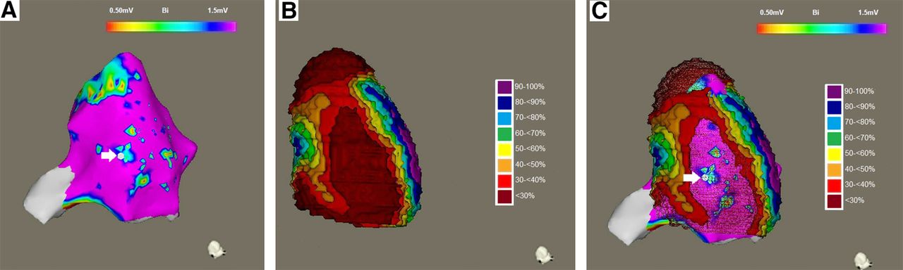
  • FIGURE 4.
    • Download figure
    • Open in new tab
    • Download powerpoint
    FIGURE 4.

    MVM. (A) EAM showing apical and inferior scars with 0.5- to 1.5-mV setting and VT channel or exit site (white point and arrow). (B) PET 3D reconstruction demonstrating larger severe PET defect (red area = MVM). (C) Coregistration of 3D PET reconstruction and EAM demonstrating VT channel or exit within MVM (arrow). Additional example is shown in supplemental materials.
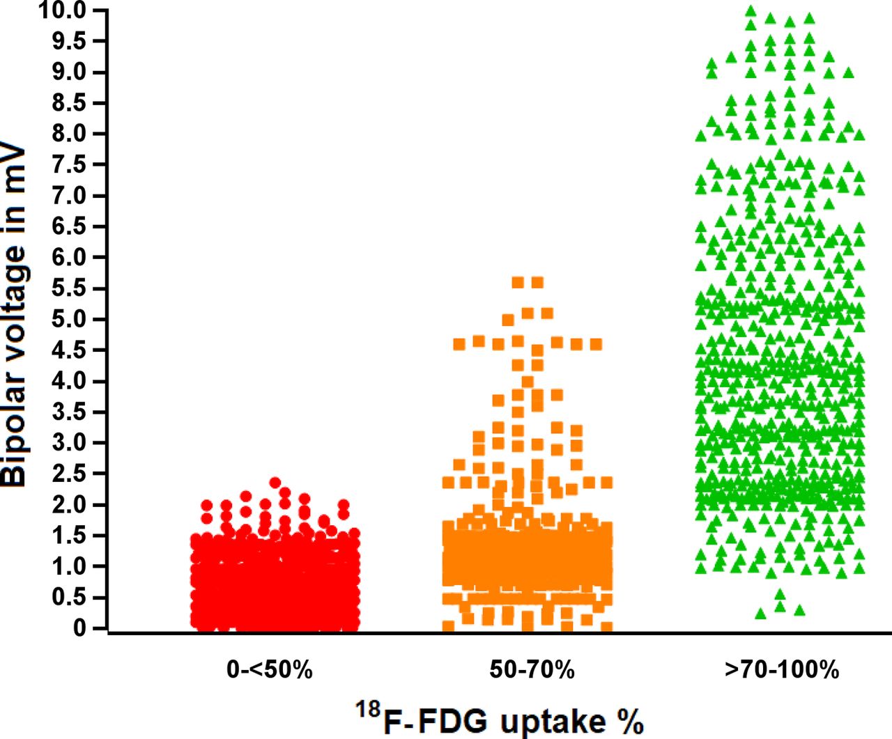
  • FIGURE 5.
    • Download figure
    • Open in new tab
    • Download powerpoint
    FIGURE 5.

    Scatterplot showing average bipolar voltage of severe PET defect, moderate PET defect, and normal PET areas. There is stepwise increase in bipolar voltage with each step in 18F-FDG PET uptake.

Tables

  • Figures
  • Additional Files
    • View popup
    TABLE 1

    Patient Characteristics

    CharacteristicData
    Mean age (±SD) at time of ablation63 ± 11 y
    Sex (male)46 (82%)
    Mean ejection fraction (±SD)29% ± 12%
    Ischemic
     Previous myocardial infarction (history, electrocardiography, or cardiac imaging)56 (100%)
     Bypass graft20 (36%)
     Coronary stenting36 (64%)
    NYHA II–III heart failure25 (45%)
    Diabetes17 (30%)
    Hypertension30 (54%)
    ICD at time of ablation35 (63%)
    Severe PET defect
     Inferior (inferolateral and inferoseptal)38 (70%)
     Anterior (anteroseptal and anterolateral)12 (23%)
     Apical3 (3.5%)
     Anterior and inferior3 (3.5%)
    EAM dense scar53 (97%)
    Antiarrhythmic drugsβ-blockers (52 [93%]), amiodarone (46 [82%]), sotalol (5 [9%])
    • NYHA = New York Heart Association; ICD = implantable cardioverter–defibrillator.

    • View popup
    TABLE 2

    Imaging Characteristics

    CharacteristicArea (cm2)Percentage of total LV EAM
    Total LV EAM270.2 ± 10.3
    Severe PET defect63.0 ± 48.423%
    Moderate to severe PET defect105.1 ± 67.239%
    EAM dense scar13.8 ± 33.15%
    EAM abnormal voltage ≤ 1.5 mV56.2 ± 62.621%
    • Data are mean ± SD.

    • View popup
    TABLE 3

    Distribution of 18F-FDG Uptake Percentage and Bipolar Voltage Characteristics of 50 Clinically Determined VT Channel or Exit Sites

    18F-FDG PET percentageEAM resultBorder zone
    Dense scar
    <30%59
    30 to <40%610
    40 to <50%18
    50%–70%05
    >70%06

Additional Files

  • Figures
  • Tables
  • Supplemental Data

    Files in this Data Supplement:

    • Supplemental Data
PreviousNext
Back to top

In this issue

Journal of Nuclear Medicine: 62 (11)
Journal of Nuclear Medicine
Vol. 62, Issue 11
November 1, 2021
  • Table of Contents
  • Table of Contents (PDF)
  • About the Cover
  • Index by author
  • Complete Issue (PDF)
Print
Download PDF
Article Alerts
Sign In to Email Alerts with your Email Address
Email Article

Thank you for your interest in spreading the word on Journal of Nuclear Medicine.

NOTE: We only request your email address so that the person you are recommending the page to knows that you wanted them to see it, and that it is not junk mail. We do not capture any email address.

Enter multiple addresses on separate lines or separate them with commas.
Metabolic Scar Assessment with18F-FDG PET: Correlation to Ischemic Ventricular Tachycardia Substrate and Successful Ablation Sites
(Your Name) has sent you a message from Journal of Nuclear Medicine
(Your Name) thought you would like to see the Journal of Nuclear Medicine web site.
Citation Tools
Metabolic Scar Assessment with18F-FDG PET: Correlation to Ischemic Ventricular Tachycardia Substrate and Successful Ablation Sites
Yousra Ghzally, Hasan Imanli, Mark Smith, Jagat Mahat, Wengen Chen, Alejandro Jimenez, Mariem A. Sawan, Mohamed Aboel-Kassem F. Abdelmegid, Hatem Abd el Rahman Helmy, Salwa Demitry, Vincent See, Stephen Shorofsky, Vasken Dilsizian, Timm Dickfeld
Journal of Nuclear Medicine Nov 2021, 62 (11) 1591-1598; DOI: 10.2967/jnumed.120.246413

Citation Manager Formats

  • BibTeX
  • Bookends
  • EasyBib
  • EndNote (tagged)
  • EndNote 8 (xml)
  • Medlars
  • Mendeley
  • Papers
  • RefWorks Tagged
  • Ref Manager
  • RIS
  • Zotero
Share
Metabolic Scar Assessment with18F-FDG PET: Correlation to Ischemic Ventricular Tachycardia Substrate and Successful Ablation Sites
Yousra Ghzally, Hasan Imanli, Mark Smith, Jagat Mahat, Wengen Chen, Alejandro Jimenez, Mariem A. Sawan, Mohamed Aboel-Kassem F. Abdelmegid, Hatem Abd el Rahman Helmy, Salwa Demitry, Vincent See, Stephen Shorofsky, Vasken Dilsizian, Timm Dickfeld
Journal of Nuclear Medicine Nov 2021, 62 (11) 1591-1598; DOI: 10.2967/jnumed.120.246413
Twitter logo Facebook logo LinkedIn logo Mendeley logo
  • Tweet Widget
  • Facebook Like
  • Google Plus One
Bookmark this article

Jump to section

  • Article
    • Visual Abstract
    • Abstract
    • MATERIALS AND METHODS
    • RESULTS
    • DISCUSSION
    • CONCLUSION
    • DISCLOSURE
    • Footnotes
    • REFERENCES
  • Figures & Data
  • Supplemental
  • Info & Metrics
  • PDF

Related Articles

  • This Month in JNM
  • PubMed
  • Google Scholar

Cited By...

  • No citing articles found.
  • Google Scholar

More in this TOC Section

  • First-in-Human Study of 18F-Labeled PET Tracer for Glutamate AMPA Receptor [18F]K-40: A Derivative of [11C]K-2
  • Detection of HER2-Low Lesions Using HER2-Targeted PET Imaging in Patients with Metastatic Breast Cancer: A Paired HER2 PET and Tumor Biopsy Analysis
  • [11C]Carfentanil PET Whole-Body Imaging of μ-Opioid Receptors: A First in-Human Study
Show more Clinical Investigation

Similar Articles

Keywords

  • functional imaging
  • 18F-FDG PET imaging
  • ventricular tachycardia substrate
  • VT channel or exit sites
SNMMI

© 2025 SNMMI

Powered by HighWire