


Article Figures & Data
Figures
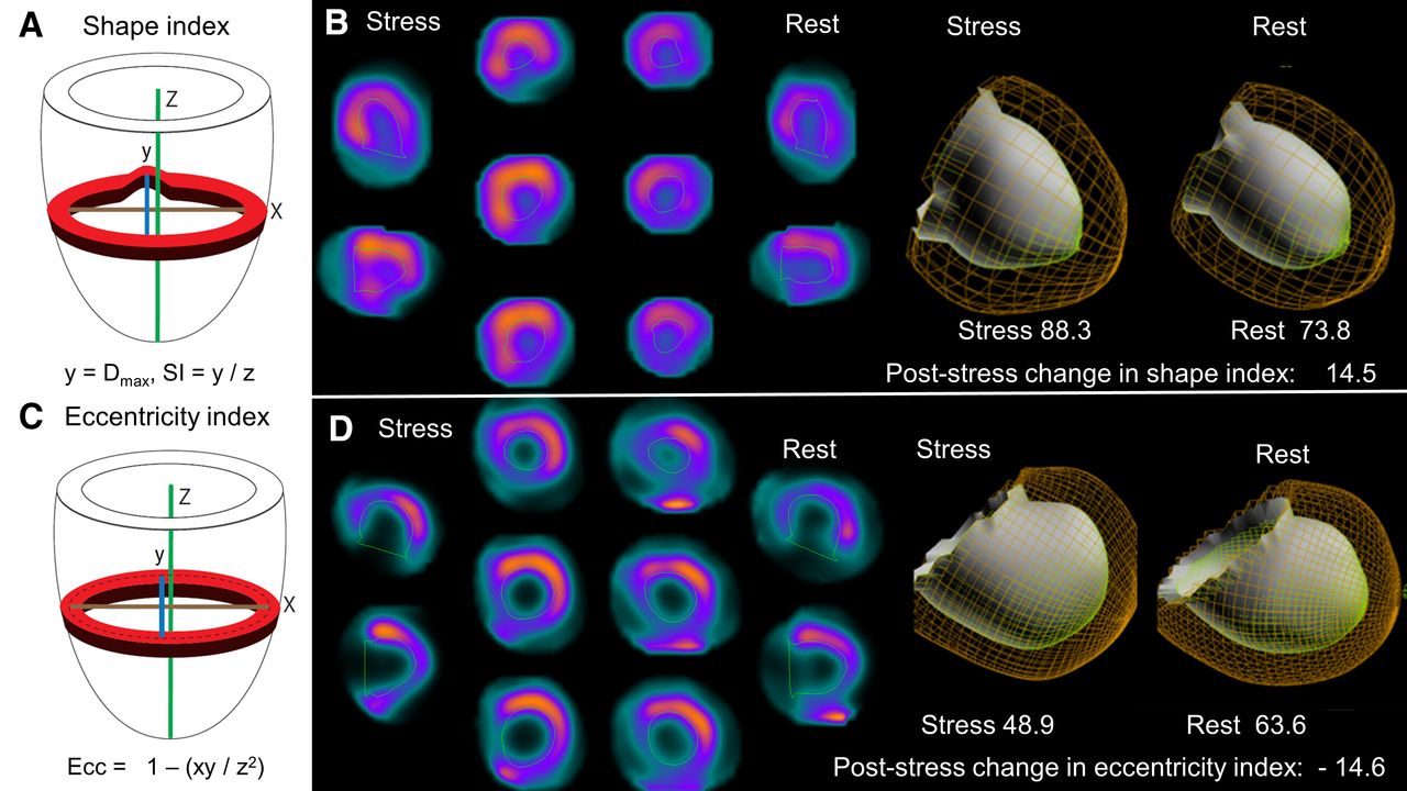
- FIGURE 1.
(A) Shape index is calculated as ratio of maximal short-axis diameter across all short-axis slices to long-axis length, from apex to mitral valve, using endocardial surface. (B) Patient with abnormal poststress change in shape index but normal poststress change in eccentricity (0.3) who was admitted for unstable angina and underwent revascularization 231 d after SPECT MPI. (C) Eccentricity index calculated from mid-myocardial surface of fitted ellipsoid and not accounting for regional anatomy. (D) Patient with abnormal poststress change in eccentricity index and mildly abnormal poststress change in shape index (0.5) who died 290 d after SPECT MPI. SI = shape index.
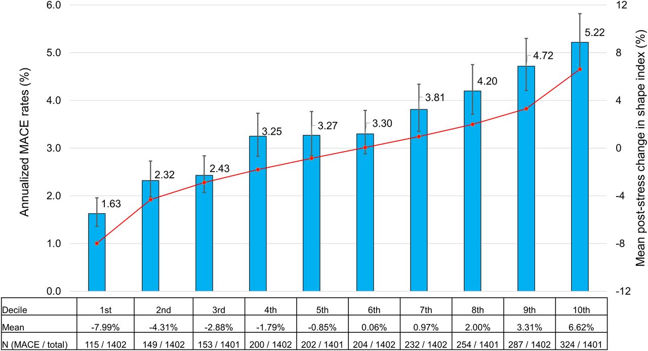
- FIGURE 2.
Annualized incidence of MACE for deciles of poststress change in shape index. Blue bars (with error bars showing 95% CI) show annualized MACE rates. Values in table reflect total number of events during follow-up. Red line shows mean poststress change in shape index for each decile; mean value is also shown in table.
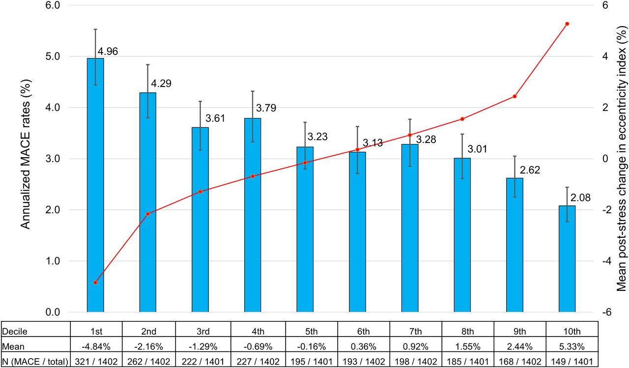
- FIGURE 3.
Annualized incidence of MACE for deciles of change in eccentricity index. Blue bars (with error bars showing 95% CI) show annualized MACE rates. Values in table reflect total number of events during follow-up. Red line shows mean poststress change in eccentricity index for each decile; mean value is also shown in table.
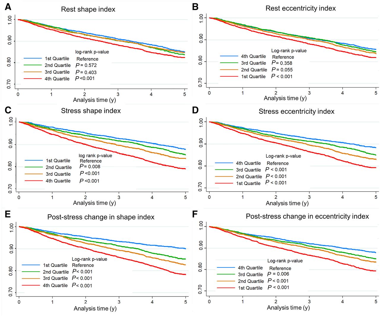
- FIGURE 4.
Kaplan–Meier survival curves for quartiles of rest, stress, and poststress change in shape index (A, C, and E, respectively) and eccentricity index (B, D, and F, respectively)


Tables
Characteristic MACE occurred (n = 2,120) No MACE (n = 11,896) P Age (y) 69.4 ± 11.8 63.3 ± 12.1 <0.001 Male 1,471 (69.4) 6,998 (58.8) <0.001 Body mass index (kg/m2) 28.1 ± 5.7 28.4 ± 6.3 0.263 Past medical history Hypertension 1,593 (75.1) 7,243 (60.9) <0.001 Diabetes 810 (38.2) 2,772 (23.3) <0.001 Dyslipidemia 1,534 (72.4) 7,219 (60.7) <0.001 Current smoker 421 (19.9) 2,712 (22.8) 0.003 PVD 462 (21.8) 1,637 (13.8) <0.001 Prior MI 549 (25.9) 1,532 (12.9) <0.001 Prior revascularization 1,088 (47.6) 2,839 (23.9) <0.001 Family history of CAD 451 (21.3) 3,274 (27.5) <0.001 Typical angina 159 (7.5) 621 (5.2) <0.001 Resting vital signs Systolic BP (mm Hg) 136.0 ± 21.2 134.2 ± 19.7 0.001 Diastolic BP (mm Hg) 77.8 ± 9.8 79.6 ± 9.3 <0.001 Heart rate (bpm) 71.4 ± 13.6 69.6 ± 13.3 <0.001 Exercise stress 523 (24.7) 5,221 (43.9) <0.001 PVD = peripheral vascular disease; MI = myocardial infarction; BP = blood pressure; bpm = beats per minute.
Qualitative data are number and percentage; continuous data are mean ± SD.
Characteristic MACE occurred (n = 2,120) No MACE (n = 11,896) P Rest shape index (%) 64.9 ± 8.3 63.9 ± 7.5 <0.001 Stress shape index (%) 65.5 ± 8.4 63.1 ± 7.1 <0.001 Poststress change in shape index(%) 0.6 ± 4.0 −0.8 ± 4.1 <0.001 Rest eccentricity index (%) 80.6 ± 4.7 81.1 ± 4.6 <0.001 Stress eccentricity index (%) 80.2 ± 5.0 81.3 ± 4.5 <0.001 Poststress change in eccentricity index (%) −0.4 ± 3.0 0.3 ± 2.8 <0.001 Resting TPD 3.8 ± 7.7 1.6 ± 4.9 <0.001 Stress TPD 8.2 ± 9.6 4.3 ± 6.6 <0.001 Ischemic TPD 4.4 ± 4.0 2.7 ± 3.0 <0.001 Resting LVEF 58.6 ± 14.8 62.8 ± 12.3 <0.001 Reduced LVEF (<40%) 251 (11.8) 482 (4.1) <0.001 Stress LVEF 56.6 ± 14.3 62.3 ± 11.9 <0.001 Poststress change in LVEF −1.9 ± 7.1 −0.5 ± 7.2 <0.001 Resting LVEDV 82.4 ± 45.5 70.7 ± 33.7 <0.001 Stress LVEDV 84.0 ± 46.4 70.2 ± 34.5 <0.001 Poststress change in LVEDV 1.6 ± 12.6 −0.57 ± 9.1 <0.001 TID 101 (4.8) 454 (3.8) 0.046 LVEDV = LV end diastolic volume.
Qualitative data are number and percentage; continuous data are mean ± SD.
Variable Adjusted HR P Rest shape index (per 10%) 1.05 (0.94–1.17) 0.370 Poststress change in shape index (per 10%) 1.38 (1.20–1.58) <0.001 Rest eccentricity index (per 10%) 0.97 (0.81–1.17) 0.763 Poststress change in eccentricity index (per 10%) 0.80 (0.66–0.98) 0.033 Age 1.02 (1.02–1.03) <0.001 Male 1.23 (1.11–1.36) <0.001 Prior myocardial infarction 1.19 (1.06–1.34) 0.004 Prior percutaneous coronary intervention 1.69 (1.53–1.87) <0.001 Prior coronary artery bypass grafting 1.14 (1.01–1.28) 0.037 Hypertension 1.15 (1.05–1.26) 0.002 Diabetes 1.35 (1.25–1.47) <0.001 Pharmacologic stress 1.40 (1.27–1.54) <0.001 Typical angina 1.52 (1.34–1.72) <0.001 Ischemic electrocardiographic response 1.43 (1.29–1.58) <0.001 Resting TPD 1.01 (1.00–1.01) 0.065 Ischemic TPD 1.12 (1.11–1.13) <0.001 Resting LVEF 0.99 (0.99–0.99) <0.001 Stress shape index and stress eccentricity index were not included in multivariable model because of inclusion of both rest and poststress change in values.
Data in parentheses are 95% CIs.
Supplemental Data
Files in this Data Supplement:
In this issue


Jump to section
Related Articles
Cited By...
- No citing articles found.