


Article Figures & Data
Figures
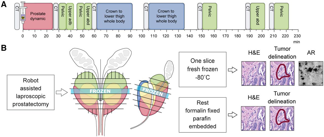
- FIGURE 1.
Schematic representation of research protocol. (A) Protocol consisting of CT scans (gray), dynamic scan (red), static images (green), and whole-body scans (blue). (B) Tissue was cut into 4-mm sections; 1 section was fresh-frozen, and remainder were formalin-fixed and paraffin-embedded. Slides were stained with hematoxylin and eosin and evaluated by pathologist. Autoradiography was performed on slides of frozen sections. abd = abdomen; AR = autoradiography; H&E = hematoxylin and eosin.
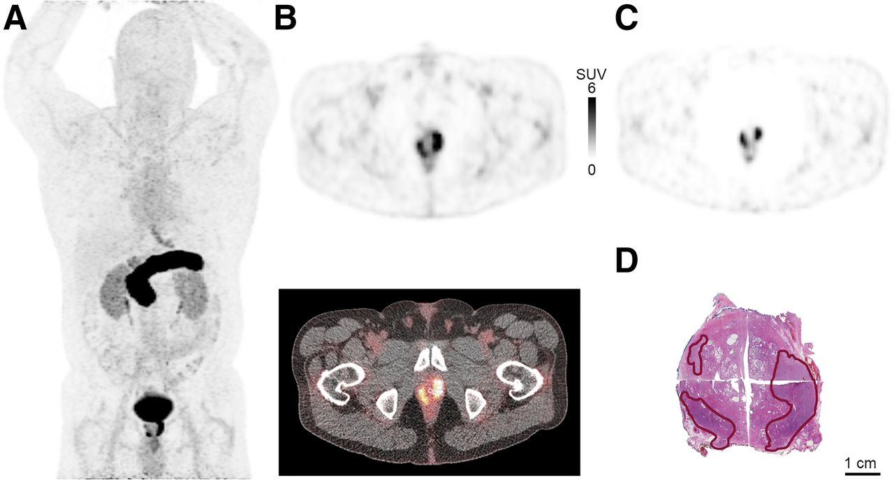
- FIGURE 2.
68Ga-SB3 PET/CT imaging of large tumor (GS 3 + 4 = 7) in primary-PCa patient. (A) Maximum-intensity projection 60 min after injection. (B) PET (top) and PET/CT (bottom) imaging 60 min after injection; tumor SUVmax, 22.7. (C) PET imaging 210 min after injection; tumor SUVmax, 20.0. (E) Corresponding histopathologic slides with tumor delineated in red.
- FIGURE 3.
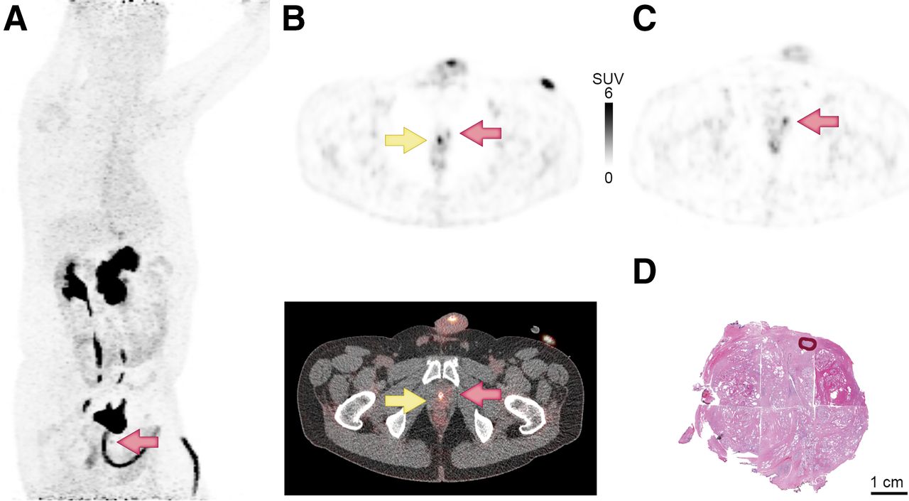
68Ga-SB3 PET/CT imaging of primary PCa patient. Very small tumor (GS 3 + 3 = 6; orange arrows) was not detected in biopsies; however, there was elevated prostate-specific antigen and family history of PCa. Catheter is indicated by yellow arrow. (A) Maximum-intensity projection 60 min after injection. (B) PET (top) and PET/CT (bottom) imaging 60 min after injection; tumor SUVmax, 4.4. (C) At 210 min after injection, with catheter removed before scan; tumor SUVmax, 4.3. (D) Corresponding histopathologic slides with tumor delineated in red.
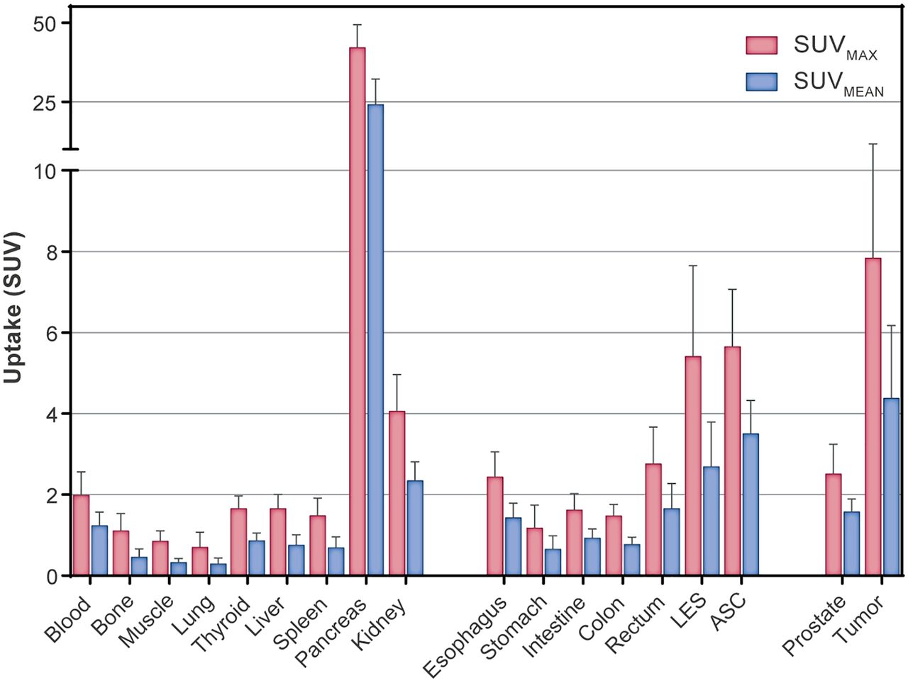
- FIGURE 4.
Biodistribution of physiologic uptake and tumor uptake of 68Ga-SB3 60 min after injection in therapy-naïve PCa patients. Mean SUVmax and SUVmean are depicted with SD. LES = lower esophageal sphincter; ASC = anal sphincter complex.
- FIGURE 5.
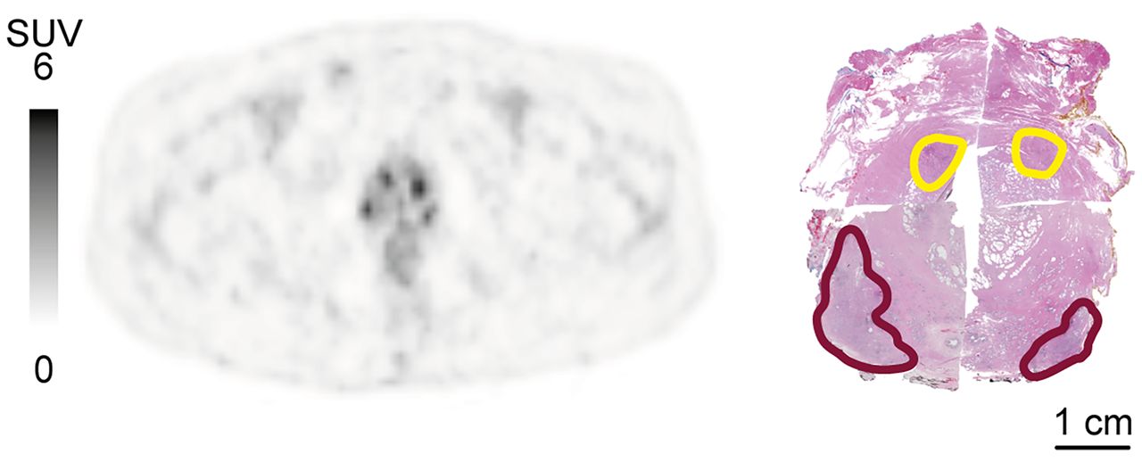
Imaging of PCa and high-grade PIN in PCa patient. (Left) SUV PET image 60 min after injection, showing almost equal uptake in PCa and PIN. (Right) Corresponding histopathology slide, with tumor and PIN delineated in red and yellow, respectively.
- FIGURE 6.
Pharmacokinetic excretion patterns of 68Ga-SB3 from pancreas, tumor, and normal prostate. Pancreas and prostate show excretion with biologic half-time of 196 and 135 min, respectively. Excretion phase of tumor shows half-time of 235 min. Fits (solid lines) and 95% CIs (dotted lines) are indicated.


Tables
Patient no. Side of prostate GS Autoradiography result PET score SUVmax PET result PI-RADS MRI result 1 L and R 3 + 4 = 7 ++ 5 22.7 TP 5 TP 5 14.4 TP* 5* TP* 4 5.9 TP* 5* TP* 2 L 3 + 4 = 7 +++ 5 7.7 TP 4 TP R 3 + 3 = 6 5 17.0 TP — FN 3 L and R 4 + 3 = 7 + 5 13.3 TP 3 TP R 4 + 3 = 7 4 4.7 TP — FN 4 L and R 3 + 4 = 7 + 5 9.9 TP 3 TP 4 6.9 TP* 3* TP* 5 L 3 + 3 = 6 − 3 5.5 TP 5 TP R 4 + 4 = 8 5 5.5 TP 5 TP 5 6.3 TP* 5* TP* 3 3.2 TP* 5* TP* 4 5.3 TP* 5* TP* 6 L 3 + 4 = 7 − 4 5.8 TP 4 TP R 3 + 4 = 7 4 5.8 TP 3 TP L PIN 5 4.9 FP TN R PIN 5 4.8 FP TN 7 L 4 + 3 = 7 + 4 4.0 TP 5 TP R 4 + 3 = 7 5 5.5 TP — FN 5 5.5 TP* —* FN* 4 3.7 TP* —* FN* 8 L 3 + 3 = 6 +++ 5 4.4 TP NA NA L 3 + 3 = 6 4 3.6 TP NA NA 9 L and R 3 + 4 = 7 − FN 4 TP 10 L and R 3 + 3 = 6 − FN NA NA * Grouped results with line above. − = GRPr-negative; + = 0%–33% GRPr-positive; ++ = 33%–66% GRPr-positive; +++ = 66%–100% GRPr-positive; TP = true positive; FN = false negative; FP = false positive; TN = true negative; NA = not available.
Supplemental Data
Files in this Data Supplement:
In this issue


Jump to section
Related Articles
Cited By...
- No citing articles found.