


Abstract
641
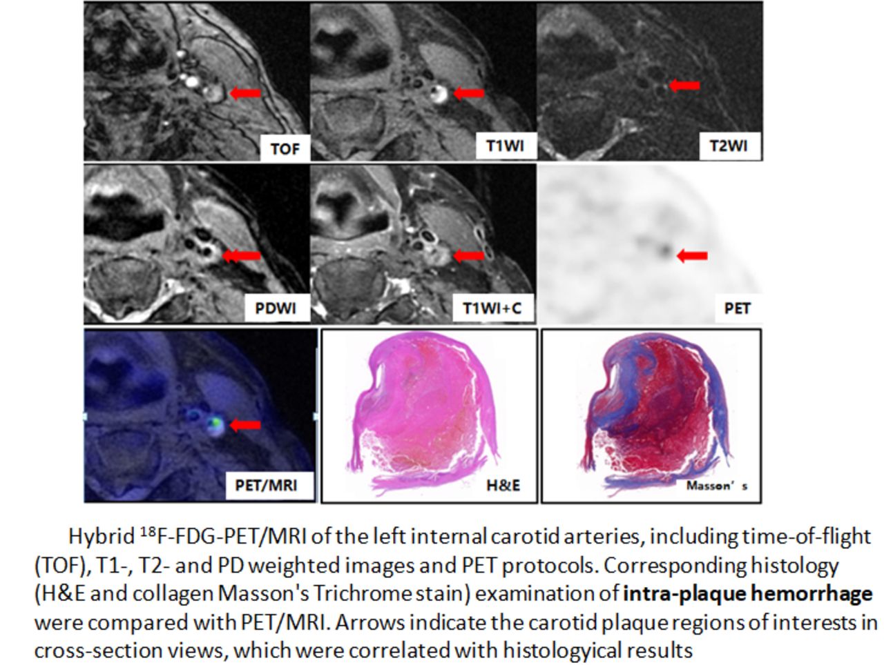
Objectives: Atherosclerosis is a chronic disease in which inflammation plays an important role at all stages. As18F-FDG-PET can be used for quantitative imaging macrophages metabolism of response to inflammation with high sensitivity and specificity. Dynamic contrast-enhanced magnetic resonance imaging (DCE-MRI) can be used for imaging neovascularization with high spatial resolution. It remains unclear whether these parameters are correlated or represent independent imaging parameters. This study determines to investigate the correlation between inflammation and neovascularization in atherosclerotic carotid plaques with hybrid 18F-FDG PET/MRI, further to verify accuracy by pathological examination, and to evaluate the clinical potential of hybrid 18F-FDG PET/MRI in carotid plaque stability. Materials and Methods: Twenty-five patients with transient ischemic attack or minor stroke in the carotid territory and ipsilateral carotid artery stenosis of 50% to 90% were included. All patients underwent hybrid PET/MR a median of 130 min after injection of 18F-FDG. 18F-FDG standard uptake values with target-to-background ratio (TBR) were determined on corresponding PET sections. Neovascularization was quantified by transfer constant (Ktrans) on corresponding DCE-MRI sections. A cohort of symptomatic patients (n=7) with carotid stenosis scheduled for carotid endarterectomy (CEA) was separately imaged with hybrid 18F-FDG PET/MRI and findings were correlated with histochemical assessment of intact carotid plaques. Spearman rank correlation coefficients between TBR and Ktrans were calculated.
Results: The correlation between TBR and Ktrans was only marginal in the whole study sample (r=0.25, p=0.043). The two variables correlated with each other in the symptomatic plaques (r=0.71, p=0.013), but were independent in the asymptomatic plaques (r=0.03, p=0.473). Neither TBR nor Ktrans was significantly higher in the symptomatic plaques, but both showed inverse relationships with time since last cerebrovascular ischemic event (r=-0.92 and -0.74 for TBR and Ktrans, respectively). Histological findings from CEA confirmed that inflammation and neovascularization expression was localized within carotid vulnerable plaques. Co-localization of cellular CD31 and CD68 expression in the plaque was observed by immunofluorescence staining.
Conclusions: The correlation between inflammation and neovascularization in carotid atherosclerotic plaques with hybrid 18F-FDG PET/MR varied with clinical conditions, pointing to a complex interplay between macrophages and neovessels under different pathophysiological conditions. Hybrid 18F-FDG PET/MR systems can help to evaluate the correlation between inflammation and neovascularization in carotid atherosclerotic plaques. Key words: Atherosclerosis, Vulnerable plaque, Inflammation, Neovascularization, Hybrid PET/MR.


In this issue


Jump to section
Related Articles
Cited By...
- No citing articles found.