
Main menu

  • Home
  • Content
    • Current
    • Ahead of print
    • Past Issues
    • JNM Supplement
    • SNMMI Annual Meeting Abstracts
  • Subscriptions
    • Subscribers
    • Institutional and Non-member
    • Rates
    • Corporate & Special Sales
    • Journal Claims
  • Authors
    • Submit to JNM
    • Information for Authors
    • Assignment of Copyright
    • AQARA requirements
  • Info
    • Permissions
    • Advertisers
    • Continuing Education
  • About
    • About Us
    • Editorial Board
    • Contact Information
  • More
    • Alerts
    • Feedback
    • Help
    • SNMMI Journals
  • SNMMI
    • JNM
    • JNMT
    • SNMMI Journals
    • SNMMI

User menu

  • Subscribe
  • My alerts
  • Log in
  • My Cart

Search

  • Advanced search
Journal of Nuclear Medicine
  • SNMMI
    • JNM
    • JNMT
    • SNMMI Journals
    • SNMMI
  • Subscribe
  • My alerts
  • Log in
  • My Cart
Journal of Nuclear Medicine

Advanced Search

  • Home
  • Content
    • Current
    • Ahead of print
    • Past Issues
    • JNM Supplement
    • SNMMI Annual Meeting Abstracts
  • Subscriptions
    • Subscribers
    • Institutional and Non-member
    • Rates
    • Corporate & Special Sales
    • Journal Claims
  • Authors
    • Submit to JNM
    • Information for Authors
    • Assignment of Copyright
    • AQARA requirements
  • Info
    • Permissions
    • Advertisers
    • Continuing Education
  • About
    • About Us
    • Editorial Board
    • Contact Information
  • More
    • Alerts
    • Feedback
    • Help
    • SNMMI Journals
  • Follow JNM on Twitter
  • Visit JNM on Facebook
  • Join JNM on LinkedIn
  • Subscribe to our RSS feeds
Meeting ReportCardiovascular

Evaluation of 18F-FGA PET/CT for specific imaging of necrotic tissue in a mouse model of coronary artery ligation

Hariprasad Gali, Alexander Mdzinarishvili, Andria Hedrick, Huining Da and Vibhudutta Awasthi
Journal of Nuclear Medicine May 2020, 61 (supplement 1) 228;
Hariprasad Gali
1University of OK Hlth Sci Ctr Oklahoma City OK United States
  • Find this author on Google Scholar
  • Find this author on PubMed
  • Search for this author on this site
Alexander Mdzinarishvili
1University of OK Hlth Sci Ctr Oklahoma City OK United States
  • Find this author on Google Scholar
  • Find this author on PubMed
  • Search for this author on this site
Andria Hedrick
1University of OK Hlth Sci Ctr Oklahoma City OK United States
  • Find this author on Google Scholar
  • Find this author on PubMed
  • Search for this author on this site
Huining Da
1University of OK Hlth Sci Ctr Oklahoma City OK United States
  • Find this author on Google Scholar
  • Find this author on PubMed
  • Search for this author on this site
Vibhudutta Awasthi
2Hexakit, Inc. Oklahoma City OK United States
  • Find this author on Google Scholar
  • Find this author on PubMed
  • Search for this author on this site
  • Article
  • Figures & Data
  • Info & Metrics
Loading

Abstract

228

Objectives: Myocardial infarction (MI) affects 735,000 Americans/year and is a leading cause of death worldwide. Its diagnosis is challenging, especially in presence of atypical symptoms, absence of chest pain, and normal ECG or cardiac troponin profile. Imaging methods also do not accurately discriminate between ischemic and necrotic regions in affected tissue. We posit that information about location and extent of infarct will improve MI diagnosis. We investigated 2-Deoxy-2-[18F]fluoroglucaric acid (FGA), the first radiopharmaceutical for PET imaging of necrosis, in a mouse model of MI.

Methods: FGA was synthesized from [18F]fluoro-2-deoxy-2-D-glucose (FDG) by a kit based procedure using TEMPO as a catalyst. MI was produced in CD-1 mice by ligation of left descending coronary artery. PET/CT images were acquired at 1 h, 6 h, 1 d, 3 d, and 4 d after occlusion; FGA (approximately 300 µCi in 300 μl) was injected via the tail vein and the images were acquired 1 h after injection. Standardized uptake values (SUVs) in heart were calculated. Separately, 1 h tissue biodistribution of FGA was assessed in both healthy control and MI mice. Myocardial injury was verified at necropsy by tissue staining with tetrazolium chloride and plasma cardiac troponin levels. For mechanistic analyses of FGA accumulation in necrosis, H9C2 cells were necrosed by H2O2 treatment (50 µM) and exposed to biotin-labeled FGA. Nuclear, mitochondrial, and cytoplasmic fractions were separated by SDS-PAGE for probing with streptavidin-HRP.

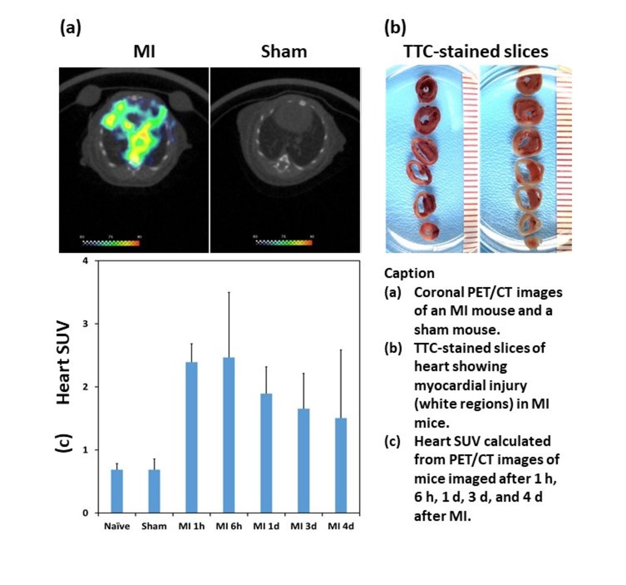
Results: Kit-based reaction converted FDG quantitatively into FGA within 7 min (radiochemical purity greater than 99%). Uptake of FGA in heart was significantly higher in MI mice than in control mice; images clearly demonstrated FGA accumulation in MI heart. Heart/skeletal muscle ratio was 2.8 in MI mice versus 1.5 in control mice. SUVs from MI mice were significantly higher than those from naïve/sham groups at all time-points of imaging post-MI. In H9C2 cells, we found enhanced uptake of biotin-FGA in nuclear fraction of H2O2-treated cells; cytoplasmic and mitochondrial fractions showed no difference.

Conclusions: Results indicate that FGA accumulates in necrotic tissue of MI heart, which could be employed for discriminatory imaging of salvageable ischemic tissue from non-viable tissue. This highly specific and positive predictive value information has potential to impact clinical decisions in management of MI patients. Mechanistically, FGA appears to target hitherto unknown nuclear proteins exposed after necrotic cell death.

Figure
  • Download figure
  • Open in new tab
  • Download powerpoint
Previous
Back to top

In this issue

Journal of Nuclear Medicine
Vol. 61, Issue supplement 1
May 1, 2020
  • Table of Contents
  • Index by author
Article Alerts
Sign In to Email Alerts with your Email Address
Email Article

Thank you for your interest in spreading the word on Journal of Nuclear Medicine.

NOTE: We only request your email address so that the person you are recommending the page to knows that you wanted them to see it, and that it is not junk mail. We do not capture any email address.

Enter multiple addresses on separate lines or separate them with commas.
Evaluation of 18F-FGA PET/CT for specific imaging of necrotic tissue in a mouse model of coronary artery ligation
(Your Name) has sent you a message from Journal of Nuclear Medicine
(Your Name) thought you would like to see the Journal of Nuclear Medicine web site.
Citation Tools
Evaluation of 18F-FGA PET/CT for specific imaging of necrotic tissue in a mouse model of coronary artery ligation
Hariprasad Gali, Alexander Mdzinarishvili, Andria Hedrick, Huining Da, Vibhudutta Awasthi
Journal of Nuclear Medicine May 2020, 61 (supplement 1) 228;

Citation Manager Formats

  • BibTeX
  • Bookends
  • EasyBib
  • EndNote (tagged)
  • EndNote 8 (xml)
  • Medlars
  • Mendeley
  • Papers
  • RefWorks Tagged
  • Ref Manager
  • RIS
  • Zotero
Share
Evaluation of 18F-FGA PET/CT for specific imaging of necrotic tissue in a mouse model of coronary artery ligation
Hariprasad Gali, Alexander Mdzinarishvili, Andria Hedrick, Huining Da, Vibhudutta Awasthi
Journal of Nuclear Medicine May 2020, 61 (supplement 1) 228;
Twitter logo Facebook logo LinkedIn logo Mendeley logo
  • Tweet Widget
  • Facebook Like
  • Google Plus One
Bookmark this article

Jump to section

  • Article
  • Figures & Data
  • Info & Metrics

Related Articles

  • No related articles found.
  • Google Scholar

Cited By...

  • No citing articles found.
  • Google Scholar

More in this TOC Section

Cardiovascular

  • Cardiac β-Adrenergic Receptor Downregulation, Evaluated by Cardiac PET, in Chronotropic Incompetence
  • Diagnostic Performance of PET Versus SPECT Myocardial Perfusion Imaging in Patients with Smaller Left Ventricles: A Substudy of the 18F-Flurpiridaz Phase III Clinical Trial
  • Quantification of Macrophage-Driven Inflammation During Myocardial Infarction with 18F-LW223, a Novel TSPO Radiotracer with Binding Independent of the rs6971 Human Polymorphism
Show more Cardiovascular

New Techniques and Tracers for Ischemic Heart Disease (Basic Science, Cardio)

  • CT-Less Attenuation Correction in Image Space Using Deep Learningfor Dedicated Cardiac SPECT: A Feasibility Study
  • In vitro characterization of a novel PET tracer for fatty acid oxidation, 18F-AS3504073-00
  • NOVEL METHODOLGY FOR MEASURING REGIONAL MYOCARDIAL EFFICIENCY
Show more New Techniques and Tracers for Ischemic Heart Disease (Basic Science, Cardio)

Similar Articles

SNMMI

© 2023 Journal of Nuclear Medicine

Powered by HighWire