


Article Figures & Data
Figures
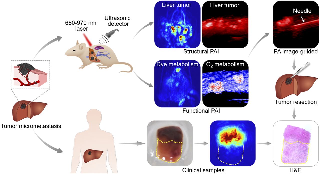
- FIGURE 1.
Workflow of PAI for melanoma hepatic-metastasis detection, profiling, and resection for animal models and clinical samples. Two kinds of hepatic metastasis models were established by injecting B16 tumor cells directly into mouse hepatic lobe and subcapsular spleen. B16 cells entered liver tissue through hepatic portal vein and developed into tumors. Tumor samples were used for PAI and were pathologically verified. H&E = hematoxylin and eosin staining; PA = photoacoustic.
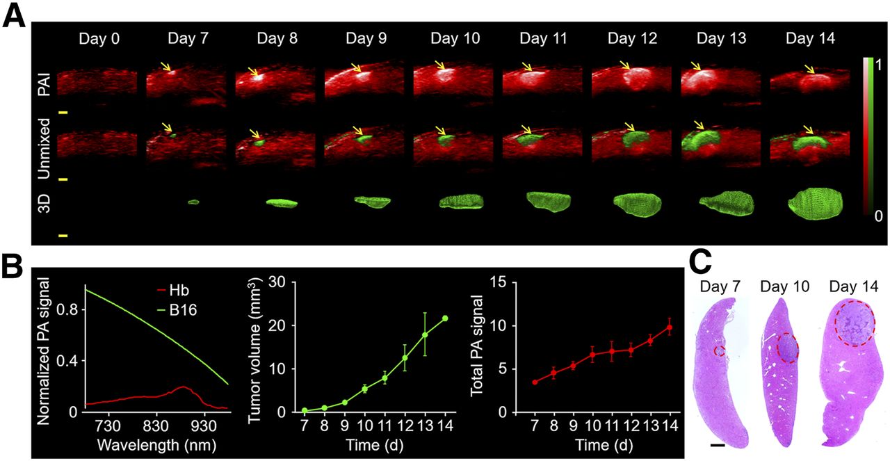
- FIGURE 2.
PAI displays changes in hepatic melanoma in situ. (A) In vivo PAI of liver before and after hepatic melanoma modeling by hepatic injection at 780 nm (n = 3). Melanoma (green) and hemoglobin (red) were unmixed by multiwavelength PAI at 680, 730, 924, and 950 nm. 3D view of tumors is shown. Scale bars in all images are 1 mm. Arrows indicate photoacoustic signal. (B) Normalized photoacoustic signal spectra of hemoglobin and B16. Measurement of tumor volume and photoacoustic signal by PAI. (C) Hematoxylin and eosin staining of tumor at different time points. Scale bars in histologic images are 1 mm. Hb = hemoglobin; PA = photoacoustic.
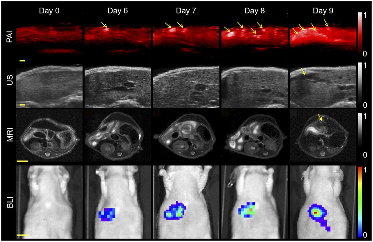
- FIGURE 3.
Comparison of various imaging modalities. PAI, ultrasound, bioluminescence imaging, and MRI of mouse on different days after tumor cell injection into spleen. Hepatic metastases could be found on PAI, ultrasound, and MRI on days 6, 9, and 9, respectively. Excitation wavelength was 780 nm. Scale bars in PAI scans and ultrasound images are 1 mm. Arrows denote metastatic tumors. Scale bars in MR and bioluminescence images are 5 and 10 mm, respectively. PA = photoacoustic; US = ultrasound.
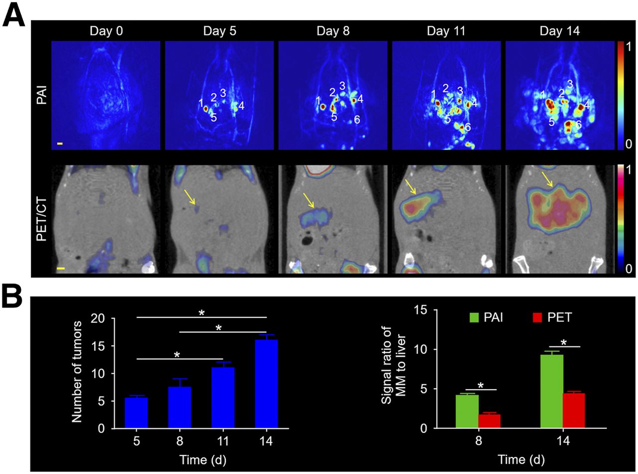
- FIGURE 4.
PAI and PET for mice with melanoma metastatic to the liver. (A) Images on different days after tumor cell injection at 3-d intervals. Scale bars in all images are 1 mm. (B) Number of tumors over time. Signal ratio for melanoma metastases to liver was quantified in PAI and PET at 8 and 14 d after tumor cell injection. MM = melanoma metastasis.
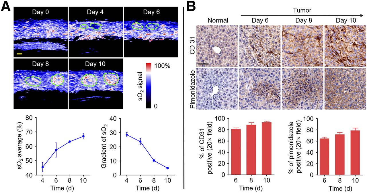
- FIGURE 5.
Monitoring of functional parameters in tumor and normal tissue. (A) Increase in tumor blood oxygenation from days 4 to 10. Region of interest represents tumor. Scale bars in sO2 images are 1 mm. sO2 average and gradient of sO2 are quantified. (B) Representative immunohistochemistry images of slices collected from normal-mouse liver and melanoma-mouse liver on days 6, 8, and 10 after injection of B16 cells. Nuclei, tumor angiogenesis, and tumor hypoxic areas are stained with hematoxylin, anti-CD31 antibody, and antipimonidazole antibody, respectively. Scale bars in immunohistochemistry images are 50 μm. Tumor angiogenesis and hypoxic areas from immunohistochemistry images are quantified.
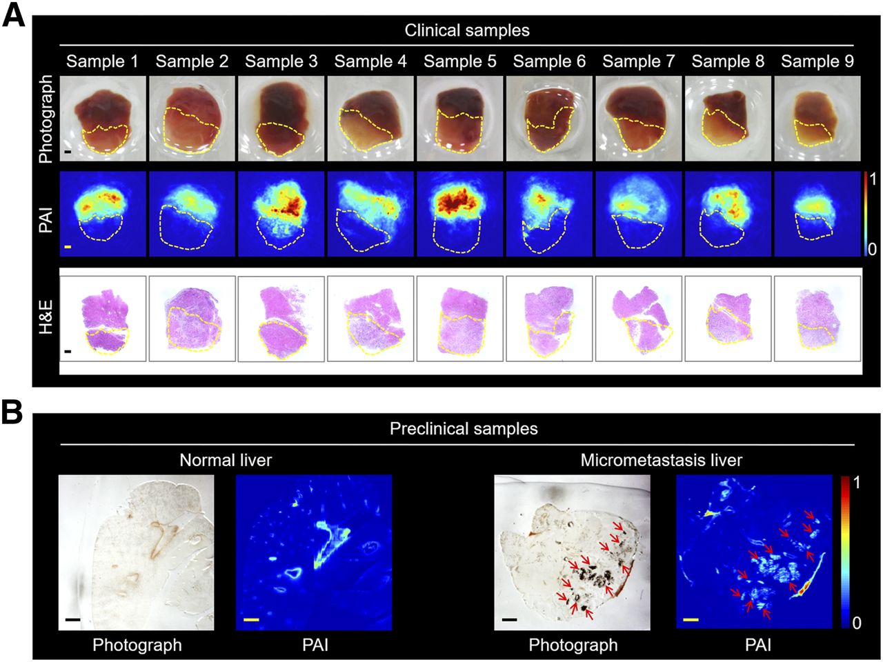
- FIGURE 6.
PAI of clinical and preclinical samples. (A) Photographs of tumor samples (in ambient normal tissue) from 9 patients, along with PAI scans and hematoxylin and eosin staining. Region of interest represents tumor. Scale bars are 1 mm. (B) Photographs and PAI of normal and melanoma micrometastasis liver slices. Arrows denote tumor. Thickness of liver slice is 50 μm. Scale bars are 1 mm. MM = melanoma metastasis.
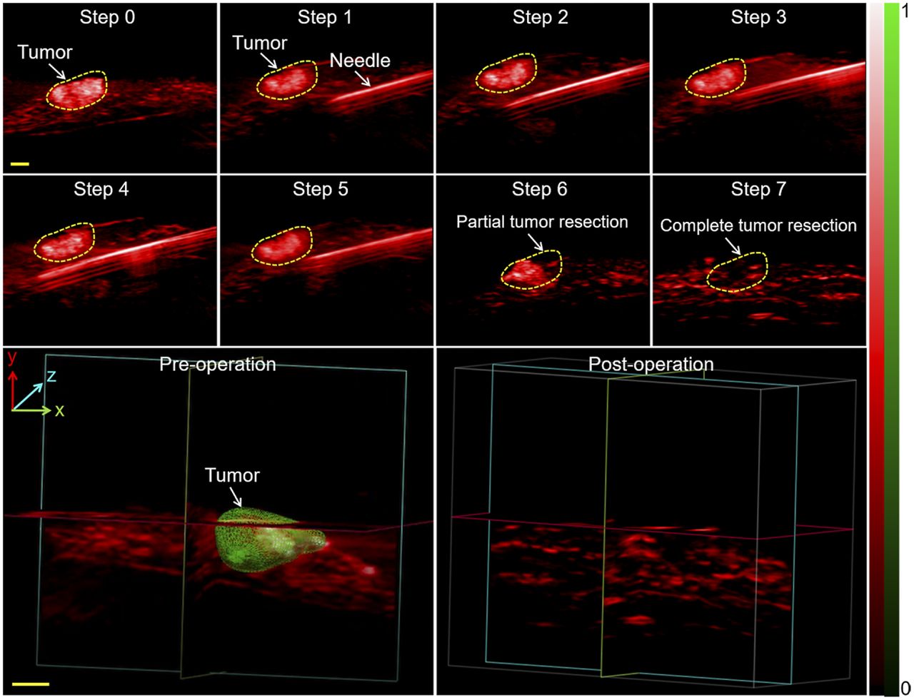
- FIGURE 7.
Needle insertion and PAI–guided tumor resection in vivo. Shown are PAI scans of metastatic tumor after laparotomy (step 1), advancement of needle toward tumor (step 2), partial resection (step 3), and complete resection (step 4) with PAI guidance. 3D PAI scans before and after resection are displayed. 3D melanoma (green) and hemoglobin (red) distributions were estimated by spectral unmixing analyses from spectroscopic acquisitions at 680, 730, 924, and 950 nm. Scale bars in all images are 1 mm. H&E = hematoxylin and eosin staining.


Additional Files
Supplemental Data
Files in this Data Supplement:


