
Main menu

  • Home
  • Content
    • Current
    • Ahead of print
    • Past Issues
    • JNM Supplement
    • SNMMI Annual Meeting Abstracts
    • Continuing Education
    • JNM Podcasts
  • Subscriptions
    • Subscribers
    • Institutional and Non-member
    • Rates
    • Journal Claims
    • Corporate & Special Sales
  • Authors
    • Submit to JNM
    • Information for Authors
    • Assignment of Copyright
    • AQARA requirements
  • Info
    • Reviewers
    • Permissions
    • Advertisers
  • About
    • About Us
    • Editorial Board
    • Contact Information
  • More
    • Alerts
    • Feedback
    • Help
    • SNMMI Journals
  • SNMMI
    • JNM
    • JNMT
    • SNMMI Journals
    • SNMMI

User menu

  • Subscribe
  • My alerts
  • Log in
  • My Cart

Search

  • Advanced search
Journal of Nuclear Medicine
  • SNMMI
    • JNM
    • JNMT
    • SNMMI Journals
    • SNMMI
  • Subscribe
  • My alerts
  • Log in
  • My Cart
Journal of Nuclear Medicine

Advanced Search

  • Home
  • Content
    • Current
    • Ahead of print
    • Past Issues
    • JNM Supplement
    • SNMMI Annual Meeting Abstracts
    • Continuing Education
    • JNM Podcasts
  • Subscriptions
    • Subscribers
    • Institutional and Non-member
    • Rates
    • Journal Claims
    • Corporate & Special Sales
  • Authors
    • Submit to JNM
    • Information for Authors
    • Assignment of Copyright
    • AQARA requirements
  • Info
    • Reviewers
    • Permissions
    • Advertisers
  • About
    • About Us
    • Editorial Board
    • Contact Information
  • More
    • Alerts
    • Feedback
    • Help
    • SNMMI Journals
  • View or Listen to JNM Podcast
  • Visit JNM on Facebook
  • Join JNM on LinkedIn
  • Follow JNM on Twitter
  • Subscribe to our RSS feeds
Review ArticleContinuing Education

Nuclear Imaging of Bacterial Infection: The State of the Art and Future Directions

Ilona Polvoy, Robert R. Flavell, Oren S. Rosenberg, Michael A. Ohliger and David M. Wilson
Journal of Nuclear Medicine December 2020, 61 (12) 1708-1716; DOI: https://doi.org/10.2967/jnumed.120.244939
Ilona Polvoy
1Department of Radiology and Biomedical Imaging, University of California, San Francisco, San Francisco, California
  • Find this author on Google Scholar
  • Find this author on PubMed
  • Search for this author on this site
Robert R. Flavell
1Department of Radiology and Biomedical Imaging, University of California, San Francisco, San Francisco, California
  • Find this author on Google Scholar
  • Find this author on PubMed
  • Search for this author on this site
Oren S. Rosenberg
2Department of Medicine, University of California, San Francisco, San Francisco, California; and
  • Find this author on Google Scholar
  • Find this author on PubMed
  • Search for this author on this site
Michael A. Ohliger
1Department of Radiology and Biomedical Imaging, University of California, San Francisco, San Francisco, California
3Department of Radiology, Zuckerberg San Francisco General Hospital, San Francisco, California
  • Find this author on Google Scholar
  • Find this author on PubMed
  • Search for this author on this site
David M. Wilson
1Department of Radiology and Biomedical Imaging, University of California, San Francisco, San Francisco, California
  • Find this author on Google Scholar
  • Find this author on PubMed
  • Search for this author on this site
  • Article
  • Figures & Data
  • Info & Metrics
  • PDF
Loading

Article Figures & Data

Figures

  • Tables
  • FIGURE 1.
    • Download figure
    • Open in new tab
    • Download powerpoint
    FIGURE 1.

    Examples of structural and functional imaging used in diagnosis of infection. (A) Plain radiography of 55-y-old man with diskitis–osteomyelitis after corpectomy. (B and C) CT and MRI of 23-y-old man with chronic diskitis–osteomyelitis. (D) Ultrasound of 4-y-old boy with perforated appendicitis and associated abscess. (E) 99mTc-methylene diphosphonate bone scan of right ulnar osteomyelitis. (Reprinted with permission of (25).) (F) 111In-WBC SPECT/CT of infected right knee arthroplasty. (Reprinted with permission of (25).) (G) 67Ga-citrate scan of 61-y-old man with infected endovascular graft of aortic arch. (Reprinted with permission of (74).) (H) 18F-FDG/PET of 66-y-old man with infected thoracic aorta endograft. (A–D) Red arrowhead indicates point of infection; (E–H) red arrowhead indicates increased tracer uptake.
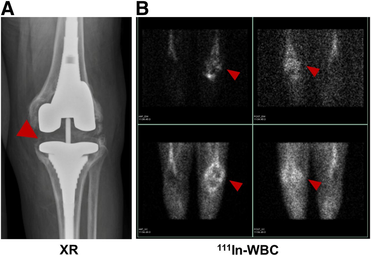
  • FIGURE 2.
    • Download figure
    • Open in new tab
    • Download powerpoint
    FIGURE 2.

    64-y-old woman with knee replacement and periprosthetic osteomyelitis (red arrowhead) as depicted via plain radiography (A) and radiolabeled 111In-leukocyte imaging (B, top row) demonstrates brightest uptake at medial aspect of tibial plateau. 99mTc-sulfur colloid imaging (B, bottom row) demonstrates no corresponding uptake in region of medial tibial plateau. Therefore, findings are consistent with osteomyelitis.
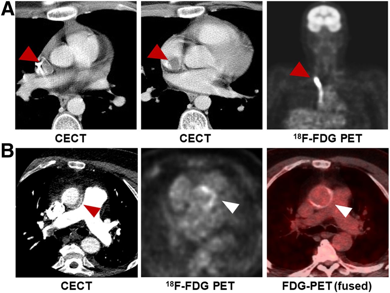
  • FIGURE 3.
    • Download figure
    • Open in new tab
    • Download powerpoint
    FIGURE 3.

    Examples of 18F-FDG PET in cardiovascular disease. (A) A 17-y-old boy with non-Hodgkin lymphoma, found to have catheter-associated thrombus consistent with infection, with arrowheads indicating increased FDG uptake in and around catheter. (B) A 59-y-old man with aortic valve prosthesis infection caused by E. faecalis, requiring surgical replacement. Arrowheads indicate increased uptake by valve. CECT = contrast-enhanced CT.

  • FIGURE 4.
    • Download figure
    • Open in new tab
    • Download powerpoint
    FIGURE 4.

    Incidental finding of infection in 65-y-old woman with fallopian tube cancer. (A) Contrast-enhanced CT shows tonsillar abscess (arrowheads). (B and C) Focal increase in uptake on 18F-FDG PET and 18F-FDG PET/CT in same location (red arrowhead).

  • FIGURE 5.
    • Download figure
    • Open in new tab
    • Download powerpoint
    FIGURE 5.

    Examples of novel non–sugar-based infection-targeted radiotracers for PET. (A and B) Increased uptake of radiotracers in infection compared with sterile inflammation in 2 rodent models: 18F-fluoropropyl-trimethoprim (FPTMP) uptake in mice infected with E. coli (arrowhead shows sterile inflammation, arrow shows infection) (A, reprinted with permission of (53)) and 18F-PABA uptake in rat infected with S. aureus (red arrows show sterile inflammation, yellow arrows show infection) (B, reprinted with permission of (64)). (C) 11C-d-Ala uptake in rat intervertebral disk infected with S. aureus (red arrowheads) and mouse lung infected with P. aeruginosa (red arrowheads). (Reprinted with permission of (73).) (D–F) Chemical structures of 18F-FPTMP (D), 18F-PABA (E), and 11C-d-Ala (F). ID = injected dose.

  • FIGURE 6.
    • Download figure
    • Open in new tab
    • Download powerpoint
    FIGURE 6.

    Examples of novel sugar-based infection-targeted radiotracers for PET/CT. (A–C) Increased uptake of radiotracers in infection compared with sterile inflammation in 3 rodent models: 18F-maltohexaose uptake in rat infected with E. coli (left arrows show infection, right arrows show sterile inflammation) (A, reprinted with permission of (57)), 18F-fluoromaltotriose uptake in mice infected with E. coli (yellow arrow shows infection site) (B, reprinted with permission of (59)), and 2-deoxy-2-18F-fluorosorbitol (18F-FDS) uptake in mice infected with E. coli (yellow arrows show infection, red arrows show sterile inflammation). (C, reprinted with permission of (61)). (D–F) Chemical structures of 18F-maltohexaose (D), 18F-fluoromaltotriose (E), and 18F-FDS (F), obtained from chemical reduction of 18F-FDG. CFU = colony-forming unit; ID = injected dose.

Tables

  • Figures
    • View popup
    TABLE 1

    Common Nuclear Imaging Techniques (15,75–77)

    RadiotracerTargetMain infectious indicationsHalf-lifeAdministered activity (MBq)AdvantagesDisadvantages
    Bone scan (99mTc-MDP)Active bone formationPBI*6 h500–1,110SensitiveLow specificity
    SOMLow costSusceptible to confounders (surgery, trauma)
    Late PJIAccessible
    Septic arthritisGood spatial resolution
    Necrotizing external otitisLow radiation dose
    WBC scan (99mTc-WBC)LeukocytesPBI† (violated bone)6 h185–370Sensitive, especially for neutrophilic induced inflammationDepends on host immune system; sensitivity decreases after antibiotic treatment
    Diabetic footBlood exposure
    Early PJIRequires sterility
    Infective endocarditisTime consuming
    Vascular graft infectionPoor resolution
    FUOHigh radiation dose
    67Ga-citrateTransferrinSOM78.3 h150–220Suitable for immunodeficiencyDelayed imaging
    Bacterial siderophoresOpportunistic infectionsPoor resolution
    Neutrophilic lactoferrinFUOHigh radiation dose
    Necrotizing external otitisExpensive
    Requires cyclotron
    18F-FDG PETEnergy consumptionPBI*110 min185–740SensitiveDepends on host immune system
    SOMSuitable for acute and chronic inflammationExpensive
    Infective endocarditisHigh resolutionLacks widespread availability
    Vascular graft infectionRelatively short scanSusceptible to confounders (e.g., surgery)
    FUOSUV quantificationRequires patient preparation
    High radiation dose
    • ↵* Nonviolated bone.

    • ↵† Violated bone.

    • MDP = methylene diphosphonate; PBI = peripheral bone infection; SOM = spinal osteomyelitis; PJI = prosthetic joint infection.

    • View popup
    TABLE 2

    Recent Bacteria-Specific Radiotracers

    TracerMechanismTarget bacterial pathogenPathogens tested in vivo (CFUsadministered)Maximum infection-to-inflammation ratioStage (published reports)
    18F-FPTMPInhibition of bacterial dihydrofolate reductaseG+, G−E. coli (106–108)∼3 (108 E. coli CFU)Preclinical
    S. aureus (108)
    P. aeruginosa (107)
    18F-FAGBacterial cell wallG+, G−E. coli (107)∼2Preclinical
    18F-maltohexaoseMaltodextrin transporterG+, G−E. coli (105–109)7 (109 CFU)Preclinical
    18F-fluoro-maltoseMaltodextrin transporterG+, G−E. coli (108)1.3Preclinical
    18F-fluoro-maltotrioseMaltodextrin transporterG+, G−E. coli (106-108)3.4 (108 E. coli CFU)Preclinical
    L. monocytogenes (2 × 10⁵)
    P. aeruginosa (106)
    S. aureus
    18F-FDSBacterial energy consumptionG−*E. coli (107)7.3 (E. coli)Clinical
    S. aureus (107–108)
    P. aeruginosa (106.5)
    11C-PABAFolic acid biosynthesisG+, G−E. coli2.6Clinical
    18F-PABAFolic acid biosynthesisG+, G−S. aureus (107–108)7.95 (108)Preclinical
    11C-d-MetBacterial cell wallG+, G−E. coli2Clinical
    S. aureus
    11C-d-AlaBacterial cell wallG+, G−E. coli (5 × 10⁶)3.5 (S. aureus)Preclinical
    S. aureus (5 × 10⁶)
    P. aeruginosa (2 × 10⁶)
    • ↵* Enterobacteriaceae.

    • CFU = colony forming units; 18F-FPTMP = 18F-fluoropropyl-trimethoprim; G+ = Gram-positive bacteria; G− = Gram-negative bacteria; 18F-FAG = 2-deoxy-2-18F-fluoroacetamido-d-glucopyranose; 18F-FDS = 2-deoxy-2-18F-fluorosorbitol; 11C-d-Met = d-methyl-11C-methionine.

PreviousNext
Back to top

In this issue

Journal of Nuclear Medicine: 61 (12)
Journal of Nuclear Medicine
Vol. 61, Issue 12
December 1, 2020
  • Table of Contents
  • Table of Contents (PDF)
  • About the Cover
  • Index by author
Print
Download PDF
Article Alerts
Sign In to Email Alerts with your Email Address
Email Article

Thank you for your interest in spreading the word on Journal of Nuclear Medicine.

NOTE: We only request your email address so that the person you are recommending the page to knows that you wanted them to see it, and that it is not junk mail. We do not capture any email address.

Enter multiple addresses on separate lines or separate them with commas.
Nuclear Imaging of Bacterial Infection: The State of the Art and Future Directions
(Your Name) has sent you a message from Journal of Nuclear Medicine
(Your Name) thought you would like to see the Journal of Nuclear Medicine web site.
Citation Tools
Nuclear Imaging of Bacterial Infection: The State of the Art and Future Directions
Ilona Polvoy, Robert R. Flavell, Oren S. Rosenberg, Michael A. Ohliger, David M. Wilson
Journal of Nuclear Medicine Dec 2020, 61 (12) 1708-1716; DOI: 10.2967/jnumed.120.244939

Citation Manager Formats

  • BibTeX
  • Bookends
  • EasyBib
  • EndNote (tagged)
  • EndNote 8 (xml)
  • Medlars
  • Mendeley
  • Papers
  • RefWorks Tagged
  • Ref Manager
  • RIS
  • Zotero
Share
Nuclear Imaging of Bacterial Infection: The State of the Art and Future Directions
Ilona Polvoy, Robert R. Flavell, Oren S. Rosenberg, Michael A. Ohliger, David M. Wilson
Journal of Nuclear Medicine Dec 2020, 61 (12) 1708-1716; DOI: 10.2967/jnumed.120.244939
Twitter logo Facebook logo LinkedIn logo Mendeley logo
  • Tweet Widget
  • Facebook Like
  • Google Plus One
Bookmark this article

Jump to section

  • Article
    • Abstract
    • IMAGING INFECTION: THE STATE OF THE ART
    • CLINICAL USES OF NM IN IMAGING INFECTION
    • NEWER APPROACHES TO MICROORGANISM-SPECIFIC IMAGING
    • CONCLUSION
    • Footnotes
    • REFERENCES
  • Figures & Data
  • Info & Metrics
  • PDF

Related Articles

  • This Month in JNM
  • PubMed
  • Google Scholar

Cited By...

  • The Development and Validation of Radiopharmaceuticals Targeting Bacterial Infection
  • Google Scholar

More in this TOC Section

  • Approaches to Imaging Immune Activation Using PET
  • Large Language Models and Large Multimodal Models in Medical Imaging: A Primer for Physicians
  • Precision Oncology in Melanoma: Changing Practices
Show more Continuing Education

Similar Articles

Keywords

  • infection
  • imaging
  • nuclear medicine
  • PET
  • SPECT
SNMMI

© 2025 SNMMI

Powered by HighWire