


Abstract
610
Purpose: In previous research, we identified a new target named α2δ1, which specifically expressed in cancer stem cells (CSCs). And a α2δ1 targeting antibody named 1B50-1 was produced at the same. These α2δ1+ tumor-initiating cells (TICs) with stem cell-like properties, might be the cell of origin for HCC recurrence. The exact mechanism was not known clearly. Herein, we reported the production of novel positron emission tomography (PET) radiotracer 64Cu-NOTA-1B50-1 and explore its feasibility of non-invasively detecting and quantitating of CSCs on hepatocellular carcinoma (HCC).
Methods: 1B50-1 antibody was labeled with 64Cu (using NOTA-NCS as bi-functional chelator), and NOTA-mIgG3 was labeled with same procedures as a control agent. Immuno-reactivity of NOTA conjugated and radiolabeled antibodies were tested by enzyme-linked immunosorbent assay (ELISA) analysis. We established a pair of HCC cell lines with the same clonal origin, named Hep-11 (CSCs negative) and Hep-12 (CSCs positive) hepatocellular carcinoma cell lines, by primary culture from the same patient’s primary and recurrent HCC tissues, respectively. These two paired cell lines were culture and/or xenografted on NOD-SCID mice for 64Cu-NOTA-1B50-1 evaluation. Binding affinity and binding specificity of 64Cu-NOTA-1B50-1 were evaluated by various cellular researches. Micro-PET imaging of 64Cu-NOTA-1B50-1 was performed in Hep-12/Hep-11 Hepatocellular Carcinoma cell xenografted tumor models at 2h, 12h and 36h post-injection. Radio-probes uptake in tumor and main organs were quantified by region of interested (ROI) analysis of the micro-PET images. Immunofluorescence (IF), immunohistochemistry (IHC) and autoradiography of tumor and main organ were carried out to confirm the expression of α2δ1.
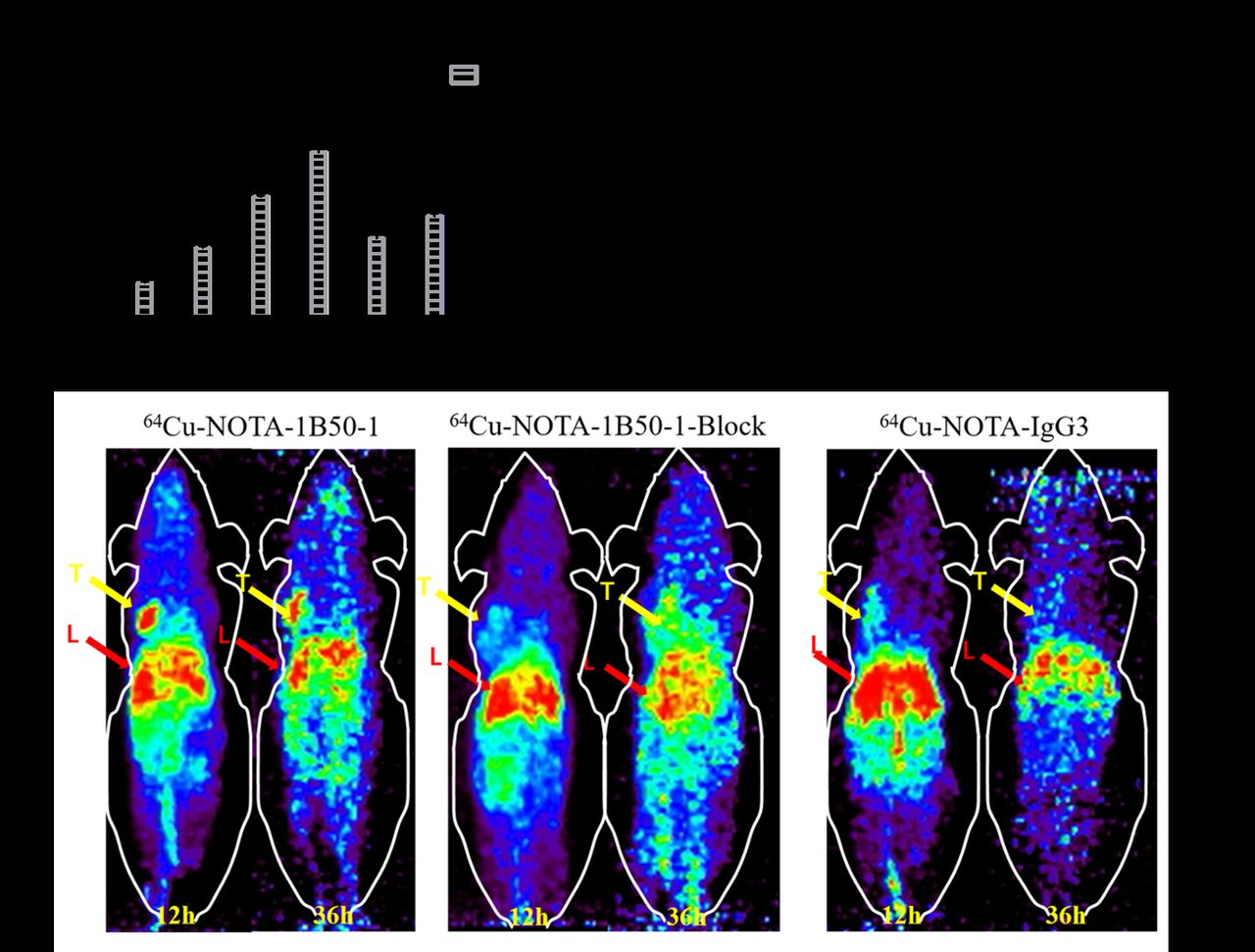
Results: The radiolabeling yield for 64Cu-NOTA-1B50-1 was 85.6 ± 2.0% (n = 6). The average specific activity of 64Cu-NOTA-1B50-1 was 5.3 GBq/µmol (37 MBq/mg) (n = 6). The radiopharmaceutical proved to be stable for up to 5 half-decay life of 64Cu both saline and 5% HAS (human serum albumin) at 4℃ incubation. In Hep-12 cellular uptakes studies, cell uptake rate of 64Cu-NOTA-1B50-1 achieved 6.5% at 2 h, which reaches 4-fold of in Hep-11 cell line, and cell uptake of 1 h and 2 h can be completely blocked (p< 0.05). In micro-PET imaging studies, the tumor uptake of 64Cu-NOTA-1B50-1 was clearly discernible at 12 and 36 h post-injection, with the T/L and T/M ratios of 3.22 ± 0.32 and 23.12 ± 5.56 at 36 h after injection. Co-injection with 0.5 mg cold 1B50-1, tumor uptake decreased significantly and 64Cu-NOTA-mIgG3 show low tumor uptake. The quantified tumor uptake of Cy5.5-1B50-1 was significantly higher than that of the Cy5.5-IgG3 at all-time points examined (P <0 .001). In bio-distribution studies, 64Cu-NOTA-1B50-1 showed a significant higher tumor uptake (6.92 ± 1.27 ID%/g) compared with 64Cu-NOTA-mIgG3 (3.55 ± 0.88 ID%/g, p=0.0012, n=5) at 12h,(14.5 ± 0.95 ID%/g) compared with (2.79 ± 1.48 ID%/g, p<0.0001, n=5) at 36h after intravenous injections. There was no significant difference in heart, liver, kidney and spleen at 12 h and 36 h after radio-probes injection. And there were good correlations between PET imaging and immunofluorescence (IF) (n = 4) and autoradiography in Hep-12 models tumor tissues.
Conclusions: We successfully synthesized a novel PET radiotracer 64Cu-NOTA-1B50-1 and application for α2δ1+ Hep12 models. And 64Cu-NOTA-1B50-1 had a great potential for non-invasive, real-time detection, staging, and follow-up supervision of the dynamic changes of α2δ1+ cells in vivo. It may be possible to early warning of recurrence of HCC at cellular level. Keywords: HCC, 1B50-1, α2δ1, Cancer stem cells, PET/CT Figure1. (A) Cell uptake of 64Cu-NOTA-1B50-1 in Hep-12 cell lines. (B) Bind affinity researches of 64Cu-NOTA-1B50-1. (C) Micro-PET images of 64Cu-NOTA-1B50-1,co-injection with 0.5 mg 1B50-1, and 64Cu-NOTA-mIgG3 in Hep-12 models.


