


Article Figures & Data
Figures
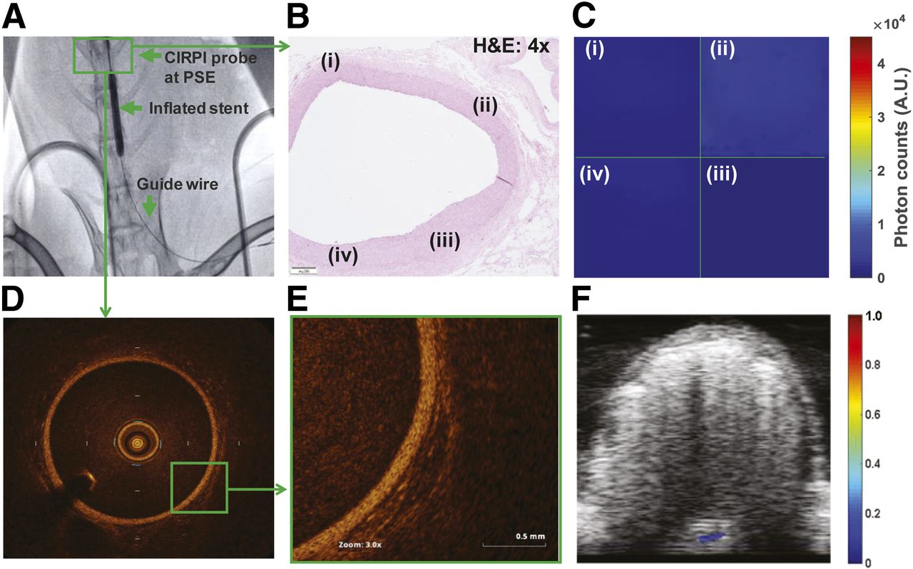
- FIGURE 1.
In vivo abdominal aorta images of the control rabbit. (A) x-ray–guided fluoroscopy image illustrates that a guide wire is followed by CIRPI probe located at the peripheral stent edge (proximal stent edge [PSE] is used as a landmark). (B) H&E-stained (2×) histologic image overviews tissue condition at which CIRPI system is used to collect radioluminescene and photoacoustic images. Further, same location was imaged with a clinical OCT system for secondary verification. Histology slide illustrates normal abdominal aortic wall thickness with no atherosclerotic plaques. (C) Radioluminescence images are collected at every 1.43° with scanning probe. Each quadrant (i), (ii), (iii), and (iv) is the representation of concatenated images between starting point of scan up to 90°. Each quadrant of radioluminescence image represents precise location of histologic slide that is highlighted with same number. None of the quadrant of radioluminescence image showed radioluminescent signal that renders an absence of macrophages. (D) Confirmatory OCT images further validate these findings by showing normal abdominal aortic wall thickness with no atherosclerotic plaques. (E) Highlighted section in OCT image shows no attenuation signals, a representation of no lipids or TCFA. (F) In photoacoustic image, no photoacoustic signals are detected at all 7 tested wavelengths, representing an absence of calcification, cholesterol cleft in form of cholesterol ester, phospholipids, cholesterol and triglyceride, and a presence of intact elastic fibers and collagen.
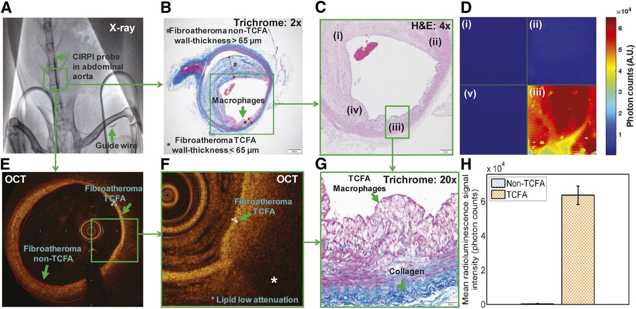
- FIGURE 2.
In vivo images of abdominal aorta with atherosclerotic plaques in WHHL rabbit. (A) x-ray–guided fluoroscopy image illustrates CIRP probe followed by guide wire marked with stent (highlighted within a green box). Histologic images are presented to give overview of plaque orientation within CRI imaging area using our CIRPI system and secondary verification with a clinical OCT system. (B) Trichrome-stained (2×) abdominal aorta highlights stable plaque, fibroatheroma non-TCFA with wall thickness > 65 μm. On the opposite wall there is clear evidence of fibroatheroma TCFA with a wall thickness < 65 μm filled with macrophages. (C) H&E-stained (4×) abdominal aorta shows 4 highlighted locations, (i), (ii), (iii), and (iv) in histology image that are same location of each quadrant of CIRPI image; (i), (ii), and (iii) represent non-TCFA, and (iv) TCFA with large macrophage accumulation. (D) Radioluminescence images are collected at every 1.43° with scanning probe. Each quadrant is representation of concatenated images between starting point of scan up to 90°. Each quadrant of radioluminescence image represents precise location of histologic slide that is highlighted with same number. Quadrant iv of radioluminescence image exhibits high radioluminescent signal that is indicative of large macrophage accumulation within TCFA. (E) Confirmatory OCT images validated presence of non-TCFA and TCFA at arterial wall of abdominal aorta. (F) Highlighted section in OCT image shows low attenuation signal, a representation of lipids within TCFA. (G) Trichrome-stained TCFA area illustrates large number of macrophages that represents same area with lipids in OCT image. (H) Statistical analysis showed 203× higher radioluminescent signal (TCFA vs. non-TCFA: 6.36 × 10⁴ ± 5.3 × 10³ vs. 3.14 × 101 ± 1.91 × 101 photon counts, P = 0.003) from area with macrophages and lipids within TCFA compared with non-TCFA.
- FIGURE 3.
Images of in vivo atherosclerotic plaques in rabbit abdominal aorta. (A) Histologic images are presented to give overview of plaque orientation within PAT imaging area using our CIRPI system and secondary verification with clinical OCT system. H&E-stained (10×) abdominal aorta highlighted plaques containing trace amount of calcification and prominent cholesterol cleft/lipids in close vicinity to macrophages, fibroatheroma TCFA with wall thickness < 65 μm. (B) Enlarged image of A shows severe cholesterol cleft and trace amount of calcification. (C) In PAT image, high photoacoustic signals were detected at area with cholesterol, calcification. (D) Confirmatory OCT images validated presence of presence of lipids at arterial wall of abdominal aorta. Highlighted section in OCT image showed low attenuation signal, a representation of lipids within TCFA. (E) EVG (20×)-stained area illustrates both broken (red arrow) and intact collagen (green arrow). (F) High photoacoustic signal was detected at area with broken collagen in PAT image collected with CIRPI system. (G) Statistical analysis showed high photoacoustic signal from (i) non-TCFA calcification (250 ± 30.33, P = 0.001, no other signal was observed); (ii) TCFA: cholesterol ester (240 ± 20.56, P = 0.003), phospholipids (110 ± 8.67, P = 0.004), cholesterol (210 ± 15.85, P = 0.002), and triglyceride (90 ± 5.75, P = 0.003), which represent the presence of severe lipids; elastin/collagen (80 ± 4.71, P = 0.0001) that elucidates broken/damaged elastic fibers and collagen.


Additional Files
Supplemental Data
Files in this Data Supplement:


Jump to section
Related Articles
Cited By...
- No citing articles found.