


Article Figures & Data
Figures
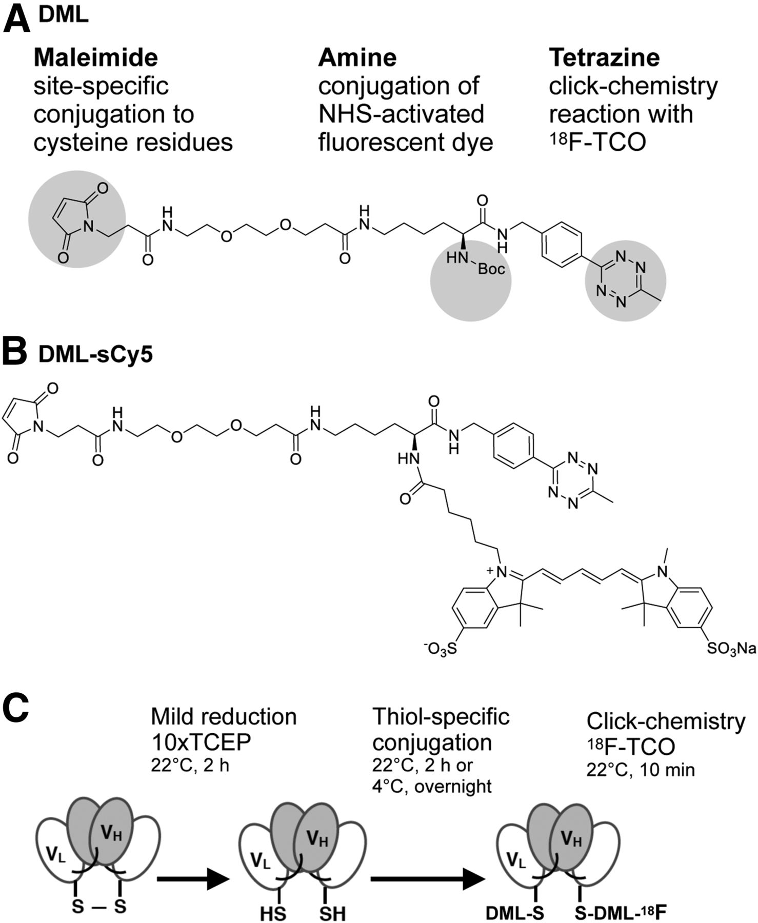
- FIGURE 1.
Concept: DML. (A) Structure of DML containing 3 functional groups. (B) Sulfo-cyanine5 NHS ester was conjugated to amine group (DML-sCy5). (C) Schematic of site-specific conjugation and radiolabeling. Reducing A2cDb C-terminal disulfide-bridge presents thiol groups for conjugation with maleimide group. Radiofluorination is achieved by click chemistry using 18F-TCO. TCEP = tris(2-carboxyethyl)phosphine; VH = heavy-chain variable domain; VL = light-chain variable domain.
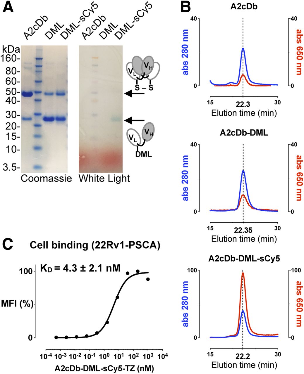
- FIGURE 2.
Biochemical characterization of DML-conjugated A2cDb. (A) Sodium dodecyl sulfate–polyacrylamide gel electrophoresis analysis of A2cDb and site-specifically conjugated A2cDb under nonreducing conditions: Coomassie-stained and unstained (white light). (B) Size exclusion chromatography of A2cDb, A2cDb-DML, and A2cDb-DML-sCy5 shows similar elution profiles for protein (280 nm). Fluorescent dye (sCy5, 650 nm) elutes at same time as protein (22.2 min), confirming conjugation to A2cDb. (C) Binding of A2cDb-DML-sCy5 to 22Rv1-PSCA cells analyzed by flow cytometry. Saturation binding curve of 1 of 3 independent experiments is shown. Apparent affinity of A2cDb-DML-sCy5 was calculated using single-site specific binding model. MFI = mean fluorescence intensity.
- FIGURE 3.
Immuno-PET imaging shows antigen-specific uptake in PSCA-positive prostate cancer xenografts. Nude mice bearing PSCA-negative (22Rv1, left shoulder) and PSCA-positive (22Rv1-PSCA, right shoulder) subcutaneous xenografts were imaged with single-modality 18F-DML-A2cDb 60-min dynamic scan (A) and 10-min static scans at 1, 2, and 4 h after injection (B) or dual-modality 18F-DMLsCy5-A2cDb 60-min dynamic scan (C) and 10-min static scans at 1, 2, and 4 h after injection (D). Depicted are representative scans (of n ≥ 6) as whole-body maximum-intensity-projection small-animal PET/CT overlays.
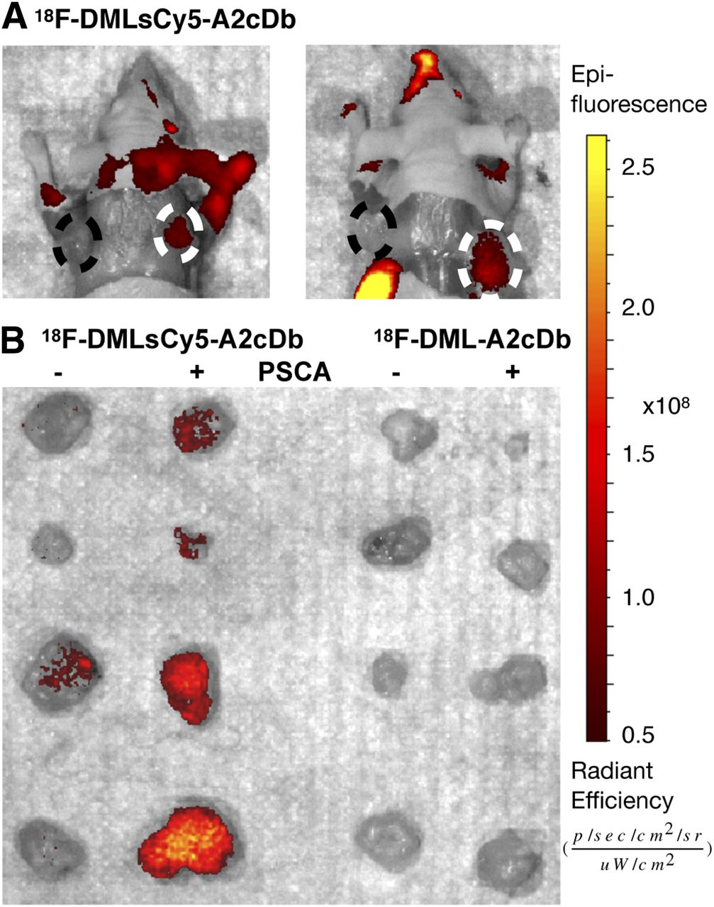
- FIGURE 4.
Same mice were assessed by optical imaging. (A) Postmortem optical imaging of mice with skin removed. Mice injected with dual-modality 18F-DMLsCy5-A2cDb show antigen-specific fluorescence signal in PSCA-positive tumor on right shoulder (dashed circle). Two representative mice (of n = 7) are shown. (B) 22Rv1 (−) and 22Rv1-PSCA (+) xenografts were analyzed ex vivo to compare relative fluorescent signal without obstruction from other organs.
- FIGURE 5.
Quantitative ROI analysis and ex vivo biodistribution. (A) Quantitative ROI analysis of blood (heart), liver, kidney, and 22Rv1-PSCA tumor. (B) Ex vivo biodistribution 4 h after injection of 18F-DML-A2cDb (n = 6) and 18F-DMLsCy5-A2cDb (n = 7). Tumors and organs were harvested and γ-counted.


Tables
18F-DML-A2cDb 18F-DMLsCy5-A2cDb Parameter Mean SD Mean SD D/P ratio ND 1.07 0.26 Labeling efficiency (%) 77.1 17.7 52.7 18.6 Specific activity (MBq/μg) 0.41 0.12 0.50 0.30 Radiochemical purity (%) 97.3 1.6 89.1 10.6 Immunoreactivity (%) 42.9 8.7 41.5 6.9 n 3 3 ND = not determined.
18F-DML-A2cDb 18F-DMLsCy5-A2cDb Site Mean SEM Mean SEM Blood 0.44 0.19 0.90 0.17 22Rv1-PSCA (+) 2.84 1.07 2.89 0.38 22Rv1 (−) 0.32 0.09 0.82 0.19 Heart 0.18 0.07 0.85 0.11 Lung 0.44 0.17 1.67 0.19 Liver 0.37 0.10 4.81 0.29 Kidney 1.71 0.56 15.2 1.1 Spleen 0.21 0.06 2.02 0.17 Stomach 0.31 0.10 0.54 0.08 Intestine 13.7 2.7 6.37 0.46 Muscle 0.05 0.01 0.30 0.04 Bone 0.25 0.06 1.53 0.17 Carcass 0.11 0.04 0.66 0.13 n 6 7 Data are %ID/g mean ± SE of mean.
Supplemental Data
Files in this Data Supplement:


Jump to section
Related Articles
Cited By...
- No citing articles found.