


Article Figures & Data
Figures
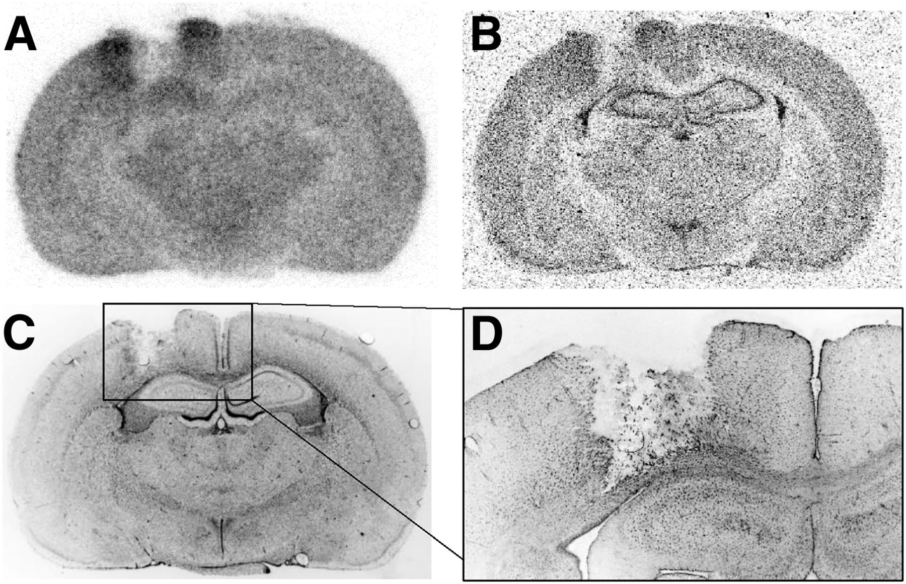
- FIGURE 1.
Coronal rat brain sections 2 d after resection of 9L glioma: 18F-FET (A) and 3H-MET (B) autoradiography, DAPI immunofluorescence staining (C), and magnified view of GFAP staining at rim of resection cavity (D). There is increased tracer uptake at rim of resection cavity and reactive astrocytosis.
- FIGURE 2.
Autoradiographic brain slices of 5 different control animals at different time points after resection of normal brain tissue demonstrating time course of treatment-related 18F-FET and 3H-MET uptake near resection cavity. There is prominent treatment-related tracer accumulation within first 7 d after resection, especially for 18F-FET, which decreases to low level 16 d after resection.
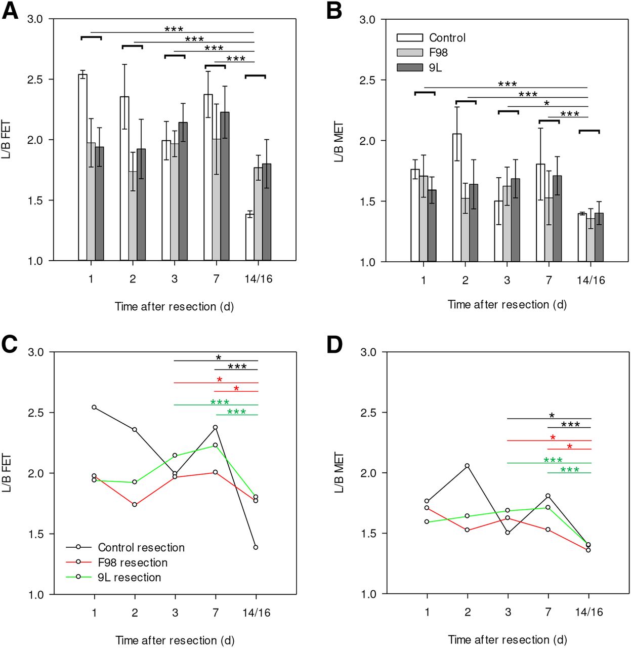
- FIGURE 3.
(A and B) Mean ± SD of L/B of treatment-related 18F-FET (A) and 3H-MET (B) uptake after resection of F98 gliomas, GS-9L tumors, and control animals. A and B show statistical difference between different time points after resection when using average value of all subgroups. Tracer uptake at rim of resection cavity decreases for both tracers on 14/16 d after resection. (C and D) Time course and statistical difference between individual subgroups (limited to comparison of day 3 or day 7 vs. 14/16 d after resection), which demonstrates that effect is present for all subgroups. *P ≤ 0.05. **P ≤ 0.01. ***P ≤ 0.001.
- FIGURE 4.
Comparison of L/B of 18F-FET and 3H-MET uptake at rim of resection cavity shows significant positive correlation. Regression line is shifted to left in relation to bisection line, demonstrating that L/B of treatment-related tracer accumulation is higher for 18F-FET than for 3H-MET.
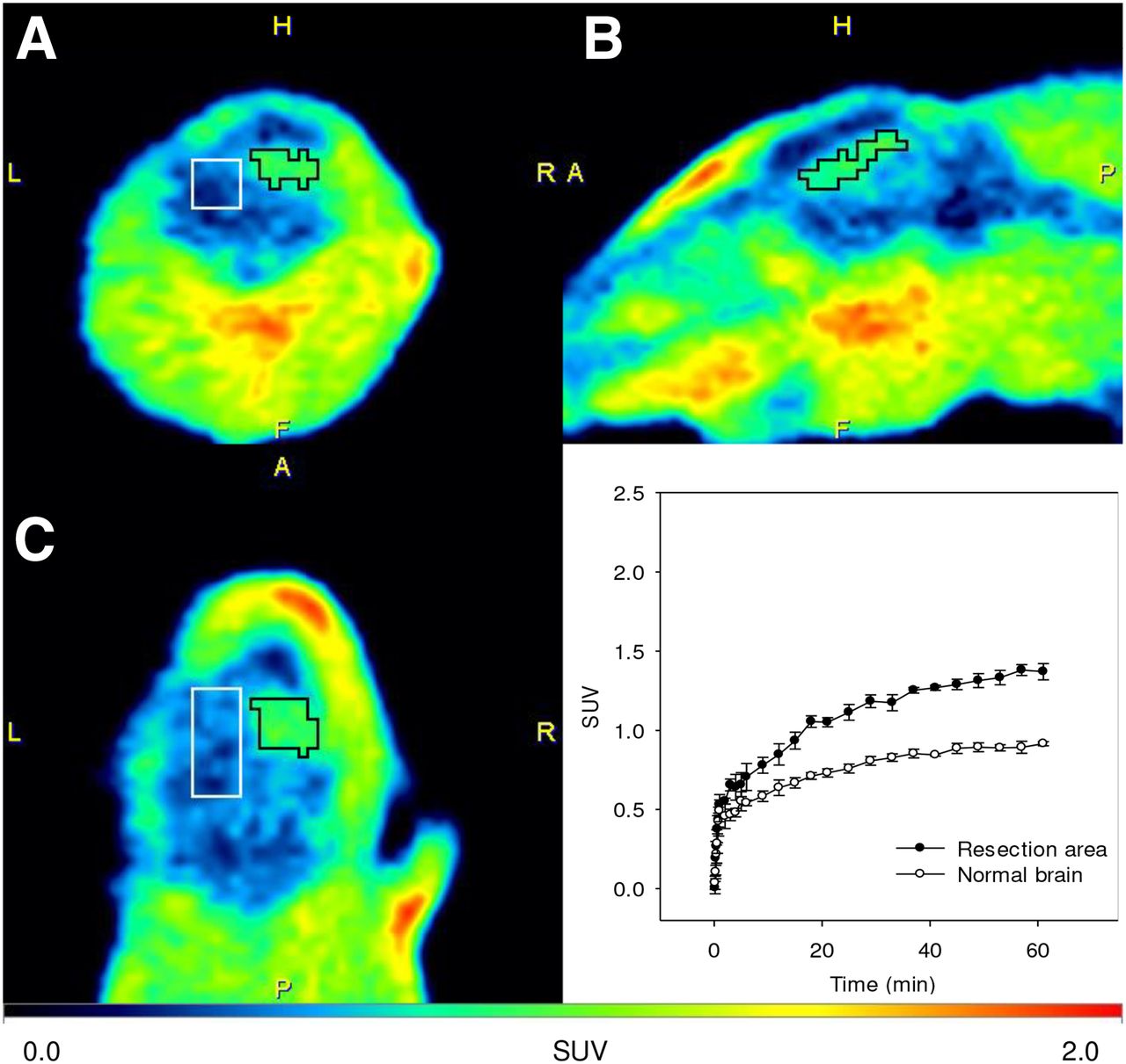
- FIGURE 5.
Examples of coronal (A), sagittal (B), and transversal (C) PET scans (18–61 min after injection) 3 d after resection of F98 rat glioma showing treatment-related 18F-FET uptake near resection area. Histology indicated no residual tumor tissue. Time–activity curves (SUV ± SD, n = 4) show treatment-related 18F-FET uptake in resection area compared with normal brain tissue of rats. Kinetic pattern shows constantly increasing 18F-FET uptake in both regions.


Tables
- TABLE 1
Data on L/B and T/B of 18F-FET and 3H-MET Uptake After Resection of F98 or GS-9L Gliomas or Normal Brain Tissue
Animal model Days after resection L/B 18F-FET T/B mean 18F-FET L/B 3H-MET T/B mean 3H-MET F98 1 1.97 ± 0.20 4.19 ± 1.00 1.71 ± 0.17 6.88 ± 0.41 F98 2 1.74 ± 0.16 NA* 1.52 ± 0.12 NA* F98 3 1.97 ± 0.11 4.77 ± 0.34 1.62 ± 0.16 5.91 ± 1.20 F98 7 2.00 ± 0.29 5.43 ± 0.39 1.53 ± 0.22 5.55 ± 0.63 F98 14 1.77 ± 0.10 4.85 ± 0.64 1.36 ± 0.08 4.66 ± 0.80 GS-9L 1 1.95 ± 0.16 4.40 ± 0.30 1.59 ± 0.11 7.36 ± 1.41 GS-9L 2 1.92 ± 0.24 4.24 ± 0.28 1.64 ± 0.20 5.77 ± 2.60 GS-9L 3 2.14 ± 0.16 4.43 ± 0.37 1.66 ± 0.16 7.06 ± 0.99 GS-9L 7 2.23 ± 0.22 4.89 ± 0.68 1.71 ± 0.16 6.41 ± 0.17 GS-9L 14 1.80 ± 0.20 4.62 ± 0.52 1.40 ± 0.09 5.84 ± 1.95 Control 1 2.54 ± 0.08 NA† 1.76 ± 0.08 NA† Control 2 2.36 ± 0.30 NA† 2.05 ± 0.22 NA† Control 3 1.99 ± 0.13 NA† 1.50 ± 0.19 NA† Control 7 2.37 ± 0.17 NA† 1.81 ± 0.30 NA† Control 16 1.38 ± 0.03 NA† 1.40 ± 0.01 NA†


