


Article Figures & Data
Figures
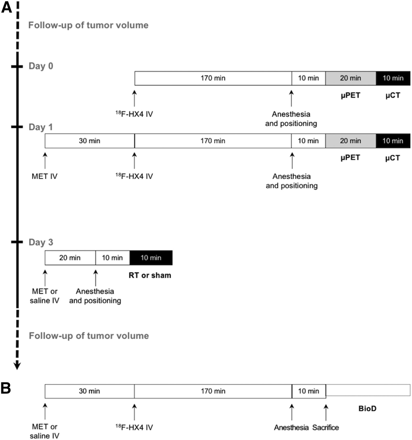
- FIGURE 1.
Experimental setup. (A) Acute metformin administration in A549 xenografts. (B) Biodistribution study. Hypoxia was quantified using 18F-flortanidazole. BioD = biodistribution study; 18F-HX4 = 18F-flortanidazole; IV = intravenous; μ = small-animal; MET = metformin; RT = radiotherapy.
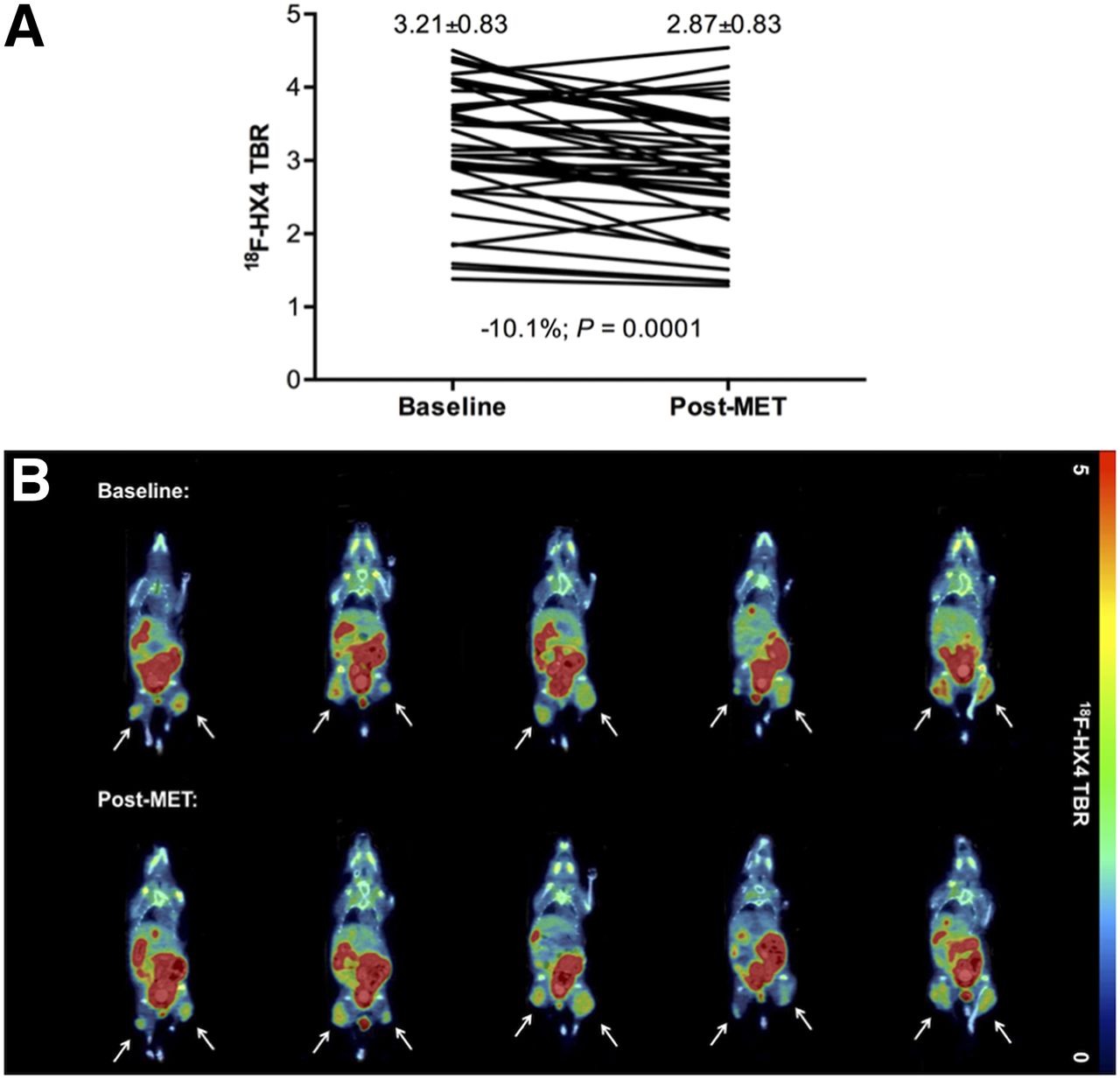
- FIGURE 2.
Metformin improves tumor oxygenation. (A) Significant decrease in 18F-flortanidazole TBR could be observed 30 min after intravenous administration of metformin (100 mg/kg; P = 0.0001). (B) Representative 18F-flortanidazole PET/CT TBR-corrected images (coronal view) of 5 mice before (upper row) and after (lower row) metformin administration. Arrows indicate tumors. 18F-HX4 = 18F-flortanidazole; MET = metformin.
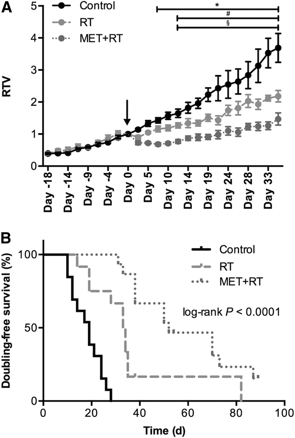
- FIGURE 3.
Metformin improves radiotherapy response in A549 tumors. (A) Tumor growth was followed, and RTVs were calculated. Arrow indicates moment of therapy administration. (B) Kaplan–Meier representation of doubling-free survival time (overall log-rank P < 0.0001). *Significant difference between metformin + radiotherapy and control. #Significant difference between metformin + radiotherapy and radiotherapy. §Significant difference between radiotherapy and control. MET = metformin; RT = radiotherapy.
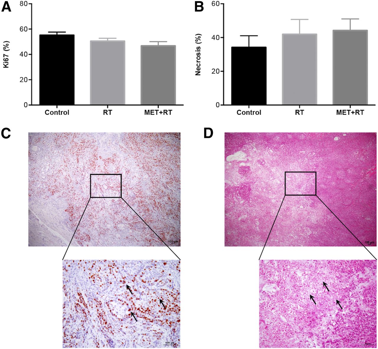
- FIGURE 4.
Tumor proliferation as assessed with Ki-67 immunohistochemistry and tumor necrosis at sacrifice. (A) No major differences in tumor proliferation could be observed between different treatment groups. (B) Same conclusions could be drawn from necrosis scoring. (C) Representative example of Ki-67 staining. Arrows indicate positively stained nuclei. (D) Representative example of hematoxylin and eosin staining. Arrows indicate areas of necrosis. MET = metformin; RT = radiotherapy.
- FIGURE 5.
Therapeutic benefit of metformin was dependent on baseline degree of tumor hypoxia and could be predicted with baseline 18F-flortanidazole PET. 18F-HX4 = 18F-flortanidazole; H0 = null hypothesis; MET = metformin; RT = radiotherapy.
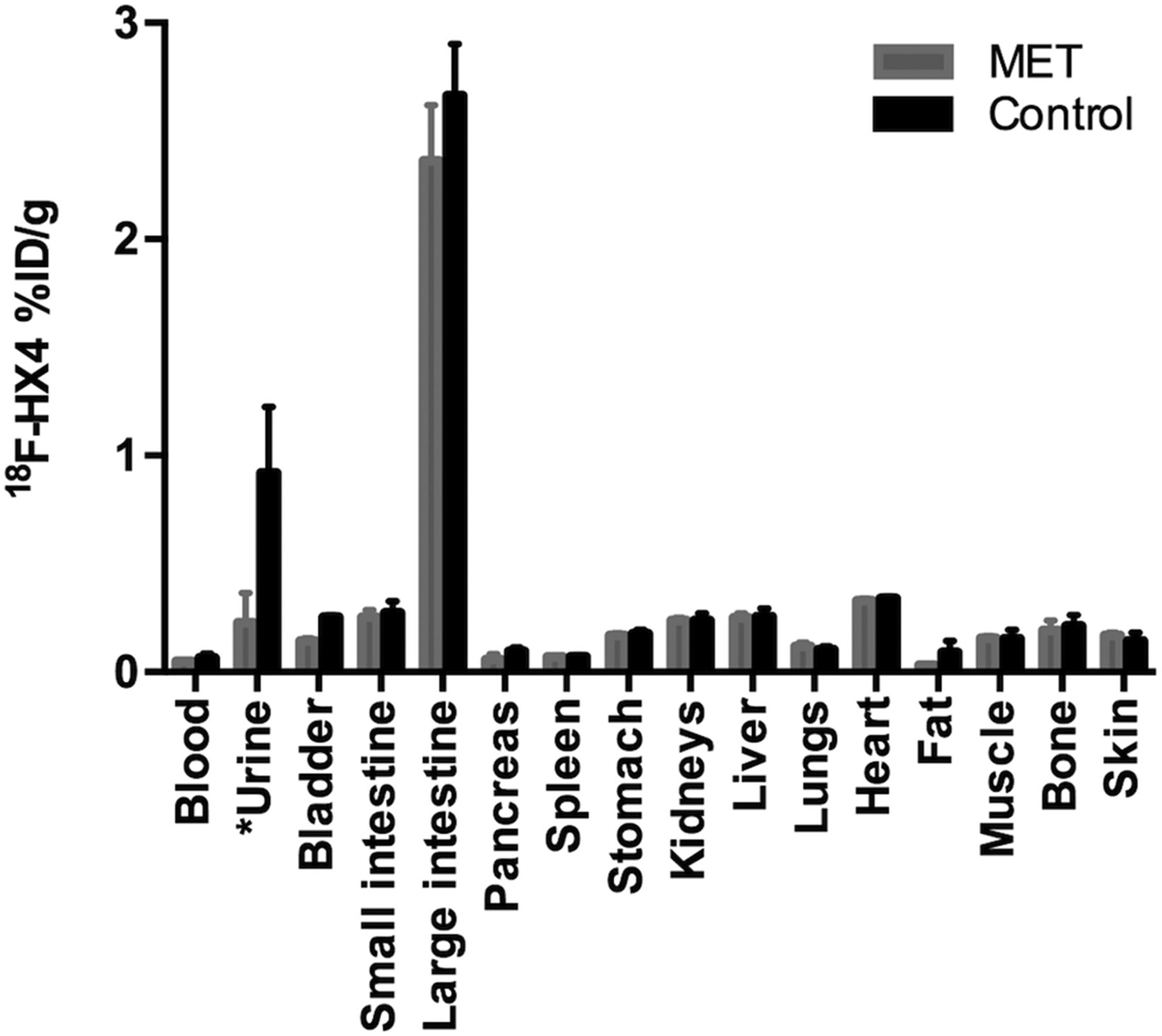
- FIGURE 6.
Results of 18F-flortanidazole biodistribution study after acute metformin administration. No major differences could be observed in 18F-flortanidazole biodistribution profile between metformin-treated mice and control group. 18F-HX4 = 18F-flortanidazole; MET = metformin. *Values are shown as %ID because of variability in urine production.


Tables
Parameter Radiotherapy Metformin + radiotherapy Control P Tumor volume (mm3) 395 ± 57 297 ± 51 329 ± 65 0.5 Animal weight (g) 29.6 ± 0.8 29.7 ± 0.9 29.4 ± 1.1 1.0 Baseline 18F-flortanidazole TBR 3.60 ± 0.17 2.74 ± 0.26 3.47 ± 0.20 0.3 Δ18F-flortanidazole TBR (%) −13 ± 4 −4 ± 4 −14 ± 3 0.2 Δ18F-flortanidazole TBR = change in 18F-flortanidazole TBR after metformin.
Data are expressed as mean ± SEM.


Jump to section
Related Articles
Cited By...
- Repurposing radiosensitising medicines for radiotherapy: an overview
- Targeting Oxidative Phosphorylation to Increase the Efficacy of Radio- and Immune-Combination Therapy
- Mitochondrial Inhibitor Atovaquone Increases Tumor Oxygenation and Inhibits Hypoxic Gene Expression in Patients with Non-Small Cell Lung Cancer