


Abstract
334
Objectives: The liver plays an important role in the excretion of xenobiotics, including many kinds of drugs, and various transporters and metabolizing enzymes are involved in its process. Since organic anion transporting polypeptide (OATP) is a component of organic anion transporter including hepatic uptake of anionic xenobiotics, it is useful to evaluate the OATP function for prediction of therapeutic efficacy non-invasively. Previously, we reported the synthesis of a novel pitavastatin derivative [18F]PTV-F1 in which a [18F]fluoroethoxy group is substituted for the [18F]fluoro group of [18F]pitavastatin. In this study, we investigated its potential as a PET probe to evaluate hepatic transport quantitatively and compared it with [18F]pitavastatin.
Methods: PTV-F1 and tosylate precursor (PTV-F1 precursor) were synthesized from isatonic anhydride. [18F]PTV-F1 was synthesized in 3 steps involving aliphatic radiofluorination in the presence of Kryptofix2.2.2 and [18F]KF, deprotection under acidic conditions using hydrochloric acid, and alkaline hydrolysis with NaOH aqueous solution of the lactone compound. Metabolite analysis in blood, bile and liver was carried out in SD rats using Radio-TLC and autoradiography. In vivo biodistribution study and PET/CT study were performed to evaluate the OATP function in SD rats. The hepatic uptake clearance of [18F]PTV-F1 and [18F]pitavastatin was calculated from the data of PET study using integration plots method. Results and discussion: PTV-F1 and PTV-F1 precursor were synthesized in each 8% and 10% yield with 11 steps. [18F]PTV-F1 was synthesized from PTV-F1 precursor in 45 ± 3% (n = 3) radiochemical yield and >99% radiochemical purity for 30 minutes in a one-pot procedure. Compared to [18F]pitavastatin using the Suzuki-coupling (28 ± 3%, 60 minutes, two-pot reaction), [18F]PTV-F1 was synthesized using a one-pot procedure in the higher radiochemical yield and the shorter operating time. [18F]PTV-F1 remained intact in the liver 40 min p.i. (>95% intact). In biodistribution study, [18F]PTV-F1 mainly distributed in the liver in the early stage and gradually into the intestine, which suggests that a part of the administrated [18F]PTV-F1 was excreted in the bile. Furthermore, PET/CT studies with [18F]PTV-F1 agreed with the biodistribution data in normal SD rats. In rifampicin-treated SD rats, radioactivity in the liver was significantly decreased and that in the blood was increased. The hepatic uptake clearance of [18F]PTV-F1 and [18F]pitavastatin was decreased by rifampicin infusion. (Table 1) These results indicated that [18F]PTV-F1 and [18F]pitavastatin were taken up by hepatic OATP. PET imaging and the hepatic uptake clearance data suggested that [18F]PTV-F1 was recognized by OATP in the same way as [18F]pitavastatin. Conclusions: [18F]PTV-F1 has the potential to be a PET probe for evaluation of OATP function in the liver. [18F]PTV-F1 can be applied to automated synthesis using necessary radioactivity of [18F]F− in the clinical study. Additionally, it can be a clinically applicable PET probe for evaluation of OATP function in human.
Table 1. The hepatic uptake clearance of [18F]PTV-F1 and [18F]pitavastatin

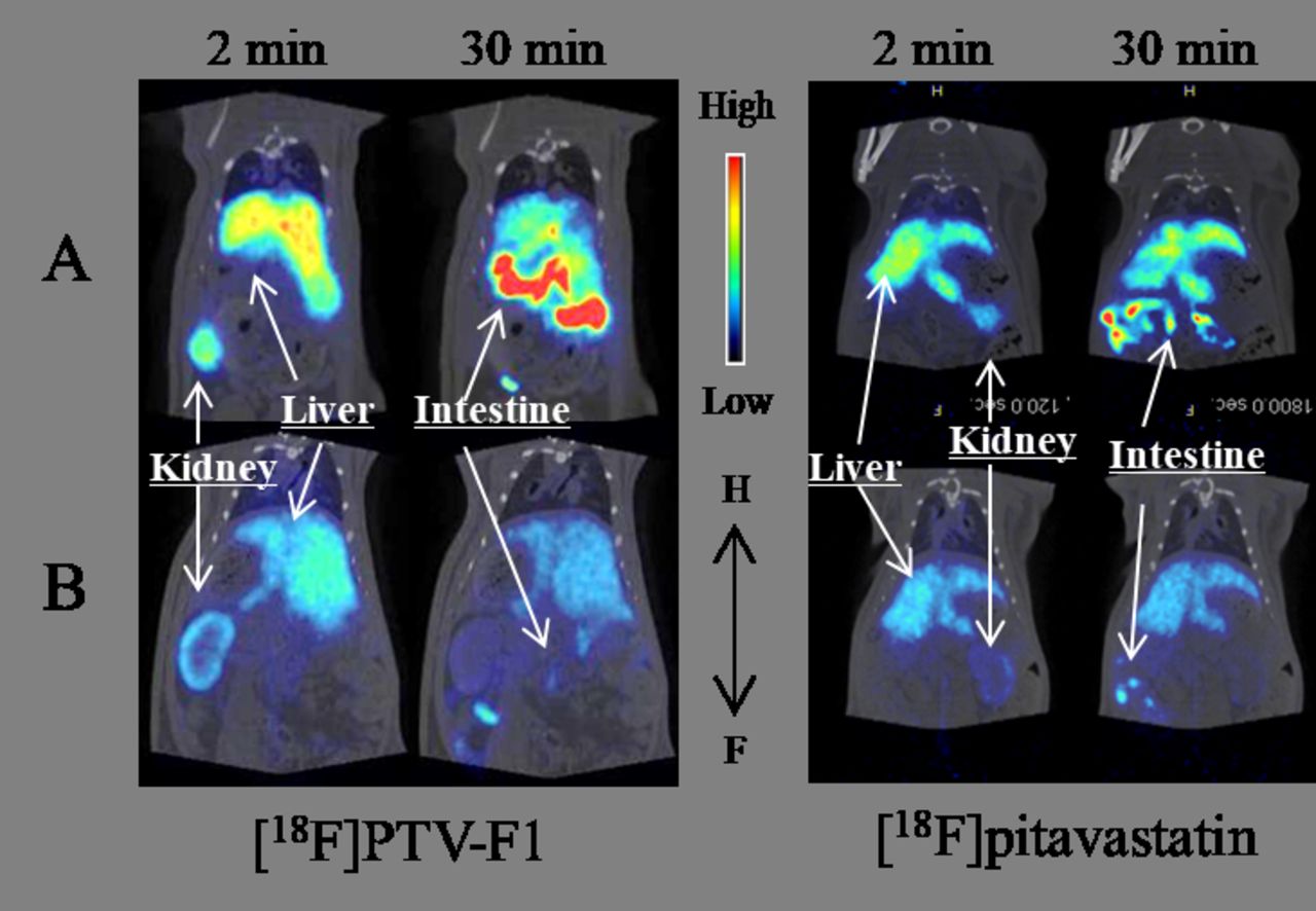
Figure 1. PET images of rat abdominal regions taken after intravenous administration of [18F]PTV-F1 and [18F]pitavastatin. PET and CT fusion image of coronal cross section in the abdominal region at 2 min and 30 min in control rats (A), and in rifampicin-treated rats at an infusion rate of 1.5 μmol/min/kg (B).
In this issue


Jump to section
Related Articles
Cited By...
- No citing articles found.