


Abstract
296
Objectives: At present, the identification of unstable atherosclerotic(AS) plaque is still lack of noninvasive detection methods. Studies have shown that 18F-NaF PET-CT can early identified unstable AS plaque by located micro-calcification in plaque at histological level, but the efficiency of 18F-NaF PET-CT for identify the unstable plaque is still controversial, so the aim of this part is to assess the ability and clinical value of 18F-NaF PET-CT for identification of unstable AS plaques with using IVUS as the golden standard.
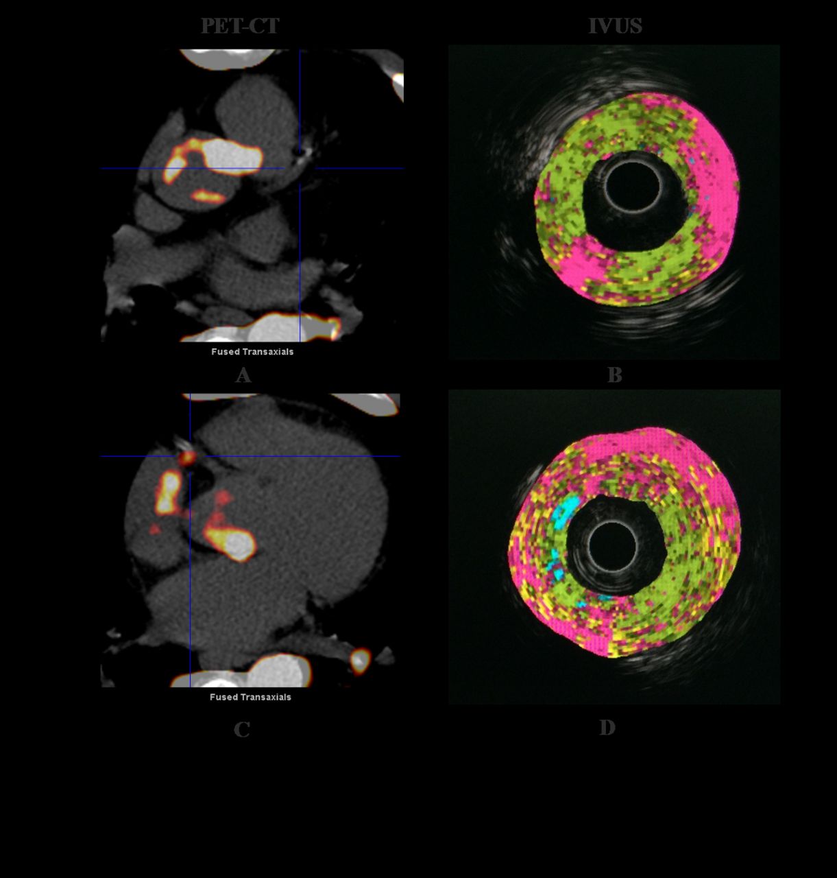
Methods: Our study screened unstable angina patients according to the established standards, and collected the clinical data including diabetes history, hypertension history , smoking history, laboratory tests, etc. 18F-NaF PET-CT, IVUS, CAG , CAC were performed in 2 weeks. IVUS results were used as golden standard for determination of unstable plaques, and then the unstable plaques were divided into 18F-NaF positive group and 18F-NaF negative group according to the results of 18F-NaF PET-CT, and the differences of SUVmax and IVUS parameters were compared between the two groups, IVUS parameters included Lumen (Area, Max, Min), Vessel (Area, Max, Min), Burden, Plaque composition (Fibrotic, Lipidic, Necrotic, Calcified) .Results: A total of 31 patients included 124 coronary arteries performed 18F-NaF PET/CT. The positive rate of 18F-NaF PET-CT for patients was 74.19%(23/31), and the positive rate of 18F-NaF PET-CT for coronary arteries was 28.23%(35/124).The differences of clinical indexes between two groups are compared, the results showed that there were significant differences in high sensitivity C reactive protein (P=0.011), but no differences in the other clinical indexes,P >0.05. In our study, 21 patients including 62 AS plaques with 38 coronary arteries underwent IVUS, 41 unstable AS plaques were determined by IVUS imaging, the positive rate of 18F-NaF PET-CT for unstable plaques was 56.1%(23/41). The sensitivity of 18F-NaF PET-CT for detection of unstable AS plaques was 56.09%, the specificity was 90.47%, the positive predictive value was 92%, the negative predictive value was 48.64% ,and the accuracy of this diagnostic test was 67.74%. The comparison results of IVUS parameters show that the minimum diameter of vascular, plaque burden, fibrosis and necrotic core in histological components of unstable AS plaques have discrepancy between the 18F-NaF positive group and 18F-NaF negative group for unstable AS plaques P<0.05, but no significant differences in other indicators.
Conclusions: 18F-NaF PET-CT imaging can identify part of unstable plaques with a higher positive predictive value, and the 18F-NaF positive plaques are more unstable than the negative ones. These results suggest that 18F-NaF PET-CT has a better ability for identification of unstable AS plaques and also can be used to stratify the unstable plaque, that would meet the needs of precise treatment. However, negative results of that can not be ruled out the high risk of MACE in patients with UA.

In this issue


Jump to section
Related Articles
Cited By...
- No citing articles found.