
Main menu

  • Home
  • Content
    • Current
    • Ahead of print
    • Past Issues
    • JNM Supplement
    • SNMMI Annual Meeting Abstracts
    • Continuing Education
    • JNM Podcasts
  • Subscriptions
    • Subscribers
    • Institutional and Non-member
    • Rates
    • Journal Claims
    • Corporate & Special Sales
  • Authors
    • Submit to JNM
    • Information for Authors
    • Assignment of Copyright
    • AQARA requirements
  • Info
    • Reviewers
    • Permissions
    • Advertisers
  • About
    • About Us
    • Editorial Board
    • Contact Information
  • More
    • Alerts
    • Feedback
    • Help
    • SNMMI Journals
  • SNMMI
    • JNM
    • JNMT
    • SNMMI Journals
    • SNMMI

User menu

  • Subscribe
  • My alerts
  • Log in
  • My Cart

Search

  • Advanced search
Journal of Nuclear Medicine
  • SNMMI
    • JNM
    • JNMT
    • SNMMI Journals
    • SNMMI
  • Subscribe
  • My alerts
  • Log in
  • My Cart
Journal of Nuclear Medicine

Advanced Search

  • Home
  • Content
    • Current
    • Ahead of print
    • Past Issues
    • JNM Supplement
    • SNMMI Annual Meeting Abstracts
    • Continuing Education
    • JNM Podcasts
  • Subscriptions
    • Subscribers
    • Institutional and Non-member
    • Rates
    • Journal Claims
    • Corporate & Special Sales
  • Authors
    • Submit to JNM
    • Information for Authors
    • Assignment of Copyright
    • AQARA requirements
  • Info
    • Reviewers
    • Permissions
    • Advertisers
  • About
    • About Us
    • Editorial Board
    • Contact Information
  • More
    • Alerts
    • Feedback
    • Help
    • SNMMI Journals
  • View or Listen to JNM Podcast
  • Visit JNM on Facebook
  • Join JNM on LinkedIn
  • Follow JNM on Twitter
  • Subscribe to our RSS feeds
Meeting ReportNeurosciences Track

Value of PET/MRI in CNS malignancies

Ahmad Shariftabrizi, Seyed Mohammadi, Frank Lieberman and James Mountz
Journal of Nuclear Medicine May 2018, 59 (supplement 1) 1693;
Ahmad Shariftabrizi
2University of Pittsburgh Medical Center Pittsburgh PA United States
  • Find this author on Google Scholar
  • Find this author on PubMed
  • Search for this author on this site
Seyed Mohammadi
4UPMC Pittsburgh PA United States
  • Find this author on Google Scholar
  • Find this author on PubMed
  • Search for this author on this site
Frank Lieberman
3Neuro-oncology University of Pittsburgh Medical Center Pittsburgh PA United States
  • Find this author on Google Scholar
  • Find this author on PubMed
  • Search for this author on this site
James Mountz
1Radiology - Nuclear Medicine University of Pittsburgh Pittsburgh PA United States
  • Find this author on Google Scholar
  • Find this author on PubMed
  • Search for this author on this site
  • Article
  • Figures & Data
  • Info & Metrics
Loading

Abstract

1693

Background: Clinical nuclear medicine services now include PET/MRI hybrid imaging: a technology that combines the delicate structural and functional characterization of tissue provided by MRI with the sensitivity of PET imaging of metabolism. However, there remain questions regarding which clinical indications are more accurately diagnosed by PET/MRI hybrid imaging versus PET plus MR performed separately. The major clinical question addressed in this study is whether there is a significant diagnostic benefit for hybrid PET/MRI imaging in patients with new or recurrent low or high-grade brain tumor and other brain malignancies.

Methods: From 2016-2017, 32 patients with CNS pathologies underwent clinical PET/MR scans at University of Pittsburgh Medical Center. Patient population included epilepsy (5), primary brain tumors (20), and metastatic brain or skull base tumor with questionable brain involvement (7). The 27 patients with primary brain tumors or metastatic brain or skull base tumors with questionable brain involvement were included in the study. All patients underwent PET/MR from a Siemens 3T mMR after injection of approximately 10 mCi F-18 FDG. Eight had fully diagnostic MR after injection with approximately 20 cc MultiHance IV. The remaining 19 had MR for attenuation correction plus T2_FLAIR, DWI, T1_MPRAGE and DWI_ADC sequences. Patients' tumor types were as follows: A-Meningioma (1), B-Maxillary sinus/nasopharyngeal (5), C-Low-grade glioma (4), D-High grade glioma (15), E- Metastatic tumor (2). An experienced nuclear medicine and radiologist physician (JMM) separately interpreted the MRI+ PET and PET/MRI images of all patients in sagittal, axial and coronal sections in a blinded manner. Patient diagnosis was first interpreted blindly from randomized PET and MR scans, separated from the original PET/MR sans. Diagnostic agreement between the separate interpretations of the PET and MRI scans versus the hybrid PET/MRI scans were scored. The added benefit from PET/MR compared to PET+MR was rated as High (benefit that changed or greatly increased the certainty of diagnosis), Moderate (benefit that increased the certainty of diagnosis), or Low (no change in certainty of diagnosis). High grade tumors were separated into positive (D+) or negative for recurrence (D-). Results: Table below shows added value of PET/MR compared with PET+MR. Figure illustrates images from a patient with suspected malignant degeneration of an oligodendroglioma (category D-) that was shown to not have high grade disease by PET/MR, but was considered as possible high grade degeneration by PET+MR.

View this table:
  • View inline
  • View popup

Added value of PET/MR compared with PET+MR

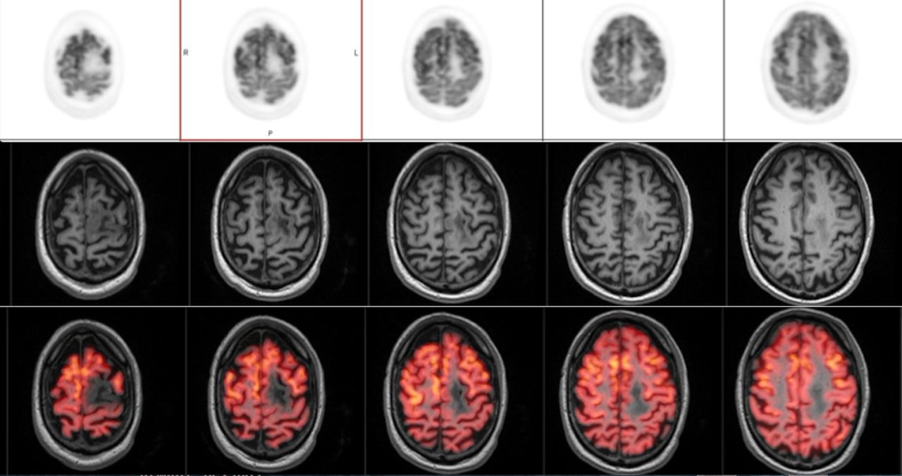
Conclusions: In summary, diagnostic accuracy in patience with questionable recurrent viable high-grade glioma improved most by PET/MR, as there was greater certainty in not overcalling normal grey matter tissue close to the tumor bed as a region of tumor recurrence. Those that benefited the least were patients with clearly definable high-grade recurrence or clearly definable high grade metastatic brain tumor, since the presence of viable tumor was unequivocally identified equally on both PET+MR and PET/MR. Figure. 58-year-old female with left frontal oligodendroglioma diagnosed 10 years ago, underwent recent RT for suspected malignant degeneration. Recent MRI (not shown) showed multiple enhancing foci in the tumor periphery. PET imaging alone (top row) shows increased uptake in the lateral left frontal lobe. PET/MR (bottom row) shows the uptake corresponds to a normal appearing gyrus on MRI (middle row). Follow-up clinical assessment and MRI scans showed tumor stability.

Figure
  • Download figure
  • Open in new tab
  • Download powerpoint
Previous
Back to top

In this issue

Journal of Nuclear Medicine
Vol. 59, Issue supplement 1
May 1, 2018
  • Table of Contents
  • Index by author
Article Alerts
Sign In to Email Alerts with your Email Address
Email Article

Thank you for your interest in spreading the word on Journal of Nuclear Medicine.

NOTE: We only request your email address so that the person you are recommending the page to knows that you wanted them to see it, and that it is not junk mail. We do not capture any email address.

Enter multiple addresses on separate lines or separate them with commas.
Value of PET/MRI in CNS malignancies
(Your Name) has sent you a message from Journal of Nuclear Medicine
(Your Name) thought you would like to see the Journal of Nuclear Medicine web site.
Citation Tools
Value of PET/MRI in CNS malignancies
Ahmad Shariftabrizi, Seyed Mohammadi, Frank Lieberman, James Mountz
Journal of Nuclear Medicine May 2018, 59 (supplement 1) 1693;

Citation Manager Formats

  • BibTeX
  • Bookends
  • EasyBib
  • EndNote (tagged)
  • EndNote 8 (xml)
  • Medlars
  • Mendeley
  • Papers
  • RefWorks Tagged
  • Ref Manager
  • RIS
  • Zotero
Share
Value of PET/MRI in CNS malignancies
Ahmad Shariftabrizi, Seyed Mohammadi, Frank Lieberman, James Mountz
Journal of Nuclear Medicine May 2018, 59 (supplement 1) 1693;
Twitter logo Facebook logo LinkedIn logo Mendeley logo
  • Tweet Widget
  • Facebook Like
  • Google Plus One
Bookmark this article

Jump to section

  • Article
  • Figures & Data
  • Info & Metrics

Related Articles

  • No related articles found.
  • Google Scholar

Cited By...

  • No citing articles found.
  • Google Scholar

More in this TOC Section

Neurosciences Track

  • Impact of cognitive reserve in frontotemporal dementia illustrated by FDG-PET.
  • Quantification of brain cholinergic denervation in dementia with Lewy bodies using PET imaging with 18F-FEOBV
  • Kinetic evaluation of [18F]MOZAT PET imaging in humans.
Show more Neurosciences Track

Neurology & Psychiatry Imaging Posters

  • comparison study of F Choline PET CT with MRI in intracranial space occupying lesions and evaluation of diagnostic accuracy for differentiating neoplastic versus non neoplastic lesions
  • Radioactive iodine labelled MIBG cardiac sympathetic imaging is less promising in differentiating PD from other parkinsonism for early onset patients
  • Dr.
Show more Neurology & Psychiatry Imaging Posters

Similar Articles

SNMMI

© 2025 SNMMI

Powered by HighWire