
Main menu

  • Home
  • Content
    • Current
    • Ahead of print
    • Past Issues
    • JNM Supplement
    • SNMMI Annual Meeting Abstracts
    • Continuing Education
    • JNM Podcasts
  • Subscriptions
    • Subscribers
    • Institutional and Non-member
    • Rates
    • Journal Claims
    • Corporate & Special Sales
  • Authors
    • Submit to JNM
    • Information for Authors
    • Assignment of Copyright
    • AQARA requirements
  • Info
    • Reviewers
    • Permissions
    • Advertisers
  • About
    • About Us
    • Editorial Board
    • Contact Information
  • More
    • Alerts
    • Feedback
    • Help
    • SNMMI Journals
  • SNMMI
    • JNM
    • JNMT
    • SNMMI Journals
    • SNMMI

User menu

  • Subscribe
  • My alerts
  • Log in
  • My Cart

Search

  • Advanced search
Journal of Nuclear Medicine
  • SNMMI
    • JNM
    • JNMT
    • SNMMI Journals
    • SNMMI
  • Subscribe
  • My alerts
  • Log in
  • My Cart
Journal of Nuclear Medicine

Advanced Search

  • Home
  • Content
    • Current
    • Ahead of print
    • Past Issues
    • JNM Supplement
    • SNMMI Annual Meeting Abstracts
    • Continuing Education
    • JNM Podcasts
  • Subscriptions
    • Subscribers
    • Institutional and Non-member
    • Rates
    • Journal Claims
    • Corporate & Special Sales
  • Authors
    • Submit to JNM
    • Information for Authors
    • Assignment of Copyright
    • AQARA requirements
  • Info
    • Reviewers
    • Permissions
    • Advertisers
  • About
    • About Us
    • Editorial Board
    • Contact Information
  • More
    • Alerts
    • Feedback
    • Help
    • SNMMI Journals
  • View or Listen to JNM Podcast
  • Visit JNM on Facebook
  • Join JNM on LinkedIn
  • Follow JNM on Twitter
  • Subscribe to our RSS feeds
Meeting ReportGeneral Clinical Specialties Track

New use for an old isotope

Di Wu, Dorina Ylli, Cristiane Jeyce Gomes Lima, Wen Lee, Kenneth Burman, Leonard Wartofsky and Douglas Van Nostrand
Journal of Nuclear Medicine May 2018, 59 (supplement 1) 1338;
Di Wu
1Medstar Health Research Institute Washington DC United States
  • Find this author on Google Scholar
  • Find this author on PubMed
  • Search for this author on this site
Dorina Ylli
1Medstar Health Research Institute Washington DC United States
  • Find this author on Google Scholar
  • Find this author on PubMed
  • Search for this author on this site
Cristiane Jeyce Gomes Lima
1Medstar Health Research Institute Washington DC United States
  • Find this author on Google Scholar
  • Find this author on PubMed
  • Search for this author on this site
Wen Lee
4Pathology MedStar Washington Hospital Center Washington DC United States
  • Find this author on Google Scholar
  • Find this author on PubMed
  • Search for this author on this site
Kenneth Burman
3Endocrinology MedStar Washington Hospital Center Washington DC United States
  • Find this author on Google Scholar
  • Find this author on PubMed
  • Search for this author on this site
Leonard Wartofsky
3Endocrinology MedStar Washington Hospital Center Washington DC United States
  • Find this author on Google Scholar
  • Find this author on PubMed
  • Search for this author on this site
Douglas Van Nostrand
2MedStar Washington Hospital Center Washington DC United States
  • Find this author on Google Scholar
  • Find this author on PubMed
  • Search for this author on this site
  • Article
  • Figures & Data
  • Info & Metrics
Loading

Abstract

1338

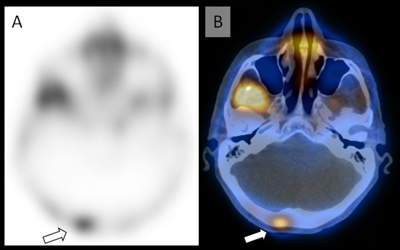
Objectives: Differentiated thyroid cancer (DTC) patients with elevated or rising serum thyroglobulin (Tg) levels and multiple other negative conventional imaging studies represent a clinically difficult group of patients. 99m Tc-sestamibi is a non-conventional imaging study for DTC that was used in the 1980s-1990s to help evaluate DTC patients before being replaced with 18F-FDG. However, 18F-FDG PET scans may also be negative in these patients. This is a preliminary analysis of our study to evaluate the diagnostic use of 99mTc-sestamibi scans to identify the source of elevated or rising Tgs in patients with negative conventional imaging including negative 18F-FDG PET scans. Methods This prospective study included DTC patients who were at least 18 years old, had at least one prior 131I therapy, and had elevated Tg>5 ng/ml or positive anti-Tg antibodies (TgAb). In addition, all of the following imaging studies must have been clinically reported as negative for recurrence or metastasis within the last 12 months: neck ultrasound, diagnostic radioiodine scan, chest x-ray, CT of the chest/abdomen with/without contrast, and 18F-FDG PET/CT. If other imaging studies were performed, such as bone scans or brain imaging, these must also have been read as negative. This study was approved by the IRB at MedStar Health and all participants signed informed consent. Participants were injected with 925 MBq (25 mCi) of 99mTc-sestamibi intravenously and whole body images were acquired after ~1h with acquisition speed 4cm/min with parallel-hole low-energy collimator. Extra spot view, pinhole view, or SPECT/CT images were performed when necessary. Two blinded nuclear medicine physicians independently interpreted the 99mTc-sestamibi images, and graded each foci 1-5 (1 = definite physiological uptake or artifact; 2 = most likely physiological uptake or artifact; 3 = indeterminate; 4 = most likely recurrence or metastasis; 5 = definite recurrence or metastasis). Discordant findings were resolved by consensus. Foci graded 3-5 were categorized as positive result and was followed-up for confirmation. Results A total of four patients completed the sestamibi scan to date. One out of four patients (Patient1) was positive on the sestamibi scan for distant metastasis. In Patient 1, the sestamibi whole-body posterior image demonstrated abnormal focal uptake in the right posterior calvarium and corresponding to an occipital lytic bone lesion on the SPECT/CT (Figure 1). The patient underwent surgical resection of the skull metastasis, and pathology confirmed metastatic thyroid cancer. Five months post-surgery the suppressed Tg was markedly reduced and stable at ~3.2ng/ml. Table 1 shows the sestamibi scan results, subsequent management and follow-up. With confirmation of the location of the metastatic foci in Patient 1, the two negative 18F-FDG PET/CT that were previously performed one year apart were reviewed again. The radiology reports did not document interpretation of the low-dose localization CT. In retrospect, the lytic skull metastasis was visible on both of the head CT, but could have been easily missed on the PET due to the normal intensive brain 18F-FDG activity.

View this table:
  • View inline
  • View popup

Table 1. Patient data.

Conclusions In this preliminary analysis, 99mTc-sestamibi was able to identify the source of elevated Tg levels in one out of four DTC patients with biochemical recurrence and negative conventional imaging studies. 99mTc-sestamibi may have a role in thyroid cancer localization when physical exam, neck ultrasound, radioiodine scan, chest/abdomen CT, and 18F-FDG PET/CT could not identify. Additional patients are being recruited. Research Support We are grateful for the generous donations from our patients.

Figure
  • Download figure
  • Open in new tab
  • Download powerpoint
Previous
Back to top

In this issue

Journal of Nuclear Medicine
Vol. 59, Issue supplement 1
May 1, 2018
  • Table of Contents
  • Index by author
Article Alerts
Sign In to Email Alerts with your Email Address
Email Article

Thank you for your interest in spreading the word on Journal of Nuclear Medicine.

NOTE: We only request your email address so that the person you are recommending the page to knows that you wanted them to see it, and that it is not junk mail. We do not capture any email address.

Enter multiple addresses on separate lines or separate them with commas.
New use for an old isotope
(Your Name) has sent you a message from Journal of Nuclear Medicine
(Your Name) thought you would like to see the Journal of Nuclear Medicine web site.
Citation Tools
New use for an old isotope
Di Wu, Dorina Ylli, Cristiane Jeyce Gomes Lima, Wen Lee, Kenneth Burman, Leonard Wartofsky, Douglas Van Nostrand
Journal of Nuclear Medicine May 2018, 59 (supplement 1) 1338;

Citation Manager Formats

  • BibTeX
  • Bookends
  • EasyBib
  • EndNote (tagged)
  • EndNote 8 (xml)
  • Medlars
  • Mendeley
  • Papers
  • RefWorks Tagged
  • Ref Manager
  • RIS
  • Zotero
Share
New use for an old isotope
Di Wu, Dorina Ylli, Cristiane Jeyce Gomes Lima, Wen Lee, Kenneth Burman, Leonard Wartofsky, Douglas Van Nostrand
Journal of Nuclear Medicine May 2018, 59 (supplement 1) 1338;
Twitter logo Facebook logo LinkedIn logo Mendeley logo
  • Tweet Widget
  • Facebook Like
  • Google Plus One
Bookmark this article

Jump to section

  • Article
  • Figures & Data
  • Info & Metrics

Related Articles

  • No related articles found.
  • Google Scholar

Cited By...

  • No citing articles found.
  • Google Scholar

More in this TOC Section

General Clinical Specialties Track

  • Quantitative Evaluation of Parathyroid Adenoma and Hyperplasia in Reference to Thyroid using Tc-99m MIBI SPECT/CT
  • A primitive study for clinical application of 18F-AlF-NOTA-octreotide PET/CT in combination with 18F-FDG PET/CT for imaging neuroendocrine neoplasms
  • Role of 18F-FDG PET/CT in detection of disease burden and response assessment in patients with myeloid sarcoma
Show more General Clinical Specialties Track

Endocrine/Neuroendocrine Posters

  • Quantitative Evaluation of Parathyroid Adenoma and Hyperplasia in Reference to Thyroid using Tc-99m MIBI SPECT/CT
  • Usefulness of glucosemetabolism of psoas muscle on 18F-FDG PET/CT in the prediction of metabolic health impairment
  • The value of 99mTc-MIBI parathyroid imaging and ultrasound in parathyroid adenoma and parathyroid hyperplasia
Show more Endocrine/Neuroendocrine Posters

Similar Articles

SNMMI

© 2025 SNMMI

Powered by HighWire