


Article Figures & Data
Figures
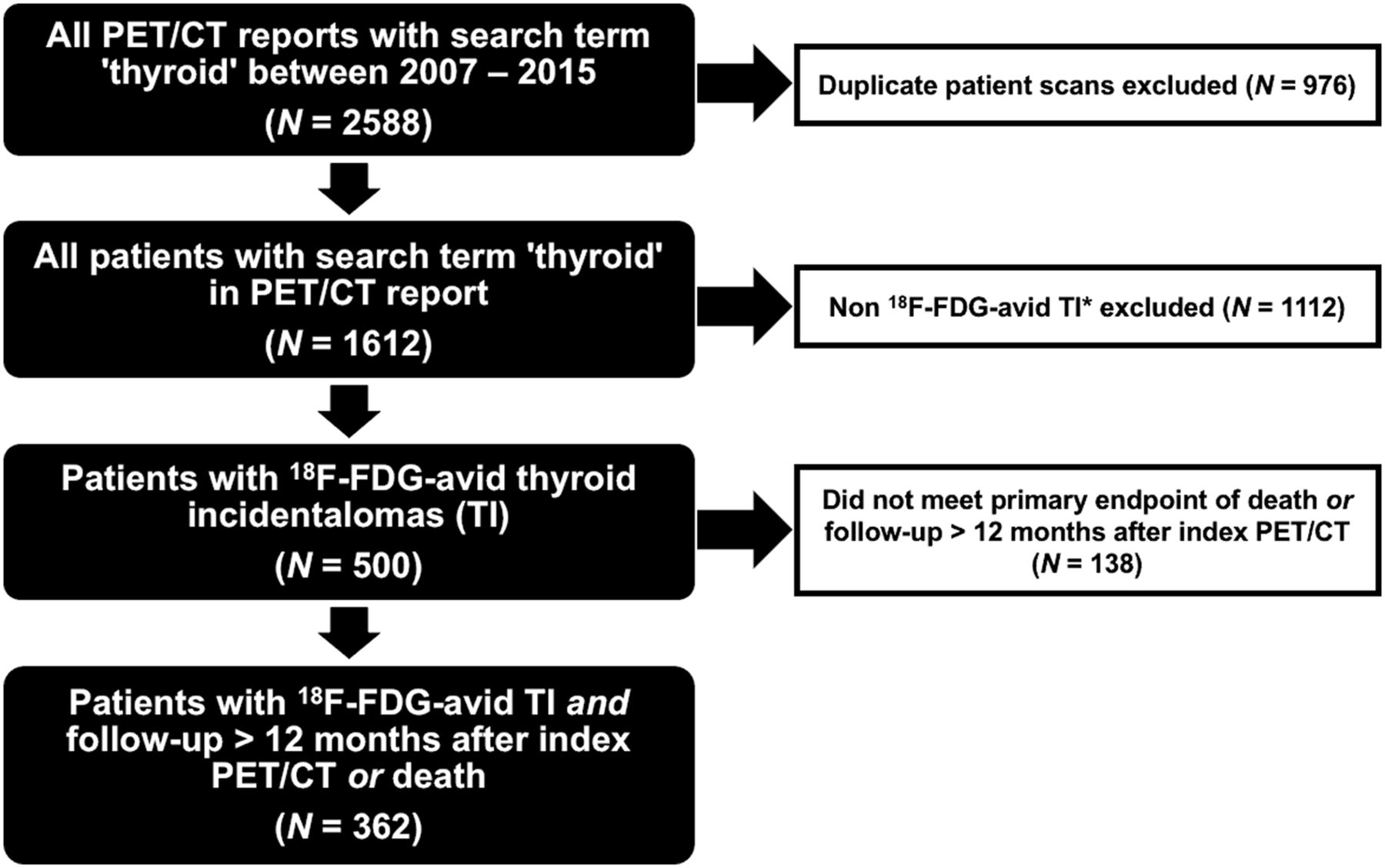
- FIGURE 1.
Consort flow diagram demonstrating identification and selection of study population. *Non–18F-FDG–avid TIs comprise nonavid thyroid nodules, diffuse thyroid 18F-FDG uptake, patients with known thyroid cancer, abnormalities adjacent to thyroid (parathyroid adenomas and lymphadenopathy), and use of non–18F-FDG radiotracers (68Ga-prostate-specific membrane antigen, 68Ga-DOTATATE).
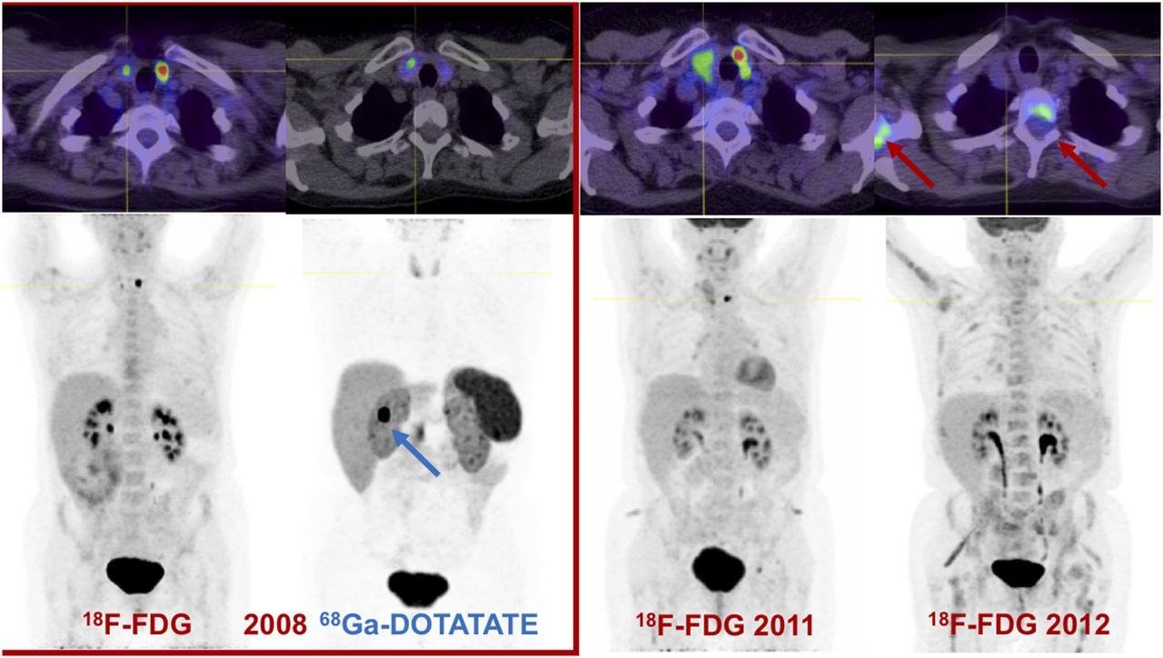
- FIGURE 2.
18F-FDG PET/CT performed on 61-y-old woman with metastatic duodenal carcinoid tumor (blue arrow; grade 2, Ki-67 of 15%) showed mildly avid (SUVmax, 4.4) right thyroid nodule and intensely avid (SUVmax, 16) left thyroid nodule. 68Ga-DOTATATE PET/CT revealed concordant mild DOTATATE uptake in right thyroid nodule, and evaluation for medullary thyroid carcinoma was recommended. Subsequent PET-directed biopsy of mildly 18F-FDG/DOTATATE–avid thyroid nodule confirmed progressive medullary thyroid carcinoma with extrathyroidal extension and lymphovascular invasion. Total thyroidectomy confirmed that intensely 18F-FDG–avid left thyroid nodule was benign follicular adenoma. Patient died approximately 18 mo later from progressive metastatic medullary thyroid cancer (red arrows).
- FIGURE 3.
(A) Kaplan–Meier survival distribution of patients who had 18F-FDG–avid (SUVmax > 3) primary disease at time of identification of 18F-FDG–avid TI on index PET/CT and patients who did not have 18F-FDG–avid primary disease. (B) Kaplan–Meier survival distribution among patients who did not undergo cytologic or histopathologic investigation of 18F-FDG–avid TI and among patients who did undergo further investigation. (C) Kaplan–Meier survival distribution of patients stratified according to American Joint Committee on Cancer stage of primary malignancy. HR = hazard ratio.
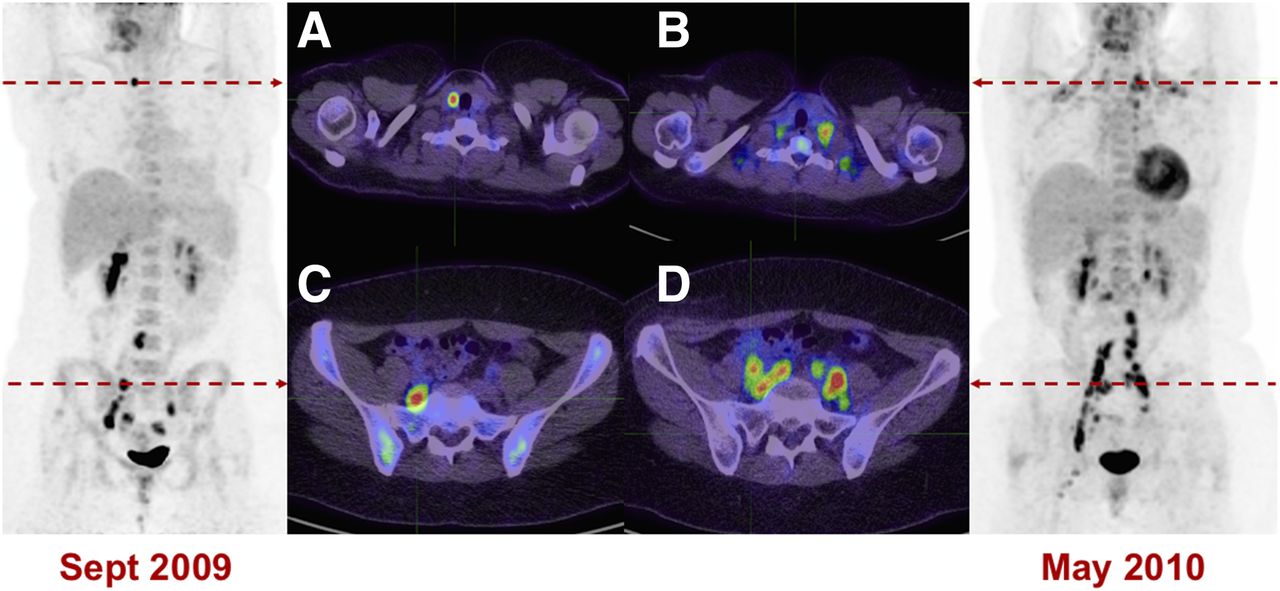
- FIGURE 4.
Intensely 18F-FDG–avid TI (SUVmax, 16) was identified on staging 18F-FDG PET/CT scan of 28-y-old woman with stage IIIB poorly differentiated cervical carcinoma and persistent 18F-FDG–avid pelvic nodal disease. Ultrasound-guided cytologic evaluation demonstrated 5-mm papillary thyroid carcinoma with no adverse features, which was treated with total thyroidectomy. (A and B) Baseline 18F-FDG–avid thyroid nodule (A) and subsequent cervical carcinoma metastases to supraclavicular nodes (B). (C and D) Baseline 18F-FDG–avid pelvic lymphadenopathy (C) with subsequent disease progression (D). Patient died 10 mo after baseline PET/CT scan from progression of metastatic poorly differentiated cervical carcinoma.
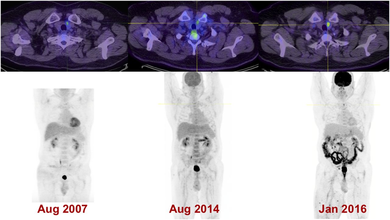
- FIGURE 5.
Mildly 18F-FDG–avid TI (SUVmax, 3.1) was initially identified on staging 18F-FDG PET/CT of 59-y-old man with metastatic melanoma in 2007. FNA (performed to exclude metastatic melanoma) confirmed incidental papillary thyroid malignancy, which remains stable under surveillance imaging and of limited clinical significance in setting of metastatic melanoma under active treatment.

Tables
Baseline parameter Data Age (y) Median 66 Range 19–96 Sex (n) Male 127 Female 235 18F-FDG–avid primary cancer on index PET/CT (n) 272 (75%) Primary malignancy (n) Lymphoma 69 (19%) Lung 59 (16%) Colorectal 43 (12%) Melanoma 33 (9%) Other 159 (44%) AJCC stage of primary malignancy (n) 1 47 (13%) 2 54 (15%) 3 100 (28%) 4 156 (43%) Occult primary tumor (n) 5 (1%) AJCC = American Joint Committee on Cancer.
- TABLE 2
Follow-up and Clinical Outcome Data in Patients with Follow-up > 12 Months or Death (n = 362)
Follow-up parameter Data Follow-up (mo) Median 24 Range 1–103 Survival (mo) Median 20 Range 0–93 Survival status at last follow-up (n) Alive 182 (50) Dead 180 (50) Primary cancer 166 (45.9) Incidental 18F-FDG–avid TI 1 (0.3) Nonmaligant etiology 13 (3.6) 18F-FDG–avid TI status at last follow-up (n) Malignant TI 47 (13) Observation 11 (3) No clinically evident disease 31 (9) Recurrent/metastatic structural disease 1 (0.3) Metastasis (from underlying malignancy) 4 (1) Nondiagnostic/indeterminate FNA 12 (3) Benign TI 72 (20) Not investigated 231 (64) Logistic regression type Independent predictor Simple (crude odds ratio) Multivariate (adjusted odds ratio) 18F-FDG avidity (nonthyroid malignancy) 8.5 (4.6–15.8) 4.0 (2.0–8.2) AJCC stage 3.0 (2.3–3.9) 2.5 (1.8–3.3) Not investigated 3.3 (2–5) 1.7 (1.04 −3.3) Nonthyroid malignancy Lymphoma 0.3 (0.1–0.5) 0.3 (0.2–0.8) Lung 1.7 (0.9–3.2) 1.3 (0.6–2.7) Colorectal 1.1 (0.5–2.2) 1.3 (0.6–3.1) Melanoma 1.5 (0.7–3.3) 1.1 (0.5–2.8) Others 1 1 Age 1.0 (0.99–1.01) 1.0 (0.98–1.02) Sex Female 1 1 Male 1.0 (0.7–1.6) 1.1 (0.6–1.8) AJCC = American Joint Committee on Cancer.
Data in parentheses are 95% CIs.
Parameter Data Malignant cases (n) Total 47 (100%) Malignant on FNA alone 11 (23%) Papillary 24 (51%) Follicular 1 (2%) Metastasis (from underlying malignancy) 4 (9%) Medullary 2 (4%) Hürthle cell cancer/oncocytic variant 5 (11%) Malignant histologic features Size (mm) Median 15 Range 2–50 Vascular invasion (n) 3 (6%) Capsule invasion (n) 9 (18%)


