


Article Figures & Data
Figures
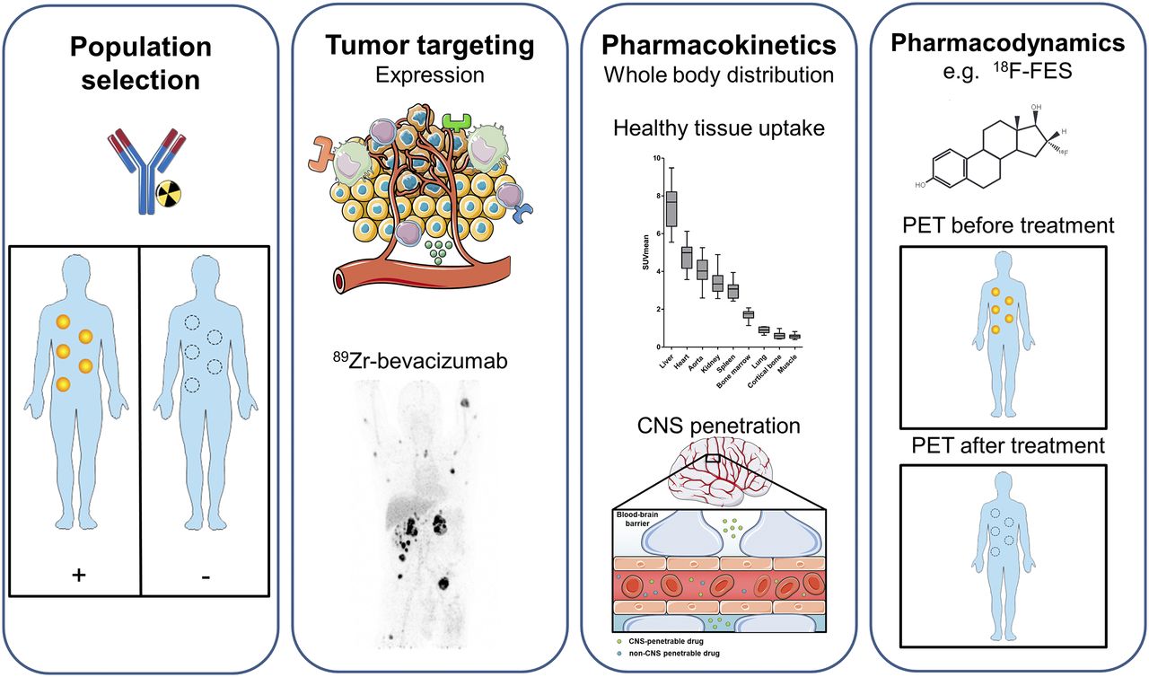
- FIGURE 1.
Information that can be extracted using molecular imaging, categorized by population selection, tumor targeting, pharmacokinetics, and pharmacodynamics (7,8). (First panel) Molecular imaging with, for instance, radiolabeled antibodies can potentially identify responders and nonresponders. (Second panel) For tumor targeting, several tumor aspects can be visualized with molecular imaging, such as tumor cell receptors, environmental factors, and immune cells. Example is PET visualization of 89Zr-bevacizumab targeting vascular endothelial growth factor A in tumor microenvironment in patient with metastatic renal cell carcinoma (bottom; adapted from (60)). (Third panel) For pharmacokinetics, molecular imaging can provide information about whole-body distribution, normal-tissue accumulation of, for instance, 89Zr-bevacizumab (top; adapted from (60)), and penetration of CNS (bottom). Data on normal-tissue uptake might explain drug behavior. (Fourth panel) Pharmacodynamic information can be obtained by performing PET before and after treatment. Example is use of 18F-FES for tumor uptake per lesion on antiestrogen therapy, resulting in less uptake. By this pharmacodynamic assessment, therapeutic dose with maximal decrease in tracer uptake can support further clinical studies. This figure was prepared using template on Servier medical art website (https://smart.servier.com/).


Jump to section
Related Articles
Cited By...
- Fluorescently labelled vedolizumab to visualise drug distribution and mucosal target cells in inflammatory bowel disease
- Using fluorescently labeled vedolizumab to visualize local drug distribution during colonoscopy and identify mucosal target cells in patients with inflammatory bowel disease
- in vivo quantitative FRET small animal imaging: intensity versus lifetime-based FRET
- in vivo quantitative FRET small animal imaging: intensity versus lifetime-based FRET
- 89Zr-DFO-Durvalumab PET/CT Before Durvalumab Treatment in Patients with Recurrent or Metastatic Head and Neck Cancer
- Imaging Androgen Receptors in Breast Cancer with 18F-Fluoro-5{alpha}-Dihydrotestosterone PET: A Pilot Study
- In vitro and in vivo NIR Fluorescence Lifetime Imaging with a time-gated SPAD camera
- First-in-Human Study of the Biodistribution and Pharmacokinetics of 89Zr-CX-072, a Novel Immunopet Tracer Based on an Anti-PD-L1 Probody
- Preclinical PET imaging with the novel human antibody 89Zr-DFO-REGN3504 sensitively detects PD-L1 expression in tumors and normal tissues
- Molecular Imaging: a Novel Tool To Visualize Pathogenesis of Infections In Situ
- The Future of Nuclear Medicine as an Independent Specialty
- Nuclear Medicine and Wall Street: An Evolving Relationship