


Article Figures & Data
Figures
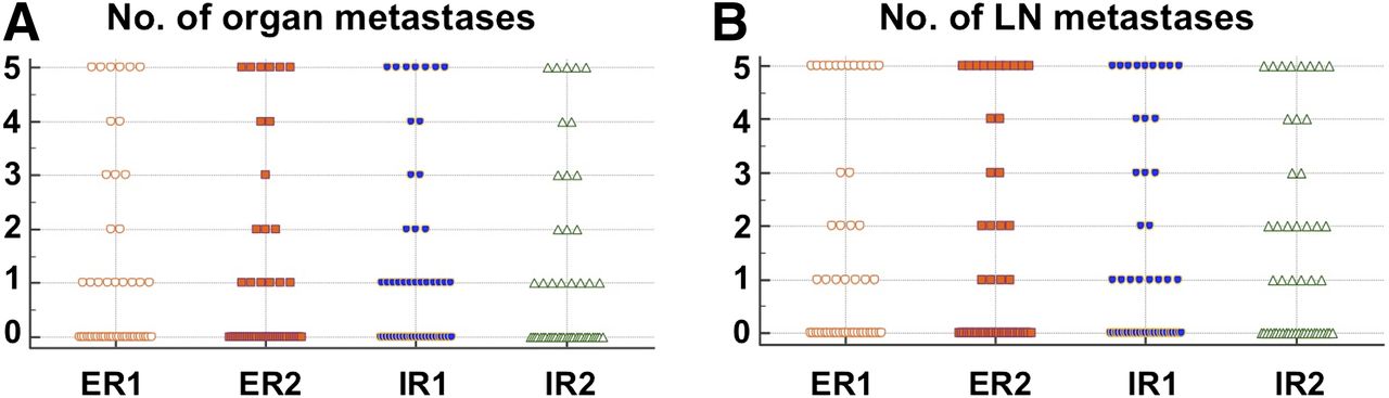
- FIGURE 1.
Distribution of number of organ (A) and LN (B) metastases for all 4 readers.
- FIGURE 2.
Target lesion assessment (identical target lesion included by all 4 readers). PSMA-RADS-1A and -1B were subsumed under PSMA-RADS-1.
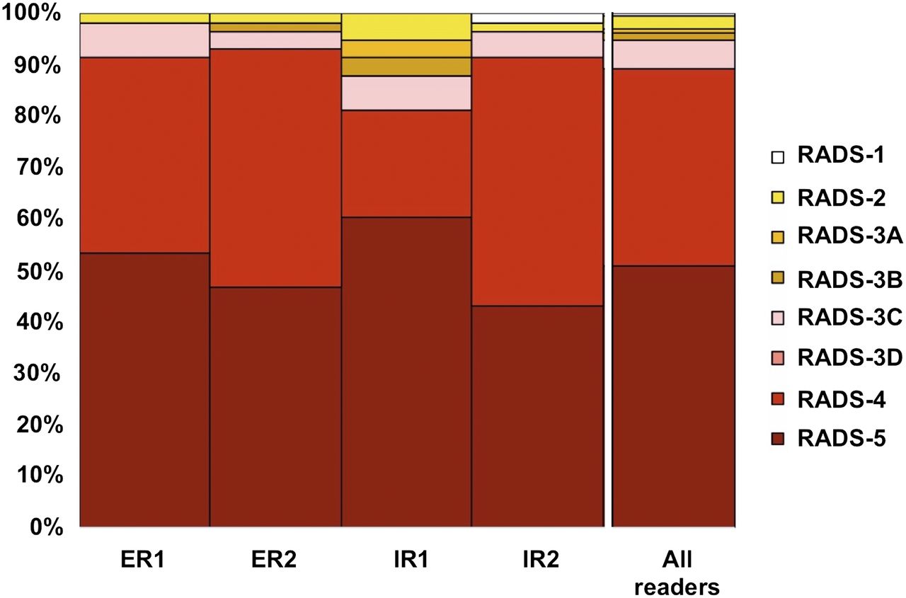
- FIGURE 3.
Overall-PSMA RADS scoring for all 4 readers. PSMA-RADS-1A and -1B were subsumed under PSMA-RADS-1.
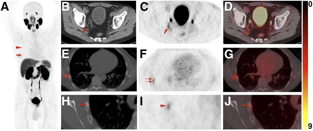
- FIGURE 4.
Images of 60-y-old man undergoing 18F-DCFPyL PET/CT for primary diagnostic assessment (prostate-specific antigen level on date of scan, 13.5; no previous therapies). (A) Whole-body maximum-intensity projection demonstrates multiple sites of suggestive radiotracer uptake (e.g., third right rib [arrowhead] and sixth right rib [arrow]). (B–D) Axial CT (B), axial 18F-DCFPyL PET (C), and axial 18F-DCFPyL PET/CT (D) show mild radiotracer uptake in right iliac LN (arrow). Although ER trained on 18F-DCFPyL PET called this lesion PSMA-RADS-4, the 2 remaining readers trained on 68Ga-PSMA PET called it PSMA-RADS-3A (i.e., suggestive but indeterminate LN) (12). One might speculate that 18F-trained reader had higher confidence in lesion interpretation on 18F-DCFPyL PET scans because of higher sensitivity in detection rate of small lesions using 18F-labeled radiotracers than using 68Ga-PSMA (27). (E–G) All 4 readers classified overall scan impression as PSMA-RADS-5, because CT (E) revealed findings corresponding to sixth right rib metastasis, with discernible radiotracer uptake on axial 18F-DCFPyL PET (F) and axial 18F-DCFPyL PET/CT (G) (doubled arrows). (H–J) Magnification of this sixth-rib suggestive site of uptake provided in axial CT (H), axial 18F-DCFPyL PET (I), and axial 18F-DCFPyL PET/CT (J) further suggested this to be malignant lesion at this uptake site (arrowhead).
- FIGURE 5.
Images of 76-y-old man undergoing 18F-DCFPyL PET/CT for staging of metastatic PCa (prostate-specific antigen level on date of scan, 0.63; prior prostatectomy). (A) Whole-body maximum-intensity projection shows radiotracer uptake in right hilar and subcarinal LNs (arrows), lung lesion (arrowhead), and right iliac bone (doubled arrow). (B–D) Axial CT (B), axial 18F-DCFPyL PET (C), and axial 18F-DCFPyL PET/CT (D) show mild to moderate radiotracer uptake in subcarinal LN (arrows). IR called this finding PSMA-RADS-4, whereas ER classified it PSMA-RADS-2 (i.e., likely benign because of low-level uptake in soft-tissue site atypical of metastatic PCa). Hilar and subcarinal LNs remained unchanged on follow-up imaging, suggesting that these are benign. All 4 readers classified overall scan impression as PSMA-RADS-5. (E–G) Axial CT (E), axial 18F-DCFPyL PET (F), and axial 18F-DCFPyL PET/CT (G) show intense radiotracer uptake in right iliac bone (doubled arrows). Apart from that, lung lesion (18F-DCFPyL whole-body maximum intensity projection in A, red arrowhead) was classified as PSMA-RADS-2 by IR. (H–J) Axial CT (H), axial 18F-DCFPyL PET (I), and axial 18F-DCFPyL PET/CT (J) of this lesion further confirm suspicion of benign lesion (arrowheads, most likely peripheral interstitial thickening). Follow-up imaging also corroborated this impression.


Tables
Parameter Characteristic Data Median age ± SD (y) 65 ± 8 Race White 38/50 (76%) Black 9/50 (18%) Asian/other 3/50 (6%) Indication for scan Staging 24/50 (48%) Biochemical recurrence 9/50 (18%) Biochemical persistence after primary surgery 6/50 (12%) Primary diagnosis 5/50 (10%) Potential withdrawal of androgen deprivation therapy 3/50 (6%) Other 3/50 (6%) Gleason score (GS) Overall median ± SD (n = 39) 8 ± 1 GS 6 1/39 (2.6%) GS 7 15/39 (38.4%) GS 8 7/39 (17.9%) GS 9 15/39 (38.5%) GS 10 1/39 (2.6%) PSA level (ng/mL) Overall median 3.2 Range 0.02–48 Prior therapies Total 41/50 (82%) Surgery 29/41 (70.7%) Hormonal therapy 21/41 (51.2%) Radiation therapy 18/41 (43.9%) Chemotherapy 6/41 (14.6%) PSA = prostate specific antigen.
Level of certainty* Parameter Parameter Parameter Parameter 0 1 2 3 4 5 Binary fashion Overall scan result (negative = 0, positive = 1) 0.75 0.64–0.83 ER 1 13 (26) 37 (74) ER 2 13 (26) 37 (74) IR 1 6 (12) 44 (88) IR 2 13 (26) 37 (74) Organ involvement (no = 0, yes = 1) 0.80 0.71–0.88 ER 1 28 (56) 22 (44) ER 2 27 (54) 23 (46) IR 1 20 (40) 30 (60) IR 2 29 (58) 21 (42) LN involvement (no = 0, yes = 1) 0.78 0.69–0.86 ER 1 25 (50) 25 (50) ER 2 27 (54) 23 (46) IR 1 21 (42) 29 (58) IR 2 24 (48) 26 (52) 5-point assessment† Affected organs (n) 0.74 0.62–0.83 ER 1 28 (56) 17 (34) 3 (6) 2 (4) ER 2 28 (56) 18 (36) 4 (8) IR 1 20 (40) 20 (40) 9 (18) 1 (2) IR 2 29 (58) 17 (34) 4 (8) Organ metastases (n) 0.92 0.89–0.95 ER 1 28 (56) 10 (20) 1 (2) 3 (6) 2 (4) 6 (12) ER 2 32 (64) 6 (12) 3 (6) 1 (2) 2 (4) 6 (12) IR 1 23 (46) 13 (26) 3 (6) 2 (4) 1 (2) 8 (16) IR 2 29 (58) 8 (16) 3 (6) 3 (6) 2 (4) 5 (10) Affected LN areas (n) 0.79 0.70–0.86 ER 1 25 (50) 11 (22) 7 (14) 5 (10) 2 (4) ER 2 27 (54) 8 (16) 7 (14) 6 (12) 2 (4) IR 1 21 (42) 19 (38) 9 (18) 1 (2) IR 2 24 (48) 11 (22) 10 (20) 3 (6) 2 (4) LN metastases (n) 0.90 0.85–0.94 ER 1 25 (50) 7 (14) 4 (8) 2 (4) 12 (24) ER 2 28 (56) 4 (8) 4 (8) 2 (4) 2 (4) 10 (20) IR 1 25 (50) 8 (16) 2 (4) 3 (6) 3 (6) 9 (18) IR 2 24 (48) 6 (12) 7 (14) 2 (4) 3 (6) 8 (16) - TABLE 3
Distribution of Target Lesions Among Compartments, with Agreement Rate and ICC Based on PSMA-RADS
Compartment-based distribution Agreement rate ICC Identical target lesions* LN Bone Prostate/local recurrence Lung Non-LN soft tissue Thyroid gland Liver For all identical target lesions For minimum agreements† For all identical target lesions for LNs All (n = 125) 64/125 (51.2) 39/125 (31.2) 11/125 (8.8) 5/125 (4.0) 3/125 (2.4) 2/125 (1.6) 1/125 (0.8) NA NA NA NA 4 (n = 58/125, 46.4%) 26/58 (44.8) 19/58 (32.8) 8/58 (13.8) 3/58 (5.2) 1/58 (1.7) 1/58 (1.7) 29/58 (50.0) 46/58 (79.3) 0.60 (0.48–0.71) 0.79 (0.66–0.89) 3 (n = 40/125, 32%) 22/40 (55.0) 12/40 (30.0) 3/40 (7.5) 2/40 (5.0) 1/40 (2.5) 21/40 (52.5) 36/40 (90.0) 0.60 (0.43–0.75) 0.66 (0.44–0.83) 2 (n = 27/125, 21.6%) 16/27 (59.3) 8/27 (29.6) 2/27 (7.4) 1/27 (3.7) 15/27 (55.6) NA 0.62 (0.32–0.81) 0.57 (0.12–0.83) ↵* All identical target lesions chosen by minimum of 2 readers, and target lesions identified by 4, 3, and 2 readers.
↵† If 4 readers selected same target lesion, minimum of 3 readers designated same PSMA-RADS score, and if 3 readers selected same target lesion, minimum of 2 readers designated same PSMA-RADS score.
NA = not applicable.
Data in parentheses are 95% CIs for ICC and percentages for all others.
- TABLE 4
Distribution of PSMA-RADS Score for 4 Identical Target Lesions and for Overall PSMA-RADS Score Among the 4 Readers
PSMA-RADS Parameter Reader 1* 2 3A 3B 3C 3D 4 5 4 identical target lesions ER 1 1/58 (1.7) 4/58 (6.9) 22/58 (37.9) 31/58 (53.5) ER 2 1/58 (1.7) 1/58 (1.7) 2/58 (3.4) 27/58 (46.6) 27/58 (46.6) IR 1 3/58 (5.2) 2/58 (3.4) 2/58 (3.4) 4/58 (6.9) 12/58 (20.7) 35/58 (60.3) IR 2 1/58 (1.7) 1/58 (1.7) 3/58 (5.2) 28/58 (48.3) 25/58 (43.1) Overall PSMA-RADS ER 1 10/50 (20) 2/50 (4) 1/50 (2) 15/50 (30) 22/50 (44) ER 2 9/50 (18) 2/50 (4) 3/50 (6) 1/50 (2) 16/50 (32) 19/50 (38) IR 1 6/50 (12) 5/50 (10) 2/50 (4) 2/50 (4) 3/50 (6) 9/50 (18) 23/50 (46) IR 2 10/50 (20) 3/50 (6) 1/50 (2) 15/50 (30) 21/50 (42) ↵* PSMA-RADS-1A and -1B were subsumed under PSMA-RADS score 1.
n = 58 target lesions and 50 scans. Data in parentheses are percentages.


