


Article Figures & Data
Figures
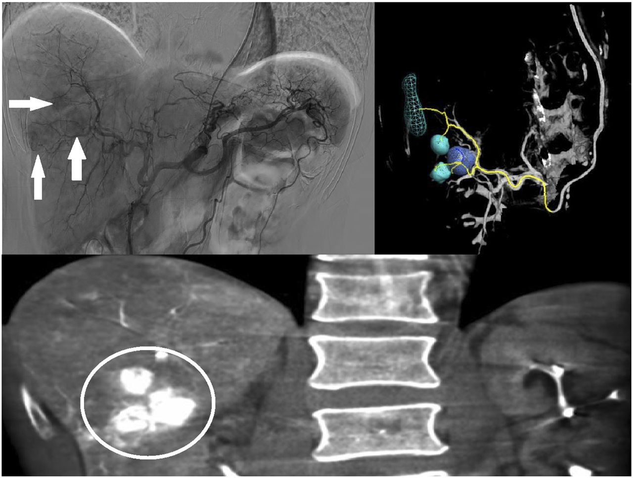
- FIGURE 1.
Images acquired during hepatic angiography of 64-y-old man scheduled for 90Y radiation lobectomy. (Top left) Catheter in celiac axis after left radial access, with multiple foci of contrast medium (arrows) in liver consistent with tumors. (Bottom) Coronal cone beam CT of liver, with multiple tumors (circle) seen in right hepatic lobe. (Top right) Three-dimensional reconstruction with targeting software, demonstrating tumors and arterial supply.
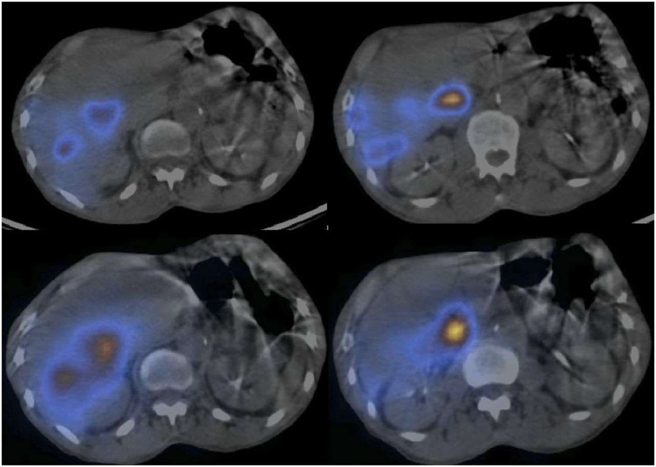
- FIGURE 2.
(Top) SPECT/CT images after injection of 99mTc-MAA in right hepatic artery of 64-y-old man. (Bottom) SPECT/CT images of same patient after 90Y microsphere radiation therapy. Radiotracer deposition is concordant between the 2 studies.
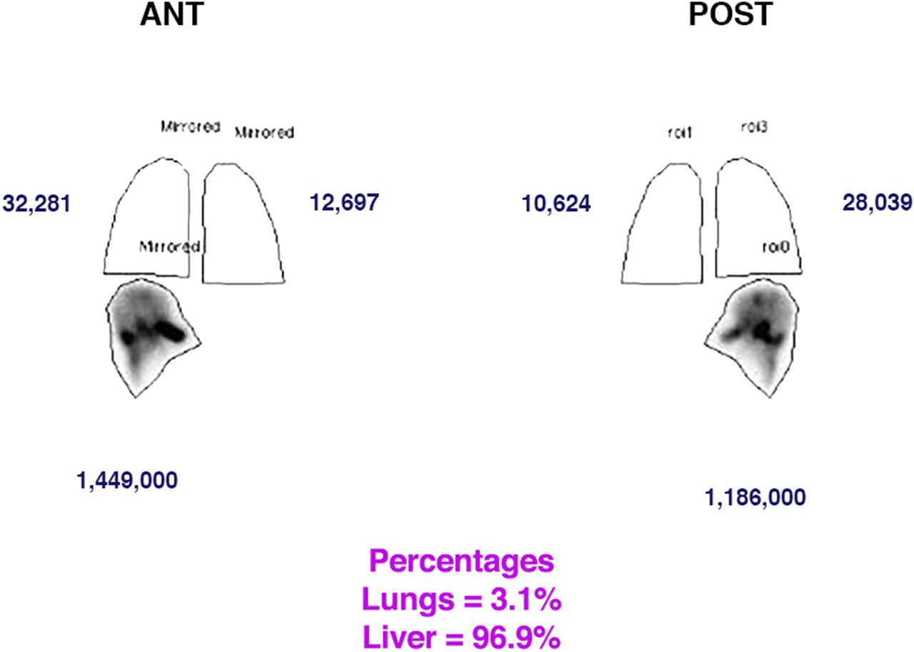
- FIGURE 3.
Anterior and posterior planar whole-body scintigraphy after 99mTc-MAA intrahepatic arterial injection. Counts in regions of interest around lung and liver reflect radiotracer deposition. From these counts, computer calculates percentage in liver and percentage in lung (lung shunt fraction). This 64-y-old man had lung shunt fraction of 3.1%, which was acceptable for 90Y microsphere radiation therapy.
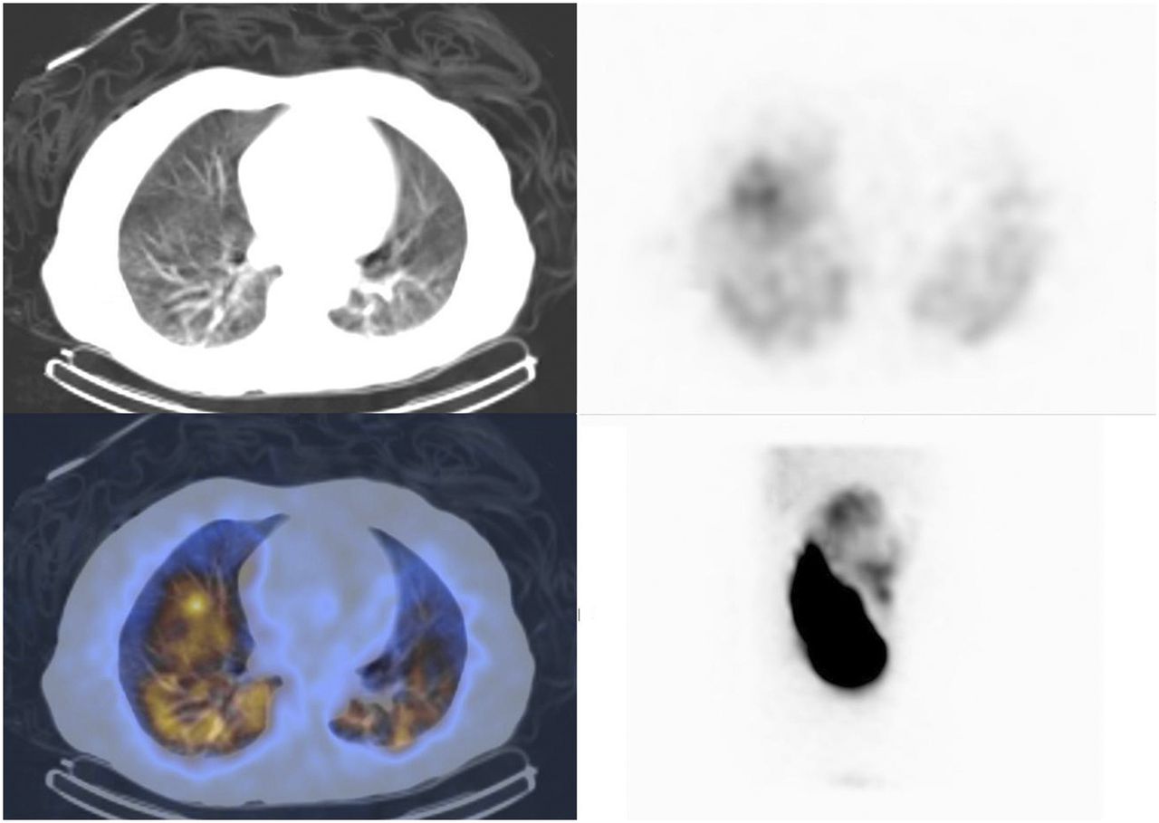
- FIGURE 4.
CT of lungs (top left), SPECT/CT of lungs (bottom left), axial scintigraphy of lungs (top right), and planar scintigraphy of lower chest and abdomen (bottom right) in 54-y-old man with hepatocellular carcinoma after his 99mTc-MAA examination demonstrated bilateral uptake within lungs after administration to liver. This patient’s lung shunt fraction was calculated to be 58% (acceptable fraction is <20%).
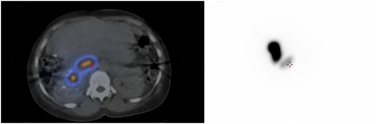
- FIGURE 5.
SPECT/CT (left) and planar scintigraphy (right) demonstrating 99mTc-MAA radiotracer deposition in small bowel, secondary to shunting from liver arteries. This 57-y-old man is no longer a candidate for 90Y radioembolization because of risk of nontarget embolization and duodenal ulceration.
- FIGURE 6.
Axial arterial-phase CT images from 68-y-old man before (left) and 1 mo after (right) 90Y therapy. Tumor (solid arrow) enhances before treatment, consistent with hepatocellular carcinoma, but after treatment shows no evidence of enhancement within treatment cavity (dashed arrow).


