


Article Figures & Data
Figures
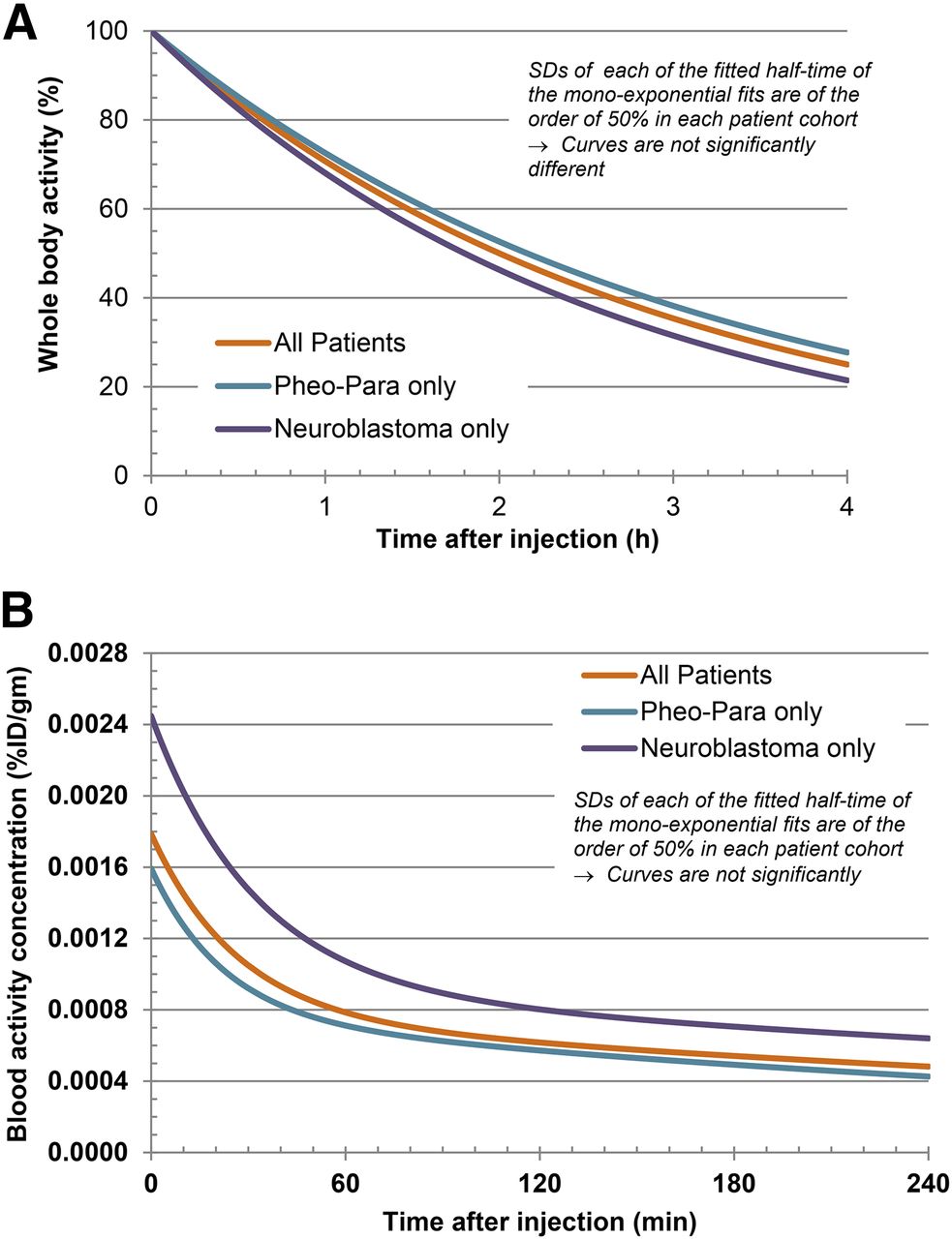
- FIGURE 1.
Whole-body (A) and -blood (B) clearance time–activity curves. Whole-body activity showed monoexponential clearance, and blood activity showed biexponential clearance. Para = paraganglioma; Pheo = pheochromocytoma.
- FIGURE 2.
Patient with metastatic pheochromocytoma. Whole-body maximum-intensity-projection scans of 18F-MFBG obtained 30–60 min after injection (A), 1–2 h after injection (B), and 3–4 h after injection (C) as against a uniform SUV scale (right bar). Lesions are distinctly seen in the liver at 1–2 h and 3–4 h after injection (B and C; arrows). Fused images show lesions more distinctly in liver (D and E; arrows). Lesion in maximum-intensity-projection image is localized to left iliac bone (F; short arrow).
- FIGURE 3.
Uptake in normal organs at various scan times after injection. (A) Uptake decreases from scan 1 (0.5–1 h after injection) to scan 2 (1–2 h after injection) and scan 3 (3–4 h after injection). (B) Prominent activity is seen in liver, which decreases over time. Focal uptake posteromedially is uptake along adrenal (SUV 5.6). (C) Cardiac activity is most prominent in early images, decreasing with time; distribution is seen along the ventricular myocardium. (D) Diffuse uptake is seen along pancreas (SUV 3.5); posteromedial uptake is physiologic uptake in adrenal gland. (E) Uptake is seen in prostate (SUV 5.6).
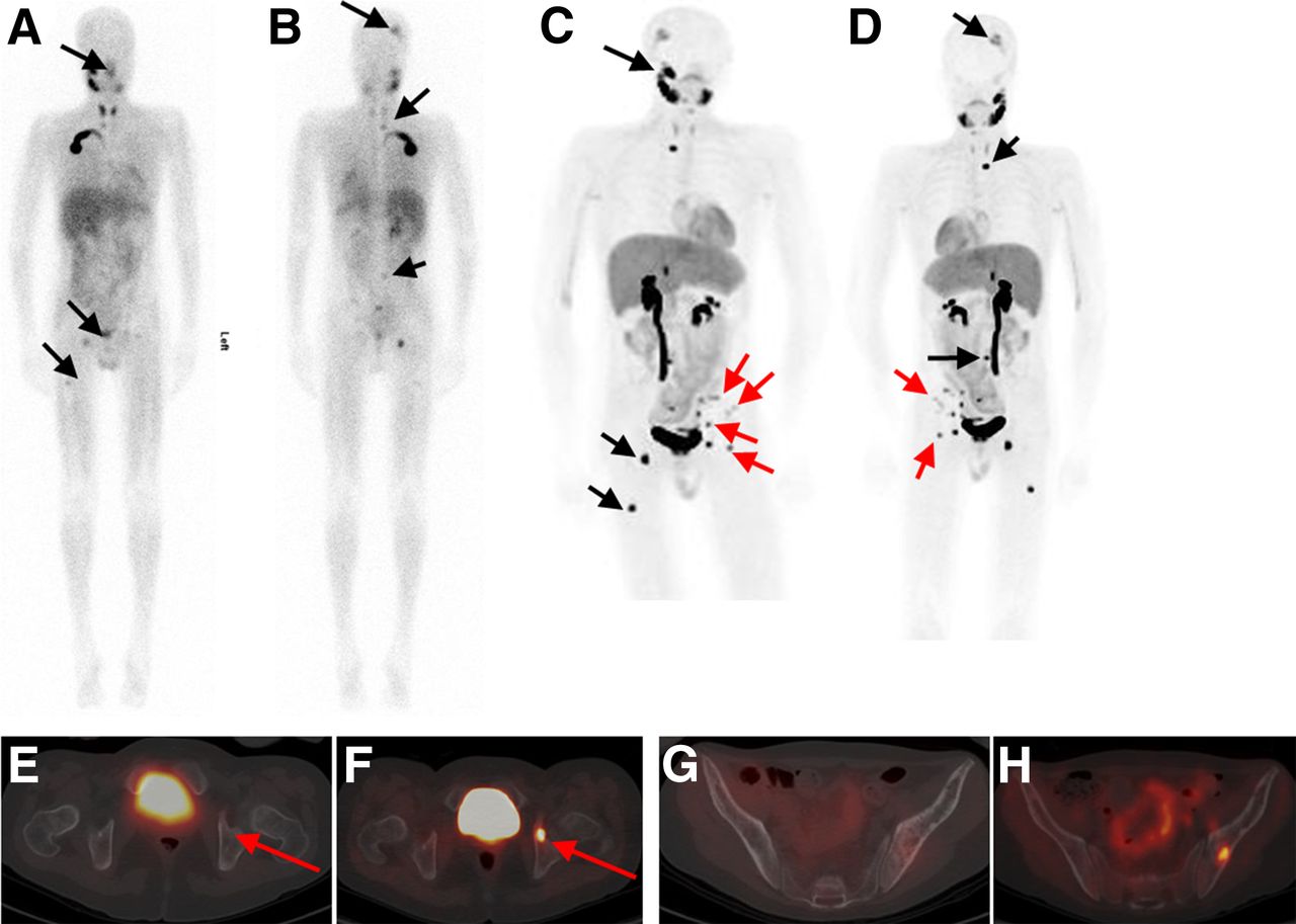
- FIGURE 4.
Patient with neuroblastoma for follow-up evaluation and possible therapy with 131I-MIBG. 123I-MIBG images (A, anterior; B, posterior) show foci of suspicious activity in skull, lumbar vertebra, right and left acetabula, and right femur (black arrows). Patient underwent imaging with 162 MBq of 18F-MFBG a wk later. Whole-body maximum-intensity-projection scans with 18F-MFBG (C and D) show all lesions seen on 123I-MIBG scan but with greater contrast and clarity (black arrows). In addition, several lesions are seen on 18F-MFBG scan only (red arrows) that are not visible on 123I-MIBG images. For example, fused PET/CT transaxial 18F-MFBG image (F) shows intense uptake in left acetabulum (red arrow), suspicious for disease, that is not seen on 123I-MIBG SPECT/CT fused transaxial image (E). Also, left iliac bone lesions are clearly avid on 18F-MFBG (H) vs. 123I-MIBG imaging (G).
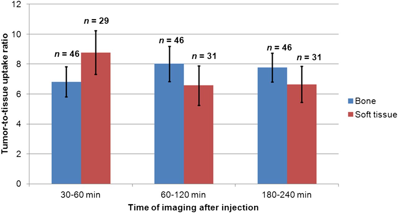
- FIGURE 5.
Tumor–to–normal bone and soft-tissue uptake ratios at different scan times after injection of 18F-MFBG (scan 1 at 30–60 min, scan 2 at 60–120 min, and scan 3 at 180–240 min). Uptake ratios were based on mean SUVs in respective tissues of 10 patients. Numbers = number of observations (i.e., lesions); error bars = SE of mean.


Tables
mGy/MBq cGy/mCi Organ Mean SD Mean SD Salivary gland 0.058 0.069 0.213 0.253 Adrenals 0.023 0.024 0.085 0.089 Brain 0.004 0.002 0.014 0.008 Breasts 0.005 0.002 0.017 0.008 Gallbladder wall 0.012 0.005 0.046 0.020 Lower large intestine wall 0.011 0.005 0.041 0.020 Small intestine 0.009 0.004 0.033 0.015 Stomach wall 0.007 0.003 0.027 0.012 Upper large intestine wall 0.009 0.004 0.032 0.014 Heart wall 0.031 0.016 0.115 0.057 Kidneys 0.028 0.025 0.105 0.092 Liver 0.046 0.026 0.171 0.097 Lungs 0.009 0.005 0.035 0.017 Muscle 0.006 0.003 0.024 0.010 Ovaries 0.011 0.005 0.041 0.019 Pancreas 0.032 0.021 0.119 0.078 Red marrow 0.006 0.002 0.022 0.008 Osteogenic cells 0.007 0.004 0.027 0.013 Skin 0.004 0.002 0.015 0.007 Spleen 0.015 0.008 0.057 0.028 Testes 0.008 0.006 0.030 0.023 Thymus 0.006 0.003 0.022 0.010 Thyroid 0.032 0.028 0.119 0.103 Urinary bladder wall 0.186 0.195 0.689 0.720 Total body 0.011 0.011 0.042 0.041 Effective dose (mSv/MBq)/*(cSv or rem/mCi) 0.023 0.012 0.085* 0.043* Patient no. 123I-MIBG + lesion no. 18F-MFBG + lesion no. Neuroblastoma 1 9 11 2 2 3 3 2 5 4 1 2 5 8 13 Pheochromocytoma/paraganglioma 1 2 6 2 14 29 3 2 4 4 9 20 5 14 29


Jump to section
Related Articles
Cited By...
- Performing [18F]MFBG Long-Axial-Field-of-View PET/CT Without Sedation or General Anesthesia for Imaging of Children with Neuroblastoma
- PET imaging of fibroblast activation protein alpha (FAP) detects incipient cardiotoxicity due to anthracycline chemotherapy
- PET/MRI in Pediatric Neuroimaging: Primer for Clinical Practice
- Radiotracers to Address Unmet Clinical Needs in Cardiovascular Imaging, Part 1: Technical Considerations and Perfusion and Neuronal Imaging
- Mars Shot for Nuclear Medicine, Molecular Imaging, and Molecularly Targeted Radiopharmaceutical Therapy
- Advances in adrenal tumors 2018