
Main menu

  • Home
  • Content
    • Current
    • Ahead of print
    • Past Issues
    • JNM Supplement
    • SNMMI Annual Meeting Abstracts
    • Continuing Education
    • JNM Podcasts
  • Subscriptions
    • Subscribers
    • Institutional and Non-member
    • Rates
    • Journal Claims
    • Corporate & Special Sales
  • Authors
    • Submit to JNM
    • Information for Authors
    • Assignment of Copyright
    • AQARA requirements
  • Info
    • Reviewers
    • Permissions
    • Advertisers
  • About
    • About Us
    • Editorial Board
    • Contact Information
  • More
    • Alerts
    • Feedback
    • Help
    • SNMMI Journals
  • SNMMI
    • JNM
    • JNMT
    • SNMMI Journals
    • SNMMI

User menu

  • Subscribe
  • My alerts
  • Log in
  • My Cart

Search

  • Advanced search
Journal of Nuclear Medicine
  • SNMMI
    • JNM
    • JNMT
    • SNMMI Journals
    • SNMMI
  • Subscribe
  • My alerts
  • Log in
  • My Cart
Journal of Nuclear Medicine

Advanced Search

  • Home
  • Content
    • Current
    • Ahead of print
    • Past Issues
    • JNM Supplement
    • SNMMI Annual Meeting Abstracts
    • Continuing Education
    • JNM Podcasts
  • Subscriptions
    • Subscribers
    • Institutional and Non-member
    • Rates
    • Journal Claims
    • Corporate & Special Sales
  • Authors
    • Submit to JNM
    • Information for Authors
    • Assignment of Copyright
    • AQARA requirements
  • Info
    • Reviewers
    • Permissions
    • Advertisers
  • About
    • About Us
    • Editorial Board
    • Contact Information
  • More
    • Alerts
    • Feedback
    • Help
    • SNMMI Journals
  • View or Listen to JNM Podcast
  • Visit JNM on Facebook
  • Join JNM on LinkedIn
  • Follow JNM on Twitter
  • Subscribe to our RSS feeds
Research ArticleInfectious Disease

The Value of 18F-FDG PET/CT in Diagnosis and During Follow-up in 273 Patients with Chronic Q Fever

Ilse J.E. Kouijzer, Linda M. Kampschreur, Peter C. Wever, Corneline Hoekstra, Marjo E.E. van Kasteren, Monique G.L. de Jager-Leclercq, Marrigje H. Nabuurs-Franssen, Marjolijn C.A. Wegdam-Blans, Heidi S.M. Ammerlaan, Jacqueline Buijs, Lioe-Fee de Geus-Oei, Wim J.G. Oyen and Chantal P. Bleeker-Rovers
Journal of Nuclear Medicine January 2018, 59 (1) 127-133; DOI: https://doi.org/10.2967/jnumed.117.192492
Ilse J.E. Kouijzer
1Division of Infectious Diseases, Department of Internal Medicine, Radboud University Medical Center, Nijmegen, The Netherlands
2Radboud Expert Centre for Q Fever, Radboud University Medical Center, Nijmegen, The Netherlands
3MIRA Institute for Biomedical Technology and Technical Medicine, University of Twente, Enschede, The Netherlands
  • Find this author on Google Scholar
  • Find this author on PubMed
  • Search for this author on this site
Linda M. Kampschreur
4Department of Internal Medicine, University Medical Center Utrecht, Utrecht, The Netherlands
  • Find this author on Google Scholar
  • Find this author on PubMed
  • Search for this author on this site
Peter C. Wever
5Department of Medical Microbiology and Infection Control, Jeroen Bosch Hospital, ’s-Hertogenbosch, The Netherlands
  • Find this author on Google Scholar
  • Find this author on PubMed
  • Search for this author on this site
Corneline Hoekstra
6Department of Nuclear Medicine, Jeroen Bosch Hospital, ’s-Hertogenbosch, The Netherlands
  • Find this author on Google Scholar
  • Find this author on PubMed
  • Search for this author on this site
Marjo E.E. van Kasteren
7Department of Internal Medicine, Elisabeth Tweesteden Hospital, Tilburg, The Netherlands
  • Find this author on Google Scholar
  • Find this author on PubMed
  • Search for this author on this site
Monique G.L. de Jager-Leclercq
8Department of Internal Medicine, Bernhoven Hospital, Uden, The Netherlands
  • Find this author on Google Scholar
  • Find this author on PubMed
  • Search for this author on this site
Marrigje H. Nabuurs-Franssen
9Department of Medical Microbiology and Infectious Diseases, Canisius Wilhelmina Hospital, Nijmegen, The Netherlands
  • Find this author on Google Scholar
  • Find this author on PubMed
  • Search for this author on this site
Marjolijn C.A. Wegdam-Blans
10Department of Medical Microbiology, Laboratory for Pathology and Medical Microbiology (PAMM), Veldhoven, The Netherlands
  • Find this author on Google Scholar
  • Find this author on PubMed
  • Search for this author on this site
Heidi S.M. Ammerlaan
11Department of Internal Medicine, Catharina Hospital, Eindhoven, The Netherlands
  • Find this author on Google Scholar
  • Find this author on PubMed
  • Search for this author on this site
Jacqueline Buijs
12Department of Internal Medicine, Zuyderland Medical Center, Heerlen, The Netherlands
  • Find this author on Google Scholar
  • Find this author on PubMed
  • Search for this author on this site
Lioe-Fee de Geus-Oei
3MIRA Institute for Biomedical Technology and Technical Medicine, University of Twente, Enschede, The Netherlands
13Department of Nuclear Medicine, Leiden University Medical Center, Leiden, The Netherlands; and
  • Find this author on Google Scholar
  • Find this author on PubMed
  • Search for this author on this site
Wim J.G. Oyen
14Institute of Cancer Research/Royal Marsden Hospital, London, U.K., and Department of Nuclear Medicine, Radboud University Medical Center, Nijmegen, The Netherlands
  • Find this author on Google Scholar
  • Find this author on PubMed
  • Search for this author on this site
Chantal P. Bleeker-Rovers
1Division of Infectious Diseases, Department of Internal Medicine, Radboud University Medical Center, Nijmegen, The Netherlands
2Radboud Expert Centre for Q Fever, Radboud University Medical Center, Nijmegen, The Netherlands
  • Find this author on Google Scholar
  • Find this author on PubMed
  • Search for this author on this site
  • Article
  • Figures & Data
  • Info & Metrics
  • PDF
Loading

Article Figures & Data

Figures

  • Tables
  • FIGURE 1.
    • Download figure
    • Open in new tab
    • Download powerpoint
    FIGURE 1.

    18F-FDG PET/CT scan of 72-y-old man with proven chronic Q fever, infected endovascular aortic graft, and psoas abscess (arrows). Patient had vascular surgery 6 mo after initiation of antibiotic therapy with doxycycline and hydroxychloroquine because of severity of infection. Four years later, patient was still on antibiotic treatment because of persisting chronic Q fever infection.

  • FIGURE 2.
    • Download figure
    • Open in new tab
    • Download powerpoint
    FIGURE 2.

    18F-FDG PET/CT scan of 67-y-old man with history of acute Q fever and analysis because of severe fatigue. Serology for C. burnetii showed increased antiphase 1 IgG; PCR on blood was negative for C. burnetii. Transesophageal echocardiography was negative for endocarditis, but 18F-FDG PET/CT showed highly increased 18F-FDG uptake of patient’s native mitral valve (arrows). Patient was treated for almost 2 y with antibiotic therapy because of chronic Q fever endocarditis.
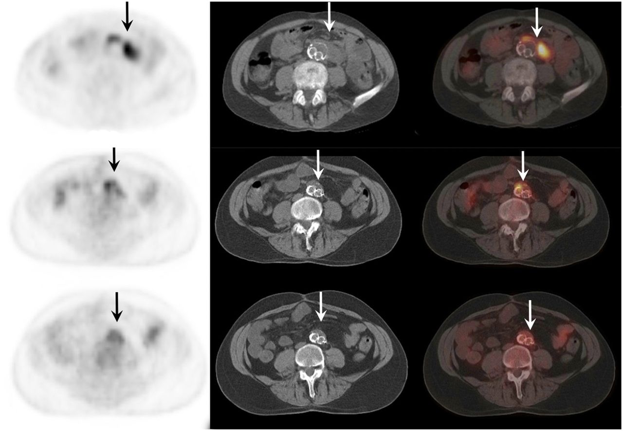
  • FIGURE 3.
    • Download figure
    • Open in new tab
    • Download powerpoint
    FIGURE 3.

    18F-FDG PET/CT scan of 64-y-old man with proven chronic Q fever, infected aneurysm, and infected vascular graft in aorta (arrows). Upper scan shows vascular infection before initiation of antibiotic treatment with doxycycline and hydroxychloroquine. Middle scan shows same area after 1 y of treatment. Lower scan, obtained 2 y after start of treatment, no longer shows vascular infection and therefore antibiotic treatment was discontinued. Four years later, patient died because of septic cholangitis. Autopsy did not show any Q fever foci, and PCR on several tissues, including aorta, was negative for C. burnetii.

Tables

  • Figures
    • View popup
    TABLE 1

    Dutch Consensus Guidelines on Chronic Q Fever Diagnosis (15)

    Chronic Q feverCharacteristics
    ProvenPositive C. burnetii PCR in blood or tissue OR IFA phase I IgG of ≥1,024 for C. burnetii phase I IgG with either definite endocarditis according to modified Duke criteria or proven large-vessel or prosthetic infection by imaging techniques (18F-FDG PET/CT, MRI, CT, ultrasound)
    ProbableIFA phase I IgG of ≥1,024 for C. burnetii phase I IgG AND valvulopathy not meeting modified Duke criteria OR known aneurysm or vascular or cardiac valve prosthesis without signs of infection by imaging techniques (TTE/TEE, 18F-FDG PET/CT, MRI, CT, ultrasound) OR suspected osteomyelitis, hepatitis, or pericarditis as manifestation of chronic Q fever OR pregnancy OR symptoms and signs of chronic infection such as fever, weight loss, night sweats, hepatosplenomegaly, and persistent ESR or CRP OR granulomatous tissue inflammation as proven by histologic examination OR immunocompromised state
    PossibleIFA phase I IgG of ≥1,024 for C. burnetii phase I IgG without manifestations meeting criteria for proven or probable chronic Q fever
    • IFA = immunofluorescence assay; TTE = transthoracic echocardiography; TEE = transesophageal echocardiography; ESR = erythrocyte sedimentation rate; CRP = C-reactive protein.

    • View popup
    TABLE 2

    Interpretation of 18F-FDG PET/CT Combining Visual Grading Score and Uptake Pattern

    ParameterDetails
    Vascular infection
     Grade 118F-FDG uptake similar to background
     Grade 2*Low 18F-FDG uptake comparable to uptake by inactive muscles and fat, and above background uptake but below liver uptake
     Grade 3*Moderate 18F-FDG uptake clearly visible and higher than uptake by inactive muscles and fat, and similar to liver uptake
     Grade 4*Strong 18F-FDG uptake clearly above liver uptake
     Interpretation
      No vascular infectionGrades 1 and 2
      InflammationGrades 3 and 4 and homogeneous uptake pattern without perigraft soft-tissue involvement
      Vascular infectionGrades 3 and 4 and inhomogeneous uptake pattern with or without perigraft soft-tissue involvement
    Endocarditis
     Grade 1No relevant 18F-FDG uptake of heart
     Grade 2†Diffuse 18F-FDG uptake in heart (reflecting normal uptake in myocardium)
     Grade 3Focal 18F-FDG uptake in heart with or without diffuse low-level uptake in myocardium
     Interpretation
      No endocarditisGrade 1
      Evaluation not possibleGrade 2
      EndocarditisGrade 3
    • ↵* Additional “a” for homogeneous 18F-FDG uptake and “b” for inhomogeneous 18F-FDG uptake was registered.

    • ↵† Evaluation of heart valves for endocarditis was not possible because of diffuse myocardial 18F-FDG uptake.

    • View popup
    TABLE 3

    Risk Factors and Outcome of All Patients with Proven, Probable, and Possible Chronic Q Fever

    ParameterProvenProbablePossible
    Number of patients1476066
    Diagnostics
     Positive PCR blood + tissue25 (17.0)00
     Positive PCR blood only58 (39.5)00
     Positive PCR tissue only27 (18.4)00
     Antiphase I IgG at diagnosis4,096 (256–131,071)4,096 (512–65,536)2,048 (1,024–32,768)
    Vascular prosthesis76 (51.7)17 (28.3)0
     Infected vascular prosthesis63 (82.9)00
    Known aneurysm44 (29.9)8 (13.3)0
     Infected aneurysm34 (77.3)00
    Cardiac valve prosthesis26 (17.7)10 (16.7)0
     Modified Duke criteria
      Definite endocarditis9 (6.1)00
      Possible endocarditis37 (25.2)19 (31.7)3 (4.5)
      Rejected endocarditis*77 (52.4)37 (61.7)55 (83.3)
     Other preexisting valvular disease13 (8.9)10 (16.7)1 (1.5)
     Pacemaker5 (3.4)1 (1.7)0
     Intracardiac defibrillator2 (1.4)2 (3.3)0
    Other infection
     Other vascular infection3 (2.0)00
     Other metastatic infection†24 (16.3)00
    Mortality
     Q fever–related18 (12.2)1 (1.7)0
     Overall33 (22.4)8 (13.3)8 (12.1)
    • ↵* In 36 patients with chronic Q fever, no echocardiography was performed.

    • ↵† Other metastatic foci were pulmonary foci (n = 4), cutaneous foci (n = 2), spinal infection (n = 3), psoas abscesses (n = 11), and both psoas abscess and spondylodiskitis (n = 4).

    • PTA = percutaneous transluminal angiography.

    • Qualitative data are expressed as numbers followed by percentages in parentheses; continuous data are expressed as mean followed by range in parentheses.

    • View popup
    TABLE 4

    Results of 221 Reevaluated 18F-FDG PET/CT Scans at Diagnosis of Chronic Q Fever

    Reevaluation
    FindingnQ fever–related mortality
    No infection: grades 1 and 21423 (2.1%)
    Inflammation: grades 3a and 4a without perigraft soft-tissue involvement160
    Infection: grades 3b and 4b with or without perigraft soft-tissue involvement6315 (23.8%)
    • View popup
    TABLE 5

    Comparison of 18F-FDG PET/CT to Modified Duke Criteria for Diagnosing Endocarditis

    DiagnosisModified Duke criteriaDuke criteria including 18F-FDG PET/CT as major criterionSignificance*
    Definite endocarditis9 (3.8)17 (7.2)0.008
    Possible endocarditis59 (24.9)57 (24.1)0.063
    Rejected endocarditis169 (71.3)163 (68.8)0.008
    Total237†237†
    • ↵* Wilcoxon test, 2-tailed.

    • ↵† In 36 patients, no echocardiography was performed.

    • Data are expressed as numbers followed by percentages in parentheses.

PreviousNext
Back to top

In this issue

Journal of Nuclear Medicine: 59 (1)
Journal of Nuclear Medicine
Vol. 59, Issue 1
January 1, 2018
  • Table of Contents
  • Table of Contents (PDF)
  • About the Cover
  • Index by author
Print
Download PDF
Article Alerts
Sign In to Email Alerts with your Email Address
Email Article

Thank you for your interest in spreading the word on Journal of Nuclear Medicine.

NOTE: We only request your email address so that the person you are recommending the page to knows that you wanted them to see it, and that it is not junk mail. We do not capture any email address.

Enter multiple addresses on separate lines or separate them with commas.
The Value of 18F-FDG PET/CT in Diagnosis and During Follow-up in 273 Patients with Chronic Q Fever
(Your Name) has sent you a message from Journal of Nuclear Medicine
(Your Name) thought you would like to see the Journal of Nuclear Medicine web site.
Citation Tools
The Value of 18F-FDG PET/CT in Diagnosis and During Follow-up in 273 Patients with Chronic Q Fever
Ilse J.E. Kouijzer, Linda M. Kampschreur, Peter C. Wever, Corneline Hoekstra, Marjo E.E. van Kasteren, Monique G.L. de Jager-Leclercq, Marrigje H. Nabuurs-Franssen, Marjolijn C.A. Wegdam-Blans, Heidi S.M. Ammerlaan, Jacqueline Buijs, Lioe-Fee de Geus-Oei, Wim J.G. Oyen, Chantal P. Bleeker-Rovers
Journal of Nuclear Medicine Jan 2018, 59 (1) 127-133; DOI: 10.2967/jnumed.117.192492

Citation Manager Formats

  • BibTeX
  • Bookends
  • EasyBib
  • EndNote (tagged)
  • EndNote 8 (xml)
  • Medlars
  • Mendeley
  • Papers
  • RefWorks Tagged
  • Ref Manager
  • RIS
  • Zotero
Share
The Value of 18F-FDG PET/CT in Diagnosis and During Follow-up in 273 Patients with Chronic Q Fever
Ilse J.E. Kouijzer, Linda M. Kampschreur, Peter C. Wever, Corneline Hoekstra, Marjo E.E. van Kasteren, Monique G.L. de Jager-Leclercq, Marrigje H. Nabuurs-Franssen, Marjolijn C.A. Wegdam-Blans, Heidi S.M. Ammerlaan, Jacqueline Buijs, Lioe-Fee de Geus-Oei, Wim J.G. Oyen, Chantal P. Bleeker-Rovers
Journal of Nuclear Medicine Jan 2018, 59 (1) 127-133; DOI: 10.2967/jnumed.117.192492
Twitter logo Facebook logo LinkedIn logo Mendeley logo
  • Tweet Widget
  • Facebook Like
  • Google Plus One
Bookmark this article

Jump to section

  • Article
    • Abstract
    • MATERIALS AND METHODS
    • RESULTS
    • DISCUSSION
    • CONCLUSION
    • DISCLOSURE
    • Acknowledgments
    • Footnotes
    • REFERENCES
  • Figures & Data
  • Info & Metrics
  • PDF

Related Articles

  • This Month in JNM
  • PubMed
  • Google Scholar

Cited By...

  • Challenging case of chronic Q fever endocarditis: usefulness of 18F-FDG PET/CT in the diagnosis and follow-up
  • Google Scholar

More in this TOC Section

Infectious Disease

  • 18F-FDG PET/CT as a Noninvasive Biomarker for Assessing Adequacy of Treatment and Predicting Relapse in Patients Treated for Pulmonary Tuberculosis
  • 18F-FDG PET/CT–Guided Treatment Duration in Patients with High-Risk Staphylococcus Aureus Bacteremia: A Proof of Principle
  • 18F-FDG PET/CT in Autosomal Dominant Polycystic Kidney Disease Patients with Suspected Cyst Infection
Show more Infectious Disease

Clinical

  • 18F-FDG PET/CT as a Noninvasive Biomarker for Assessing Adequacy of Treatment and Predicting Relapse in Patients Treated for Pulmonary Tuberculosis
  • 18F-FDG PET/CT–Guided Treatment Duration in Patients with High-Risk Staphylococcus Aureus Bacteremia: A Proof of Principle
  • 18F-FDG PET/CT in Autosomal Dominant Polycystic Kidney Disease Patients with Suspected Cyst Infection
Show more Clinical

Similar Articles

Keywords

  • Q fever
  • 18F-FDG PET/CT
  • vascular infection
  • endocarditis
SNMMI

© 2025 SNMMI

Powered by HighWire