
Main menu

  • Home
  • Content
    • Current
    • Ahead of print
    • Past Issues
    • JNM Supplement
    • SNMMI Annual Meeting Abstracts
    • Continuing Education
    • JNM Podcasts
  • Subscriptions
    • Subscribers
    • Institutional and Non-member
    • Rates
    • Journal Claims
    • Corporate & Special Sales
  • Authors
    • Submit to JNM
    • Information for Authors
    • Assignment of Copyright
    • AQARA requirements
  • Info
    • Reviewers
    • Permissions
    • Advertisers
  • About
    • About Us
    • Editorial Board
    • Contact Information
  • More
    • Alerts
    • Feedback
    • Help
    • SNMMI Journals
  • SNMMI
    • JNM
    • JNMT
    • SNMMI Journals
    • SNMMI

User menu

  • Subscribe
  • My alerts
  • Log in
  • My Cart

Search

  • Advanced search
Journal of Nuclear Medicine
  • SNMMI
    • JNM
    • JNMT
    • SNMMI Journals
    • SNMMI
  • Subscribe
  • My alerts
  • Log in
  • My Cart
Journal of Nuclear Medicine

Advanced Search

  • Home
  • Content
    • Current
    • Ahead of print
    • Past Issues
    • JNM Supplement
    • SNMMI Annual Meeting Abstracts
    • Continuing Education
    • JNM Podcasts
  • Subscriptions
    • Subscribers
    • Institutional and Non-member
    • Rates
    • Journal Claims
    • Corporate & Special Sales
  • Authors
    • Submit to JNM
    • Information for Authors
    • Assignment of Copyright
    • AQARA requirements
  • Info
    • Reviewers
    • Permissions
    • Advertisers
  • About
    • About Us
    • Editorial Board
    • Contact Information
  • More
    • Alerts
    • Feedback
    • Help
    • SNMMI Journals
  • View or Listen to JNM Podcast
  • Visit JNM on Facebook
  • Join JNM on LinkedIn
  • Follow JNM on Twitter
  • Subscribe to our RSS feeds
Meeting ReportGeneral Clinical Specialties Track

Preliminary Study of PET/MR in Pancreatic Cystic Lesions: Compared with PET/CT

Wenjia Zhu, Haiqun Xing, Guilan Hu, Tong Wang, Li Huo and Fang Li
Journal of Nuclear Medicine May 2017, 58 (supplement 1) 377;
Wenjia Zhu
1Peking Union Medical College Hospital Beijing China
  • Find this author on Google Scholar
  • Find this author on PubMed
  • Search for this author on this site
Haiqun Xing
1Peking Union Medical College Hospital Beijing China
  • Find this author on Google Scholar
  • Find this author on PubMed
  • Search for this author on this site
Guilan Hu
1Peking Union Medical College Hospital Beijing China
  • Find this author on Google Scholar
  • Find this author on PubMed
  • Search for this author on this site
Tong Wang
1Peking Union Medical College Hospital Beijing China
  • Find this author on Google Scholar
  • Find this author on PubMed
  • Search for this author on this site
Li Huo
1Peking Union Medical College Hospital Beijing China
  • Find this author on Google Scholar
  • Find this author on PubMed
  • Search for this author on this site
Fang Li
1Peking Union Medical College Hospital Beijing China
  • Find this author on Google Scholar
  • Find this author on PubMed
  • Search for this author on this site
  • Article
  • Figures & Data
  • Info & Metrics
Loading

Abstract

377

Objectives: PET/MR has demonstrated significant influence in the characterization of pancreatic malignancies. This preliminary study aimed to prospectively evaluate TOF (Time-of-flight) PET/MR in the application of pancreatic cystic lesions.

Methods: Twenty-four patients with pancreatic cystic lesions were recruited in the study. After at least 6 hours of fasting, each patient underwent a torso non-TOF 18F-FDG PET/CT scan (Biograph 64, Siemens Healthcare) and subsequently a dedicated pancreas TOF PET/MR scan (SIGNA PET/MR, GE Healthcare). The MR sequences included RTr Cor SSFSE T2, RTr Ax fs T2 Propeller, RTr Ax DWI (b=0, 20, 50, 100, 200, 400, 600, 800 s/mm2), BH Ax LAVA-Flex and MRCP (magnetic resonance cholangiopancreatography). One experienced nuclear medicine physician assessed the PET/CT and PET images of PET/MR. One experienced radiologist analyzed the MR images. Neither interpreter was aware of the clinical history or histopathological results. On 18F-FDG PET scans, any focal tracer accumulation exceeding normal pancreas parenchyma uptake was considered positive. SUVmax and Standard ADCmean (mean apparent diffusion coefficient) of positive lesions were measured based on PET/MR images. LPR (lesion-to-pancreas ratio) was calculated comparing SUVmax of positive lesions with that of normal pancreas parenchyma and was set to one in patients without visible focal uptake. The image parameters were compared to histopathological results. Tumors with invasive carcinoma or poorly differentiated neuroendocrine tumors (G3) were considered as malignancy.

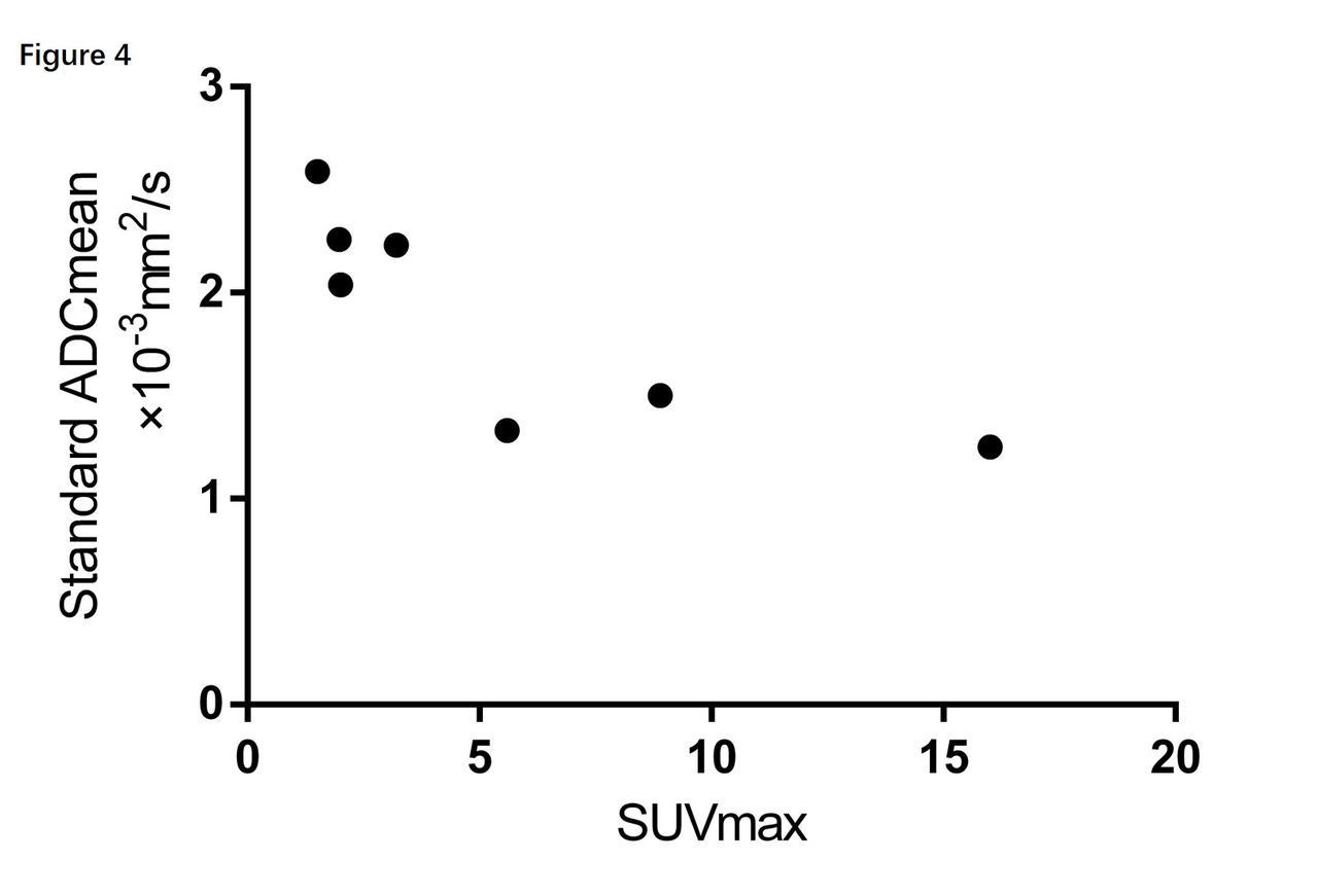
Results: Ten patients were ruled out since they were considered to have benign lesions and didn’t undergo surgery. Fourteen patients with histopathological confirmation remained in the study (Table 1). Based on 18F-FDG accumulation, six patients had positive PET scans. PET/MR revealed 3 more lesions than PET/CT on these patients (11 lesions vs. 8 lesions, respectively), which turned out to be the uptake of one mural nodule and two paraaortic lymph nodes (Figure 1,). Both PET/CT and PET/MR had good negative predictive value for benign lesions (Figure 2). In patients with intraductal papillary mucinous neoplasm, those with invasive carcinoma have a much higher LPR than those with dysplasia . A similar result was also shown in malignancies compared with benign lesions (Figure 3). Both SUVmax and LPR of positive lesions were correlated with Standard ADCmean (Figure 4). In one patient, both CT and MRI showed a large cyst with a mural nodule. But the uptake of the mural nodule was comparable to normal pancreas parenchyma. It turned out to be benign mucinous cystadenoma with intermediate dysplasia. T2-weighted images and MRCP were helpful in illustration of pancreatic duct dilation, intracystic septations, mural nodules, fluid heterogeneity, and connection to the main pancreatic duct. These anatomical features are critical in the differential diagnosis of pancreatic cystic lesions.

Conclusion: Both PET/CT and PET/MR do well in differentiating between benign and malignant pancreatic cystic lesions. However, TOF PET images could reveal more positive lesions than non-TOF images. Furthermore, PET/MR could provide more diagnostic anatomical information.

Figure
  • Download figure
  • Open in new tab
  • Download powerpoint
Figure
  • Download figure
  • Open in new tab
  • Download powerpoint
Figure
  • Download figure
  • Open in new tab
  • Download powerpoint
Figure
  • Download figure
  • Open in new tab
  • Download powerpoint
Figure
  • Download figure
  • Open in new tab
  • Download powerpoint
Previous
Back to top

In this issue

Journal of Nuclear Medicine
Vol. 58, Issue supplement 1
May 1, 2017
  • Table of Contents
  • Index by author
Article Alerts
Sign In to Email Alerts with your Email Address
Email Article

Thank you for your interest in spreading the word on Journal of Nuclear Medicine.

NOTE: We only request your email address so that the person you are recommending the page to knows that you wanted them to see it, and that it is not junk mail. We do not capture any email address.

Enter multiple addresses on separate lines or separate them with commas.
Preliminary Study of PET/MR in Pancreatic Cystic Lesions: Compared with PET/CT
(Your Name) has sent you a message from Journal of Nuclear Medicine
(Your Name) thought you would like to see the Journal of Nuclear Medicine web site.
Citation Tools
Preliminary Study of PET/MR in Pancreatic Cystic Lesions: Compared with PET/CT
Wenjia Zhu, Haiqun Xing, Guilan Hu, Tong Wang, Li Huo, Fang Li
Journal of Nuclear Medicine May 2017, 58 (supplement 1) 377;

Citation Manager Formats

  • BibTeX
  • Bookends
  • EasyBib
  • EndNote (tagged)
  • EndNote 8 (xml)
  • Medlars
  • Mendeley
  • Papers
  • RefWorks Tagged
  • Ref Manager
  • RIS
  • Zotero
Share
Preliminary Study of PET/MR in Pancreatic Cystic Lesions: Compared with PET/CT
Wenjia Zhu, Haiqun Xing, Guilan Hu, Tong Wang, Li Huo, Fang Li
Journal of Nuclear Medicine May 2017, 58 (supplement 1) 377;
Twitter logo Facebook logo LinkedIn logo Mendeley logo
  • Tweet Widget
  • Facebook Like
  • Google Plus One
Bookmark this article

Jump to section

  • Article
  • Figures & Data
  • Info & Metrics

Related Articles

  • No related articles found.
  • Google Scholar

Cited By...

  • No citing articles found.
  • Google Scholar

More in this TOC Section

General Clinical Specialties Track

  • Quantitative Evaluation of Parathyroid Adenoma and Hyperplasia in Reference to Thyroid using Tc-99m MIBI SPECT/CT
  • A primitive study for clinical application of 18F-AlF-NOTA-octreotide PET/CT in combination with 18F-FDG PET/CT for imaging neuroendocrine neoplasms
  • Role of 18F-FDG PET/CT in detection of disease burden and response assessment in patients with myeloid sarcoma
Show more General Clinical Specialties Track

Gastrointestinal Nuclear Medicine

  • Clinical and histopathologic features of chronic liver disease associated with hepatic fluorine-18 fluorocholine uptake
  • Are the temporal Inconsistencies of proximal stomach counts Indicative of Intragastric food redistribution?
  • A New Normal. Evaluating normal values for functional gallbladder imaging with the use of fatty meal substitute for ejection fraction
Show more Gastrointestinal Nuclear Medicine

Similar Articles

SNMMI

© 2025 SNMMI

Powered by HighWire