
Main menu

  • Home
  • Content
    • Current
    • Ahead of print
    • Past Issues
    • JNM Supplement
    • SNMMI Annual Meeting Abstracts
    • Continuing Education
    • JNM Podcasts
  • Subscriptions
    • Subscribers
    • Institutional and Non-member
    • Rates
    • Journal Claims
    • Corporate & Special Sales
  • Authors
    • Submit to JNM
    • Information for Authors
    • Assignment of Copyright
    • AQARA requirements
  • Info
    • Reviewers
    • Permissions
    • Advertisers
  • About
    • About Us
    • Editorial Board
    • Contact Information
  • More
    • Alerts
    • Feedback
    • Help
    • SNMMI Journals
  • SNMMI
    • JNM
    • JNMT
    • SNMMI Journals
    • SNMMI

User menu

  • Subscribe
  • My alerts
  • Log in
  • Log out
  • My Cart

Search

  • Advanced search
Journal of Nuclear Medicine
  • SNMMI
    • JNM
    • JNMT
    • SNMMI Journals
    • SNMMI
  • Subscribe
  • My alerts
  • Log in
  • Log out
  • My Cart
Journal of Nuclear Medicine

Advanced Search

  • Home
  • Content
    • Current
    • Ahead of print
    • Past Issues
    • JNM Supplement
    • SNMMI Annual Meeting Abstracts
    • Continuing Education
    • JNM Podcasts
  • Subscriptions
    • Subscribers
    • Institutional and Non-member
    • Rates
    • Journal Claims
    • Corporate & Special Sales
  • Authors
    • Submit to JNM
    • Information for Authors
    • Assignment of Copyright
    • AQARA requirements
  • Info
    • Reviewers
    • Permissions
    • Advertisers
  • About
    • About Us
    • Editorial Board
    • Contact Information
  • More
    • Alerts
    • Feedback
    • Help
    • SNMMI Journals
  • View or Listen to JNM Podcast
  • Visit JNM on Facebook
  • Join JNM on LinkedIn
  • Follow JNM on Twitter
  • Subscribe to our RSS feeds
Meeting ReportCardiovascular Track

Harnessing Radioluminescence and Sound to Reveal Molecular Pathology of Atherosclerotic Plaques

Raiyan Zaman, Siavash Yousefi, Steven Long, Christopher Contag, Sanjiv Gambhir, Butras Khuri-Yakub and Lei Xing
Journal of Nuclear Medicine May 2017, 58 (supplement 1) 31;
Raiyan Zaman
3Medicine, Division of Cardiovascular Medicine Stanford University School of Medicine Palo Alto CA United States
  • Find this author on Google Scholar
  • Find this author on PubMed
  • Search for this author on this site
Siavash Yousefi
2Department of Radiation Oncology Stanford University School of Medicine Palo Alto CA United States
  • Find this author on Google Scholar
  • Find this author on PubMed
  • Search for this author on this site
Steven Long
4Pathology Stanford University School of Medicine Stanford CA United States
  • Find this author on Google Scholar
  • Find this author on PubMed
  • Search for this author on this site
Christopher Contag
5Pediatrics (Neonatology) Stanford University School of Medicine Stanford CA United States
  • Find this author on Google Scholar
  • Find this author on PubMed
  • Search for this author on this site
Sanjiv Gambhir
7Radiology Stanford University School of Medicine Stanford CA United States
  • Find this author on Google Scholar
  • Find this author on PubMed
  • Search for this author on this site
Butras Khuri-Yakub
1Electrical Engineering Stanford University Stanford CA United States
  • Find this author on Google Scholar
  • Find this author on PubMed
  • Search for this author on this site
Lei Xing
6Radiation Oncology Stanford University School of Medicine Stanford CA United States
  • Find this author on Google Scholar
  • Find this author on PubMed
  • Search for this author on this site
  • Article
  • Figures & Data
  • Info & Metrics
Loading

Abstract

31

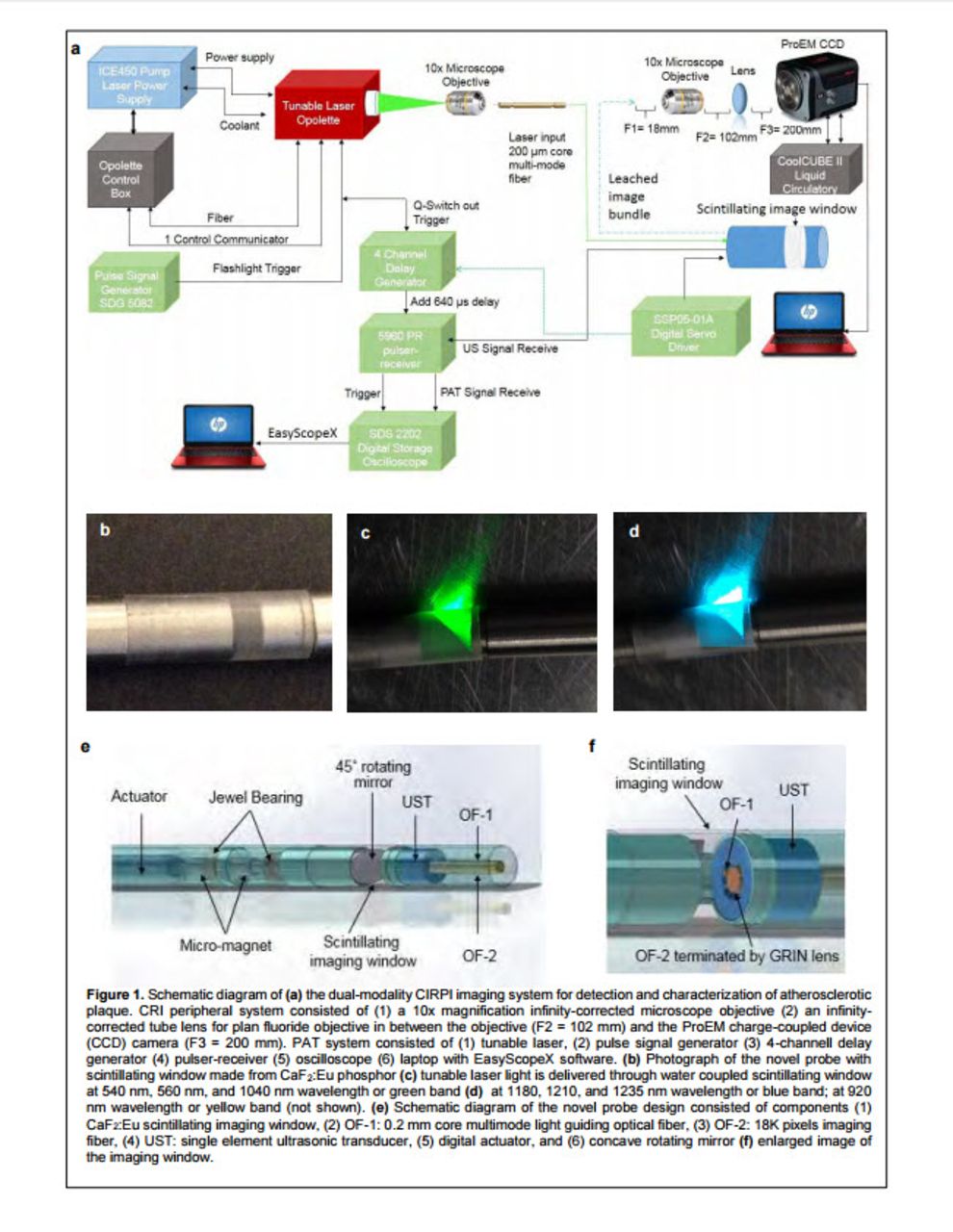
Objectives: Thin-cap fibro atheroma (TCFA) are the unstable lesions in coronary artery disease (CAD) that are prone to rupture resulting in substantial morbidity and mortality worldwide. Early clinical diagnosis and effective risk stratification of these lesions has the potential to dramatically impact management of CAD and prevent progression to catastrophic events. However, their small size, and complex morphological/biological features make early detection and risk assessment difficult. To enable detection and characterization of vulnerable plaque structure and biology, we developed a Circumferential-Intravascular-Radioluminescence-Photoacoustic-Imaging (CIRPI) system (Fig. 1).

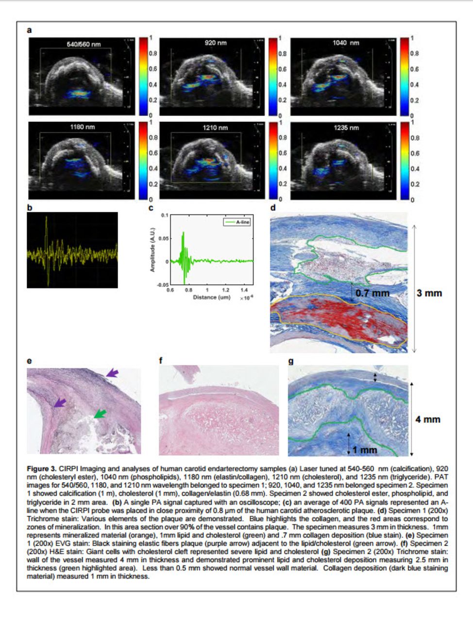
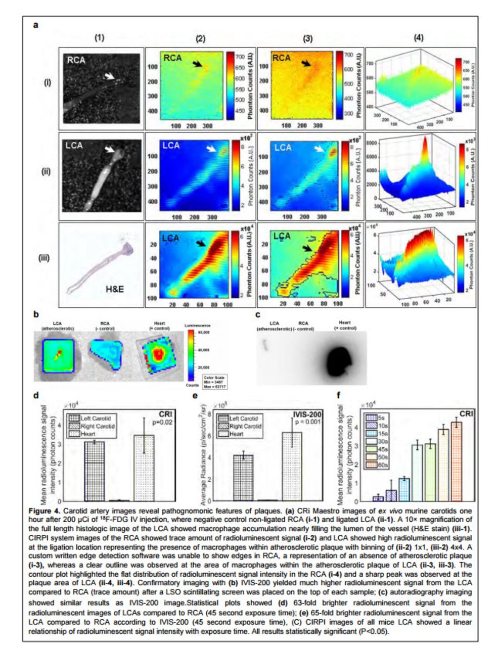
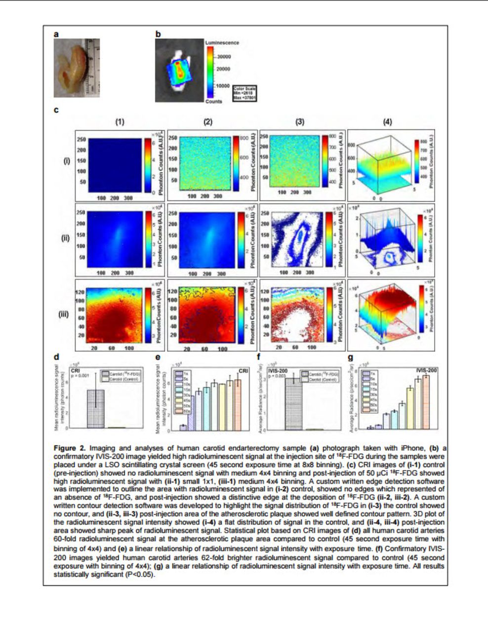
Methods: The CIRPI system includes a novel optical probe combined circumferential radioluminescence imaging (CRI) and photoacoustic tomography (PAT). The probe’s CaF2:Eu based scintillating imaging window captures a 360° view of human (n=7) and murine carotid (n=10) arterial plaques by detecting optical radiation from the energy deposited by β-particles during 18F-FDG decay. A tunable laser-based photoacoustic imaging system characterized tissue constituents of TCFA at 7 different wavelengths—540-560 nm (calcification), 920 nm (cholesteryl ester), 1040 nm (phospholipids), 1180 nm (elastin/collagen), 1210 nm (cholesterol), and 1235 nm (triglyceride). Each A-line computed from an average of 400 A-lines; 330 A-lines created a single B-scan. Human carotid endarterectomy samples (25-28 mm length) were imaged with the CRI system pre (control) and post 18F-FDG (50 µCi) injection within ~1h of tissue collection followed by PAT imaging. For FVB mice, carotid lesions were created by 4 weeks of high-fat diet and diabetes induction, followed by left carotid ligation. An hour after 18F-FDG (200 Ci)IV injection left, right (control) carotid arteries, and heart (+ control) were harvested for imaging intact with the CRi Maestro before imaging with the CIRPI system. Results were verified with IVIS-200, autoradiography, histology, and ultrasound imaging.

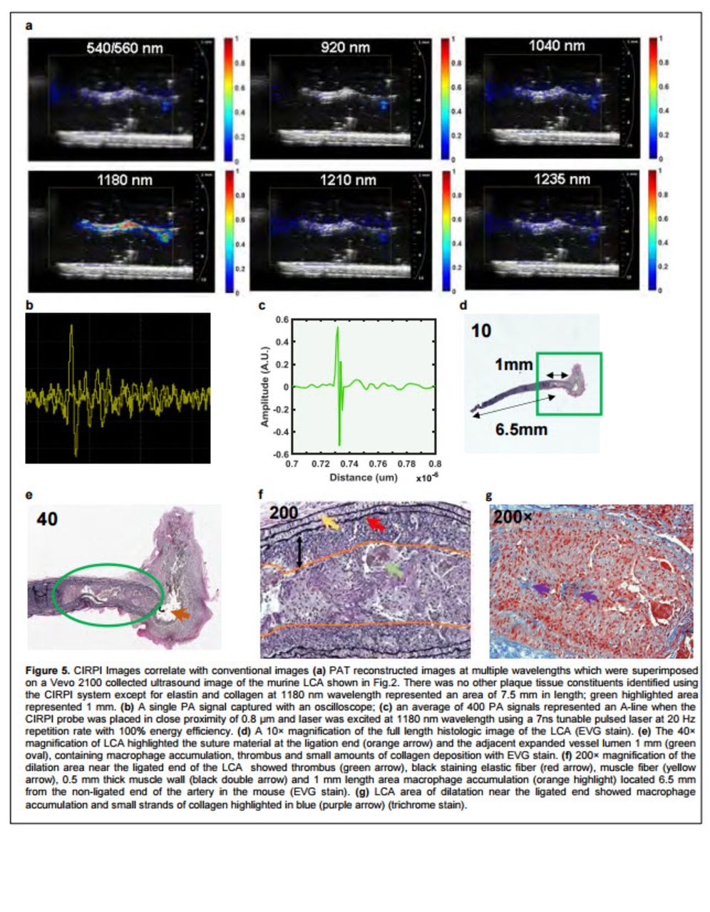
Results: This dual-modality hybrid imaging tool was able to detect and characterize human and murine atherosclerotic plaques. The catheter-based-CRIPI-probe detected data on human plaque inflammation, through a 60x higher (4.96×10⁴±2.25×10⁴ vs. 8.37×10²±6.63×101 photon counts, p=0.001) 18F-FDG radioluminescent signal (Fig. 2c-d), which was enriched by analyses of the plaque tissue constituents and morphologic characterization provided by photoacoustic imaging (Fig. 3a). Confirmatory IVIS-200 images exhibited similar results (62x). PAT imaging system detected TCFA constituents such as calcification, severe lipid/fatty acid in the form of mostly cholesteryl ester, phospholipid, cholesterol, and triglyceride in human samples. Also, we have observed the presence of elastin/collagen. Murine carotid plaques from ligated left carotid artery (LCA) showed 63x (3.10×104±9.41×10² vs. 4.90×10²±2.40×10² photon counts, p=0.02) higher radioluminescent signal to the control, non-ligated right carotid artery (RCA), due to plaque macrophages (Fig. 4). PAT images of murine LCA over a range of wavelengths showed no prominent photoacoustic signals except at 1180 nm, which elucidated the presence of elastin/collagen throughout the length of the vessel (Fig. 5a). All human plaque burdens ranged from 20-55%, unlike 10% or less for mice. The CIRPI images of human and murine samples were highly correlated with histological analyses (Fig. 3d-g, 5d-g).

Conclusion: The CIRPI system will drive a paradigm shift in the diagnosis and risk stratification of CAD by uniting cellular, molecular, and morphologic data for a more complete pathologic and prognostic characterization of vulnerable plaques enabling clinical detection and early diagnosis of TCFA, and improving risk assessment. Research Supports: Authors gratefully acknowledge the financial support from an NIH K99/R00 award (1 K99 HL127180-01).

Figure
  • Download figure
  • Open in new tab
  • Download powerpoint
Figure
  • Download figure
  • Open in new tab
  • Download powerpoint
Figure
  • Download figure
  • Open in new tab
  • Download powerpoint
Figure
  • Download figure
  • Open in new tab
  • Download powerpoint
Figure
  • Download figure
  • Open in new tab
  • Download powerpoint
Previous
Back to top

In this issue

Journal of Nuclear Medicine
Vol. 58, Issue supplement 1
May 1, 2017
  • Table of Contents
  • Index by author
Article Alerts
Sign In to Email Alerts with your Email Address
Email Article

Thank you for your interest in spreading the word on Journal of Nuclear Medicine.

NOTE: We only request your email address so that the person you are recommending the page to knows that you wanted them to see it, and that it is not junk mail. We do not capture any email address.

Enter multiple addresses on separate lines or separate them with commas.
Harnessing Radioluminescence and Sound to Reveal Molecular Pathology of Atherosclerotic Plaques
(Your Name) has sent you a message from Journal of Nuclear Medicine
(Your Name) thought you would like to see the Journal of Nuclear Medicine web site.
Citation Tools
Harnessing Radioluminescence and Sound to Reveal Molecular Pathology of Atherosclerotic Plaques
Raiyan Zaman, Siavash Yousefi, Steven Long, Christopher Contag, Sanjiv Gambhir, Butras Khuri-Yakub, Lei Xing
Journal of Nuclear Medicine May 2017, 58 (supplement 1) 31;

Citation Manager Formats

  • BibTeX
  • Bookends
  • EasyBib
  • EndNote (tagged)
  • EndNote 8 (xml)
  • Medlars
  • Mendeley
  • Papers
  • RefWorks Tagged
  • Ref Manager
  • RIS
  • Zotero
Share
Harnessing Radioluminescence and Sound to Reveal Molecular Pathology of Atherosclerotic Plaques
Raiyan Zaman, Siavash Yousefi, Steven Long, Christopher Contag, Sanjiv Gambhir, Butras Khuri-Yakub, Lei Xing
Journal of Nuclear Medicine May 2017, 58 (supplement 1) 31;
Twitter logo Facebook logo LinkedIn logo Mendeley logo
  • Tweet Widget
  • Facebook Like
  • Google Plus One
Bookmark this article

Jump to section

  • Article
  • Figures & Data
  • Info & Metrics

Related Articles

  • No related articles found.
  • Google Scholar

Cited By...

  • No citing articles found.
  • Google Scholar

More in this TOC Section

Cardiovascular Track

  • To Evaluated the Cardiac Function of Patients with Acute Myocardial Infarction by the Volume and Filling Curve of 99mTc-MIBI SPECT Myocardial Perfusion Imaging
  • Standard versus low-dose rubidium-82 dynamic positron emission tomography imaging with scanner-dependent bias correction for myocardial perfusion imaging and blood flow quantification
  • Evaluation of sympathetic function with PET 11C-hydroxyephedrine (HED) and ammonia (13N-NH3) in a canine pacing model of atrial fibrillation
Show more Cardiovascular Track

Cardiovascular Young Investigator Award Symposium

  • Vesicular storage and release of the novel sympathetic nerve tracer 18F-LMI1195
  • Common Carotid Artery Molecular Calcification Assessed by 18F-NaF PET/CT is Associated with Increased Cardiovascular Disease Risk: Results from the CAMONA Study
  • Comparative evaluation of F-18 FLT PET/CT and F-18 FDG PET/CT in patients with newly diagnosed thoracic sarcoidosis
Show more Cardiovascular Young Investigator Award Symposium

Similar Articles

SNMMI

© 2025 SNMMI

Powered by HighWire