
Main menu

  • Home
  • Content
    • Current
    • Ahead of print
    • Past Issues
    • JNM Supplement
    • SNMMI Annual Meeting Abstracts
    • Continuing Education
    • JNM Podcasts
  • Subscriptions
    • Subscribers
    • Institutional and Non-member
    • Rates
    • Journal Claims
    • Corporate & Special Sales
  • Authors
    • Submit to JNM
    • Information for Authors
    • Assignment of Copyright
    • AQARA requirements
  • Info
    • Reviewers
    • Permissions
    • Advertisers
  • About
    • About Us
    • Editorial Board
    • Contact Information
  • More
    • Alerts
    • Feedback
    • Help
    • SNMMI Journals
  • SNMMI
    • JNM
    • JNMT
    • SNMMI Journals
    • SNMMI

User menu

  • Subscribe
  • My alerts
  • Log in
  • Log out
  • My Cart

Search

  • Advanced search
Journal of Nuclear Medicine
  • SNMMI
    • JNM
    • JNMT
    • SNMMI Journals
    • SNMMI
  • Subscribe
  • My alerts
  • Log in
  • Log out
  • My Cart
Journal of Nuclear Medicine

Advanced Search

  • Home
  • Content
    • Current
    • Ahead of print
    • Past Issues
    • JNM Supplement
    • SNMMI Annual Meeting Abstracts
    • Continuing Education
    • JNM Podcasts
  • Subscriptions
    • Subscribers
    • Institutional and Non-member
    • Rates
    • Journal Claims
    • Corporate & Special Sales
  • Authors
    • Submit to JNM
    • Information for Authors
    • Assignment of Copyright
    • AQARA requirements
  • Info
    • Reviewers
    • Permissions
    • Advertisers
  • About
    • About Us
    • Editorial Board
    • Contact Information
  • More
    • Alerts
    • Feedback
    • Help
    • SNMMI Journals
  • View or Listen to JNM Podcast
  • Visit JNM on Facebook
  • Join JNM on LinkedIn
  • Follow JNM on Twitter
  • Subscribe to our RSS feeds
Meeting ReportNeurosciences Track

The Effect of Spatial Normalization of Brain 18F-FDG PET-MR Images Using SPM and Different PET Templates

Umit Akdemir, Uguray Aydos, Mahsun Ozcelik, Seda Gulbahar, Nese Ilgin Karabacak and Lutfiye Atay
Journal of Nuclear Medicine May 2017, 58 (supplement 1) 1260;
Umit Akdemir
1Department of Nuclear Medicine Gazi University, Faculty of Medicine Ankara Turkey
  • Find this author on Google Scholar
  • Find this author on PubMed
  • Search for this author on this site
Uguray Aydos
1Department of Nuclear Medicine Gazi University, Faculty of Medicine Ankara Turkey
  • Find this author on Google Scholar
  • Find this author on PubMed
  • Search for this author on this site
Mahsun Ozcelik
1Department of Nuclear Medicine Gazi University, Faculty of Medicine Ankara Turkey
  • Find this author on Google Scholar
  • Find this author on PubMed
  • Search for this author on this site
Seda Gulbahar
1Department of Nuclear Medicine Gazi University, Faculty of Medicine Ankara Turkey
  • Find this author on Google Scholar
  • Find this author on PubMed
  • Search for this author on this site
Nese Ilgin Karabacak
1Department of Nuclear Medicine Gazi University, Faculty of Medicine Ankara Turkey
  • Find this author on Google Scholar
  • Find this author on PubMed
  • Search for this author on this site
Lutfiye Atay
1Department of Nuclear Medicine Gazi University, Faculty of Medicine Ankara Turkey
  • Find this author on Google Scholar
  • Find this author on PubMed
  • Search for this author on this site
  • Article
  • Figures & Data
  • Info & Metrics
Loading

Abstract

1260

Objectives: We aimed to compare the results of different methods of spatial normalization of brain 18F-FDG PET data acquired on a hybrid PET-MRI system.

Methods: For the development of an institutional normal brain 18F-FDG PET-MR database we acquired additional brain PET and T1-weighted MR images in a clinical adult patient group who had whole-body 18-FDG PET examinations for pulmonary nodule evaluation. Exclusion criteria were history of neuropsychiatric disease, presence of visually apparent abnormalities and imaging artefacts on the acquired brain PET-MR images or any treatments that might confound brain metabolism. Brain PET-MR images were processed and analyzed using spm8. After manually correcting the image orientations in reference to the T1 MR template provided with spm8, T1 images were spatially normalized to the T1 MR template and the calculated deformation maps of patients were used for spatial normalization of the corresponding PET images. The spatially normalized images were smoothed using an isotropic 8 mm Gaussian filter. These data constituted the reference and used for the creation of an institutional 18F-FDG PET template. An additional patient group of mixed neurodegenerative disorders was selected to further characterize the effects of normalization template on quantitative results. All PET images were spatially normalized applying three PET templates (the 15O-water PET template provided with spm8, a neurodegenerative disease specific 18F-FDG PET template provided by Della Rosa P.A., et al. and the newly created institutional PET template) yielding three sets of PET data from the normal and neurodegenerative patient groups. Paired t-test was used to compare the three sets of normal group data with the reference normal database and independent samples t-test was used to compare the three sets of neurodegenerative group data with the reference normal database.

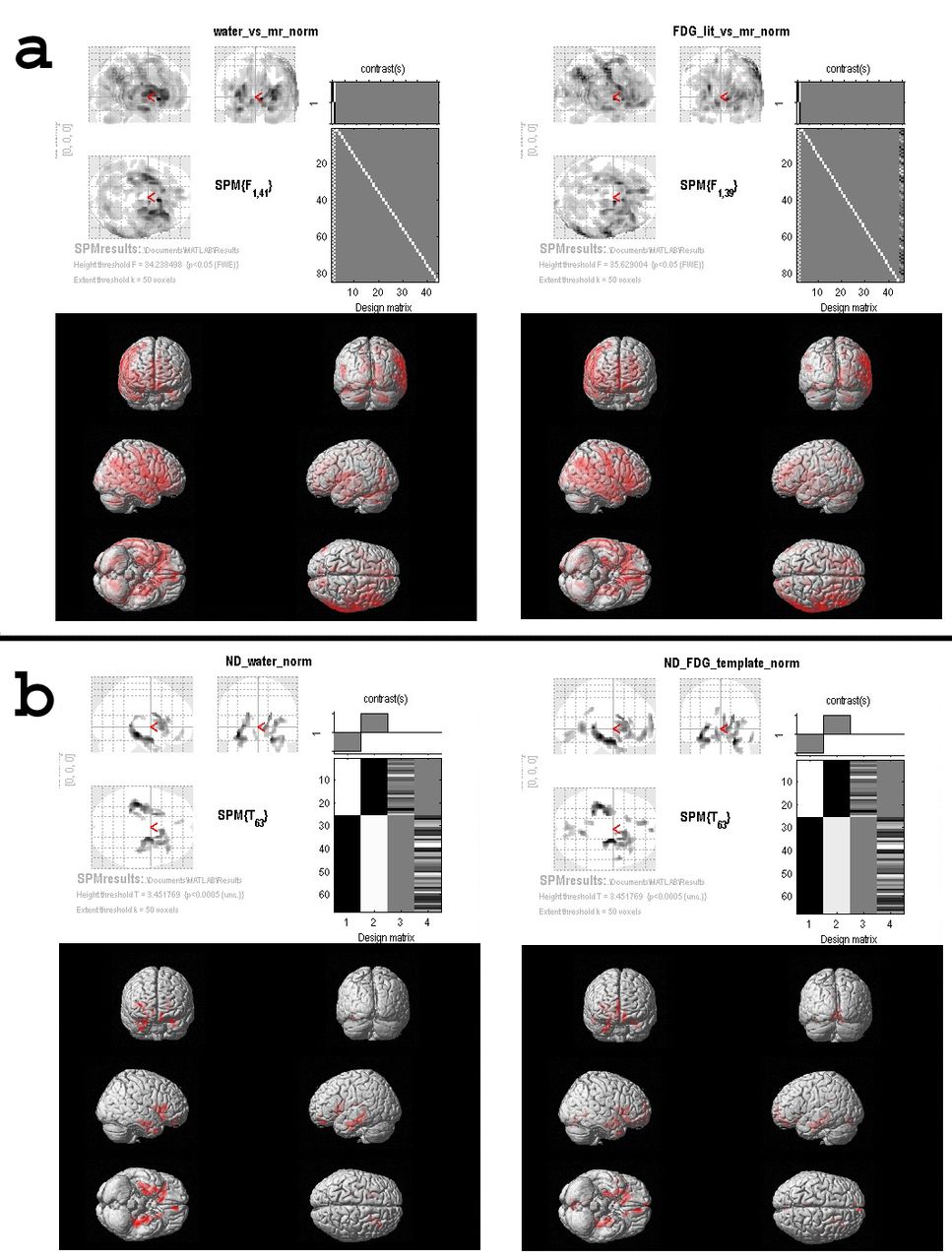
Results: Forty-two brain 18F-FDG PET-MR image sets were eligible for the creation of the institutional normal database and we had 25 patients in the neurodegenerative group. In the normal group no significant difference in metabolism was observed when institutional PET template was used for normalization (FWE-corrected p = 0.05 and extend threshold = 50). However, when water and neurodegenerative disease specific 18F-FDG PET templates were used for spatial normalization, significant differences were observed in extensive cortical regions and subcortical structures with a similar distribution (Figure 1a). In the neurodegenerative patient group, no significant metabolic differences in comparison to the normal database were detected when the institutional PET template was used for spatial normalization. However, the water PET template normalized data showed lower metabolism bilaterally in mesial temporal and orbitofrontal regions (Uncorrected p = 0.001 and extend threshold = 50). A similar result was observed when the neurodegenerative disease specific 18F-FDG PET template was used for normalization (Figure 1b).

Conclusion: Brain 18F-FDG PET images acquired on a hybrid PET-MR camera showed significant differences in metabolism when spatial normalization was done with different templates. The differences observed when PET data of the neurodegenerative patient group was compared with the normal database might be specific to hybrid PET-MR, since the distribution of hypometabolism corresponded to brain regions close to bony structures at the skull-base where the differences due to MR-based attenuation correction were expected to be maximum. Therefore, accurate quantitative analysis of brain 18F-FDG PET images acquired on a hybrid PET-MR system requires a separate PET template for spatial normalization and an institutional normal database for statistical analysis. Research Support: The PET-MR system used in this study was provided by Republic of Turkey Ministry of Development.

Figure
  • Download figure
  • Open in new tab
  • Download powerpoint
Previous
Back to top

In this issue

Journal of Nuclear Medicine
Vol. 58, Issue supplement 1
May 1, 2017
  • Table of Contents
  • Index by author
Article Alerts
Sign In to Email Alerts with your Email Address
Email Article

Thank you for your interest in spreading the word on Journal of Nuclear Medicine.

NOTE: We only request your email address so that the person you are recommending the page to knows that you wanted them to see it, and that it is not junk mail. We do not capture any email address.

Enter multiple addresses on separate lines or separate them with commas.
The Effect of Spatial Normalization of Brain 18F-FDG PET-MR Images Using SPM and Different PET Templates
(Your Name) has sent you a message from Journal of Nuclear Medicine
(Your Name) thought you would like to see the Journal of Nuclear Medicine web site.
Citation Tools
The Effect of Spatial Normalization of Brain 18F-FDG PET-MR Images Using SPM and Different PET Templates
Umit Akdemir, Uguray Aydos, Mahsun Ozcelik, Seda Gulbahar, Nese Ilgin Karabacak, Lutfiye Atay
Journal of Nuclear Medicine May 2017, 58 (supplement 1) 1260;

Citation Manager Formats

  • BibTeX
  • Bookends
  • EasyBib
  • EndNote (tagged)
  • EndNote 8 (xml)
  • Medlars
  • Mendeley
  • Papers
  • RefWorks Tagged
  • Ref Manager
  • RIS
  • Zotero
Share
The Effect of Spatial Normalization of Brain 18F-FDG PET-MR Images Using SPM and Different PET Templates
Umit Akdemir, Uguray Aydos, Mahsun Ozcelik, Seda Gulbahar, Nese Ilgin Karabacak, Lutfiye Atay
Journal of Nuclear Medicine May 2017, 58 (supplement 1) 1260;
Twitter logo Facebook logo LinkedIn logo Mendeley logo
  • Tweet Widget
  • Facebook Like
  • Google Plus One
Bookmark this article

Jump to section

  • Article
  • Figures & Data
  • Info & Metrics

Related Articles

  • No related articles found.
  • Google Scholar

Cited By...

  • No citing articles found.
  • Google Scholar

More in this TOC Section

Neurosciences Track

  • Impact of cognitive reserve in frontotemporal dementia illustrated by FDG-PET.
  • Quantification of brain cholinergic denervation in dementia with Lewy bodies using PET imaging with 18F-FEOBV
  • Kinetic evaluation of [18F]MOZAT PET imaging in humans.
Show more Neurosciences Track

Neurology Posters

  • FBPA PET/CT: to distinguish radiation necrosis from recurrent brain tumor
  • The use of 4’-[methyl-11C]thiothymidine PET/CT to differentiate recurrence from treatment-induced necrosis in high-grade gliomas.
  • A new diagnostic marker for differentiating multi centric gliomas from multiple intracranial diffuse large B-cell lymphomas on 18F-FDG PET images
Show more Neurology Posters

Similar Articles

SNMMI

© 2025 SNMMI

Powered by HighWire