


Article Figures & Data
Figures
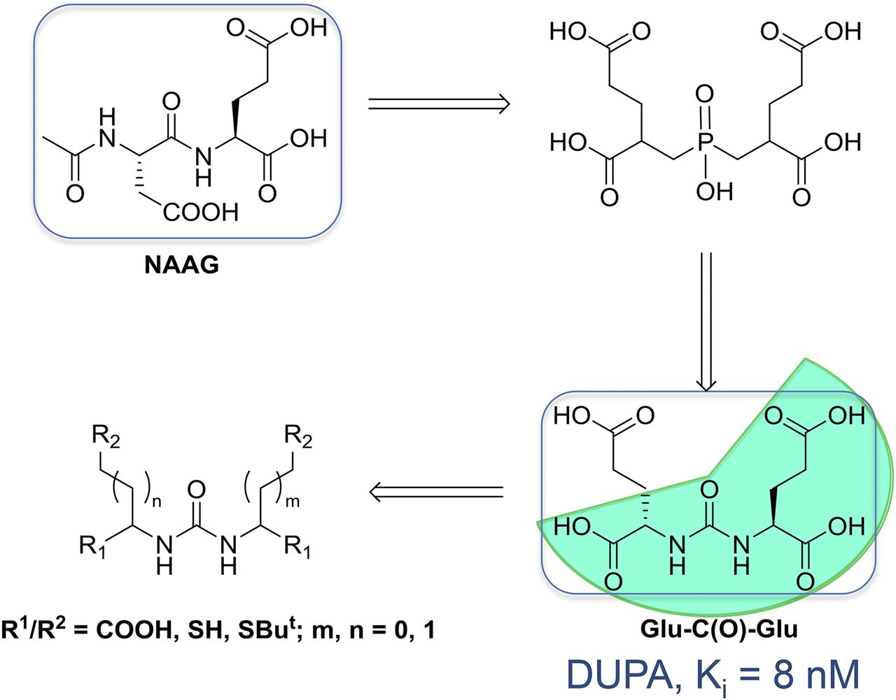
- FIGURE 1.
Glu-ureido–based PSMA inhibitor exemplifying rational design of urea-based glutamate carboxypeptidase II inhibitors. DUPA = 2-[3-(1,3-dicarboxypropyl)ureido]pentanedioic acid; NAAG = N-acetyl-l-aspartyl-l-glutamate. (Reprinted with permission of (9).)
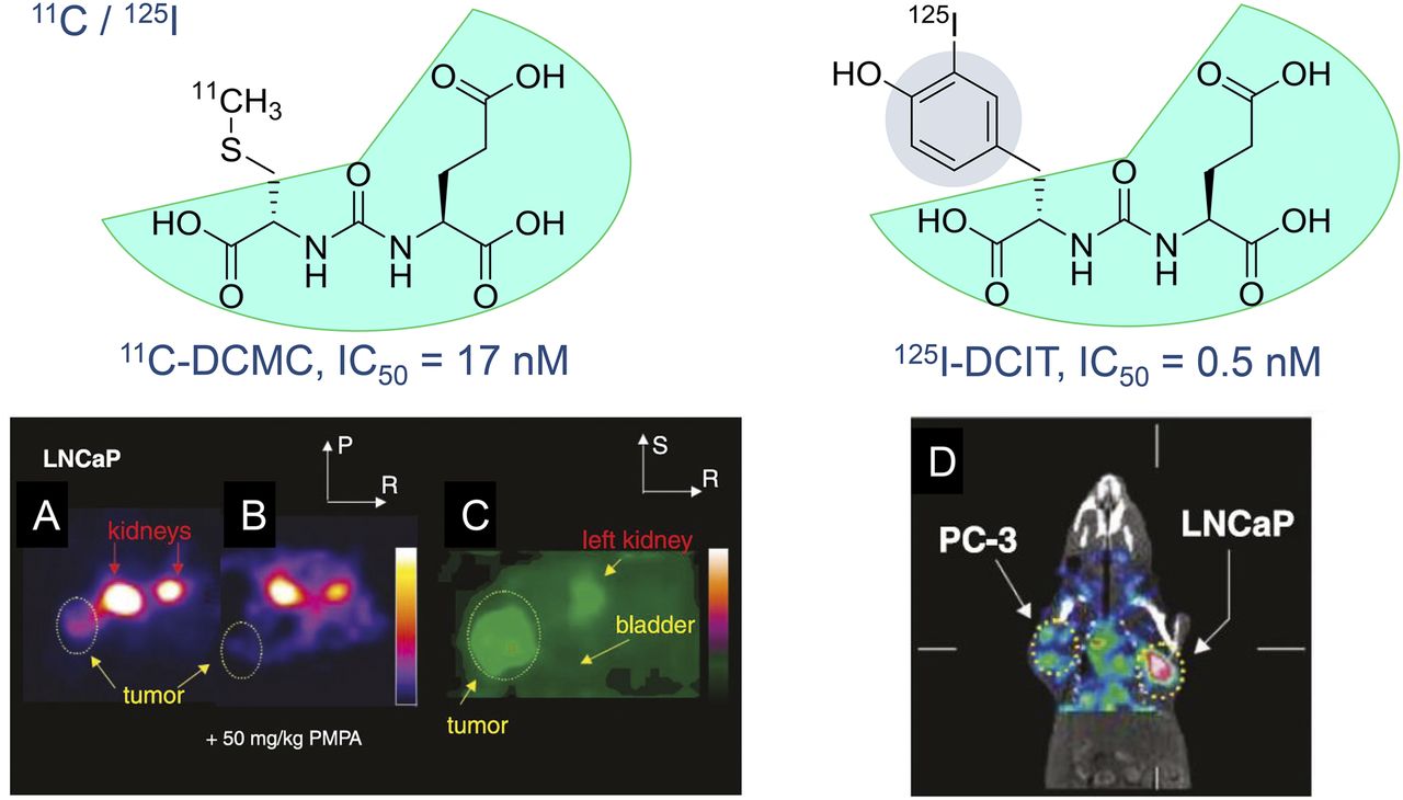
- FIGURE 2.
In vivo examinations of radiolabeled small-molecule PSMA inhibitors in experimental models of PC. (A and B) Axial PET images of N-[N-[(S)-1,3-dicarboxypropyl]carbamoyl]-S-11C-methyl-l-cysteine (11C-DCMC) in mouse kidney and LNCaP tumors without (A) and with (B) coinjection of 2-(phosphonomethyl)pentane-1,5-dioic acid (PMPA) at 50 mg/kg to block PSMA. (C) Magnified, coronal reconstructed image of animal in A outside plane of kidneys immediately after urination indicates lack of specific binding to bladder. Tumor and portion of left kidney were clearly visualized. Images in A and B were scaled to same maximum. IC50 = 50% inhibitory concentration; P = posterior, R = right, S = superior. (D) SPECT with CT overlay shows uptake of N-[N-[(S)-1,3-dicarboxypropyl]carbamoyl]-S-3-125I-iodo-l-tyrosine (125I-DCIT) in LNCaP tumor, whereas PC-3 tumor retains only minimal radiotracer. (Reprinted with permission of (13).)
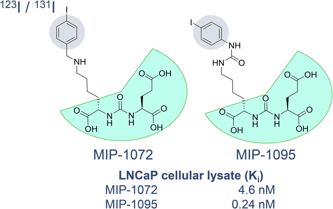
- FIGURE 3.
Radioiodinated (“haloaromatic”) Glu-ureido–based PSMA inhibitors (MIP compounds)—MIP-1072: 2-(3-(1-carboxy-5-(4-iodo-benzylamino)pentyl)ureido)pentanedioic acid; MIP-1095: (S)-2-(3-((R)-1-carboxy-5-(3-(4-iodophenyl)ureido)pentyl)ureido)pentanedioic acid (5,6,18,19).
- FIGURE 4.
Glu-ureido–based PSMA radioligands of clinical relevance. Cpd. = compound; Ref. = reference; RN = radionuclide.
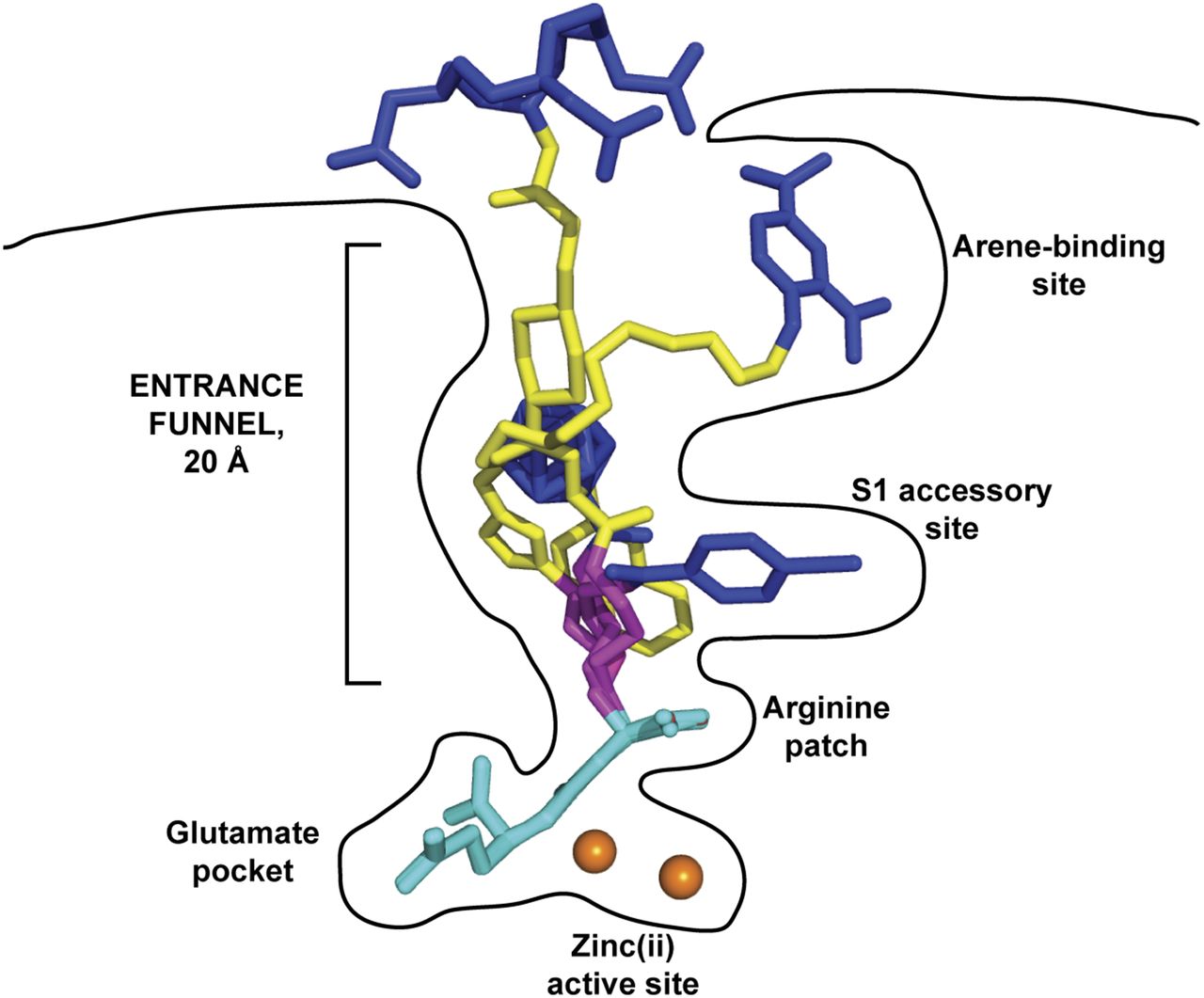
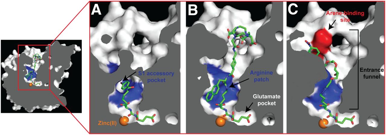
- FIGURE 5.
Internal cavity of PSMA. Cross-section of PSMA showing internal inhibitor-binding cavity comprising S1′ glutamate recognition pocket, dinuclear zinc(II) active site, and irregularly shaped entrance funnel. Although S1′ glutamate recognition pocket is restricted in size and shape, spacious entrance funnel can accommodate functional groups of different sizes and physicochemical characteristics. Within entrance funnel, arginine patch, S1 accessory hydrophobic pocket, and arene-binding site—prominent structural features used for inhibitor design—are highlighted. Zinc ions are shown as orange spheres, and PSMA ligands are shown as stick representations: DCIBzL (A) (PDB code 3D7H), PSMA-617 (B) (unpublished data), and ARM-P4 (C) (PDB code 2XEG).
- FIGURE 6.
Glu-ureido–based ligands within binding cavity of PSMA. Complexes between PSMA and 4 Glu-ureido–based ligands are superimposed on corresponding Cα atoms of protein. Although there is complete structural overlap of pharmacophore modules (cyan), positioning of flexible proximal linker (magenta), functional spacer (yellow), and effector moiety (blue) is divergent within (and outside) amphipathic entrance funnel. Zinc ions are shown as orange spheres. PSMA–inhibitor complexes used were DCIBzL (PDB code 3D7H) (48), ARM-P4 (PDB code 2XEG) (52), carborane (PDB code 4OME) (55), and PSMA-617 (unpublished data).
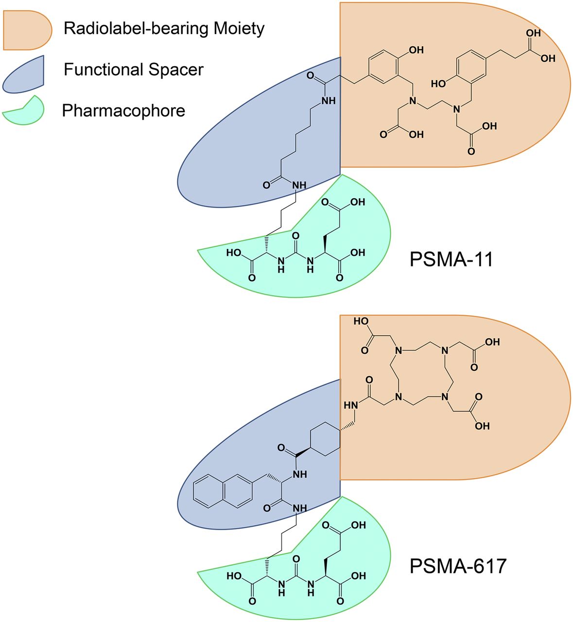
- FIGURE 7.
Dependence of transformation of diagnostic tracer PSMA-11 into theranostic variant PSMA-617 on structural arrangement of pharmacophore, functional spacer, and radiolabel-bearing/effector moiety.
- FIGURE 8.
PET/CT and SPECT/CT imaging of PC-3 PIP/flu tumor-bearing mice. Tumor-targeting efficacy and pharmacokinetic properties were evaluated with 44Sc-PSMA-617 (A), 177Lu-PSMA-617 (B), 68Ga-PSMA-617 (C), and 68Ga-PSMA-11 (D) 2 h after injection. Bl or bl = bladder; ki = kidney. (Reprinted with permission of (39).)
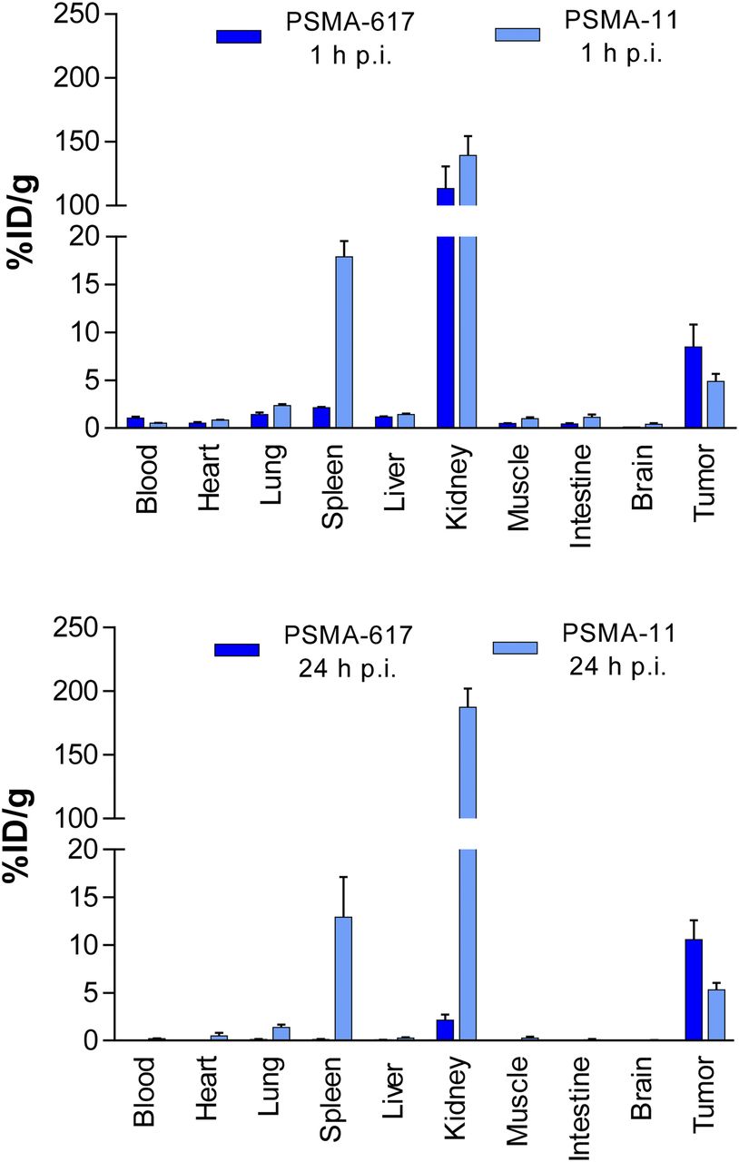
- FIGURE 9.
Organ distribution of 68Ga-PSMA-617 and 68Ga-PSMA-11 1 h after injection (p.i.) and of 177Lu-PSMA-617 and 67Ga-PSMA-11 24 h after injection. (Reprinted with permission of (21).)


Tables
- TABLE 1
PSMA Inhibition Potency (Ki) and Cellular Internalization of PSMA-11 and PSMA-617, as Determined with LNCaP Cells (21)
Compound Ki (nM) Internalization 68Ga-PSMA-11 12.0 ± 2.8 9.47 ± 2.56 68Ga-PSMA-617 2.34 ± 2.94 17.67 ± 4.34 Data are reported as mean ± SD. Internalization is reported as percentage applied activity/106 LNCaP cells.


Jump to section
- Article
- Abstract
- PROLOG
- LESSON 1: RETROSPECTIVE ON GLU-UREIDO–BASED PSMA RADIOLIGANDS OF CLINICAL RELEVANCE
- LESSON 2: NEED FOR STRUCTURE-AIDED DESIGN OF GLU-UREIDO–BASED PSMA RADIOLIGANDS
- LESSON 3: ELUCIDATING STRUCTURE–PROPERTY RELATIONSHIPS WITH IN VITRO, IN VIVO, AND EX VIVO ASSAYS
- LESSON 4: CONSIDERATION OF HOW TO TRANSFER PSMA RADIOLIGAND APPROACH TO OTHER TARGETED THERANOSTIC APPROACHES
- EPILOG
- DISCLOSURE
- Acknowledgments
- REFERENCES
- Figures & Data
- Info & Metrics
Related Articles
Cited By...
- Targeting prostate cancer by new bispecific monocyte engager directed to prostate-specific membrane antigen
- Radionuclide Therapy in Prostate Cancer: From Standalone to Combination PSMA Theranostics
- Intraindividual Comparison of 18F-PSMA-1007 and 18F-DCFPyL PET/CT in the Prospective Evaluation of Patients with Newly Diagnosed Prostate Carcinoma: A Pilot Study
- A Perspective on the Evolving Story of PSMA Biology, PSMA-Based Imaging, and Endoradiotherapeutic Strategies
- Theranostic Concepts: More Than Just a Fashion Trend--Introduction and Overview