


Article Figures & Data
Figures
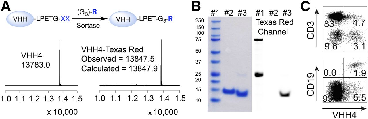
- FIGURE 1.
Characterization of anti–human class II MHC VHH (VHH4). (A) Schematic representation of sortase reaction to site-specifically labeled VHHs followed by liquid chromatography–mass spectrometry analysis. Sortase recognizes LPXTG motif and replaces G residue with substrate containing a NH2-G3-R moiety, where R represents tag of interest. Liquid chromatography–mass spectrometry analysis on VHH4 and VHH4-Texas Red confirms efficient labeling for VHH4. (B) Sodium dodecyl sulfate polyacrylamide gel electrophoresis characterization of VHH4: lane 1, marker; lane 2, VHH4; lane 3, VHH4-Texas Red (left); and same sodium dodecyl sulfate polyacrylamide gel electrophoresis gel scanned for Texas Red fluorophore (right). (C) Freshly isolated human peripheral blood lymphocytes cells were stained with VHH4-Texas Red and B and T cell lineage-specific antibodies. Representative data of 1 of 3 donors.
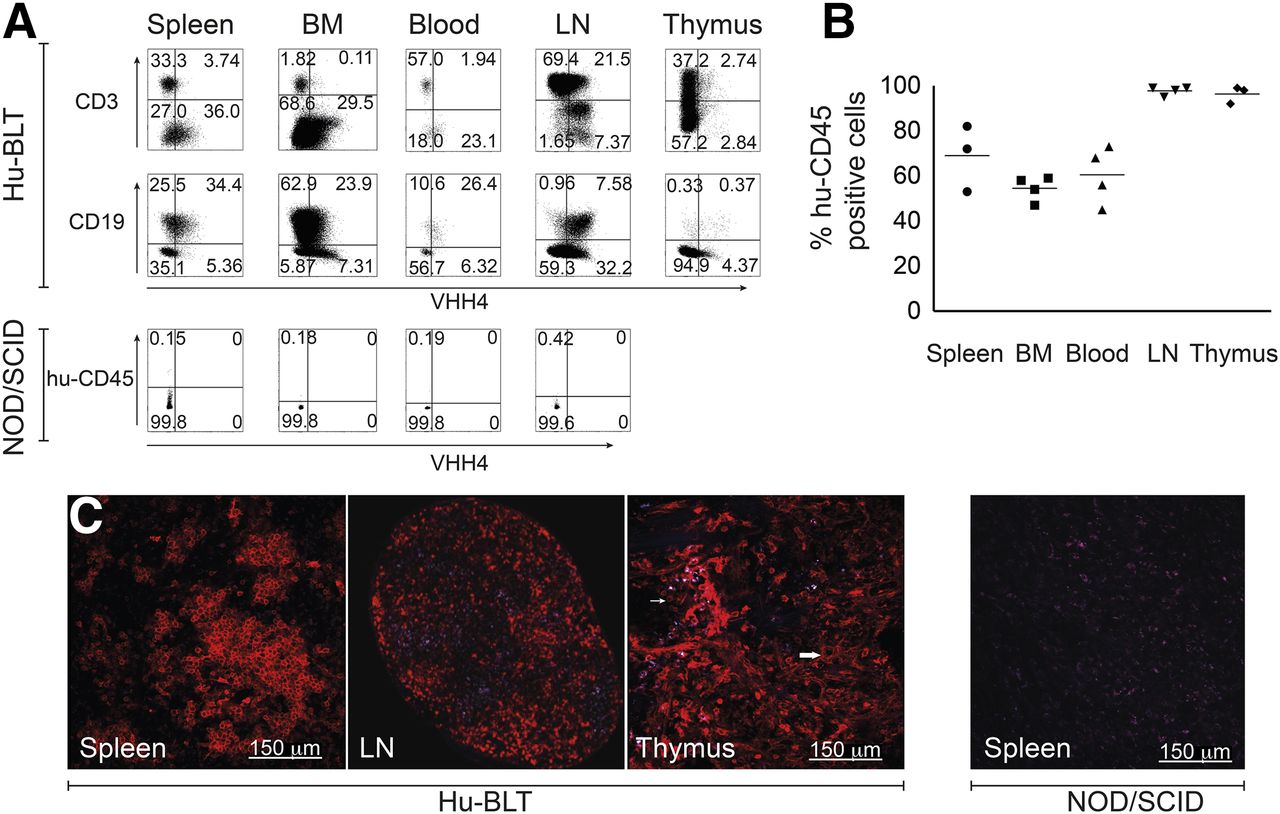
- FIGURE 2.
Human reconstitution of BLT mice. BLT mice were injected with 10 μg of VHH4-Texas Red via tail vein injection. Two hours after injection, mice were euthanized and organs isolated. (A) Single cell suspensions were prepared. By flow cytometry, cells were gated on human CD45 and analyzed for expression of class II MHC (VHH4) and B and T cell lineage markers; data representative of 5 individual experiments. (B) Percentages of human CD45-positive cells in different organs as analyzed by flow cytometry. (C) Analysis of VHH4 staining in spleen, lymph node, and thymus by 2-photon microscopy on Hu-BLT mice (n = 3) or in spleen of nonreconstituted NOD/SCID mouse, analyzed by 2-photon microscopy (n = 3). BM = bone marrow; LN = lymph node.
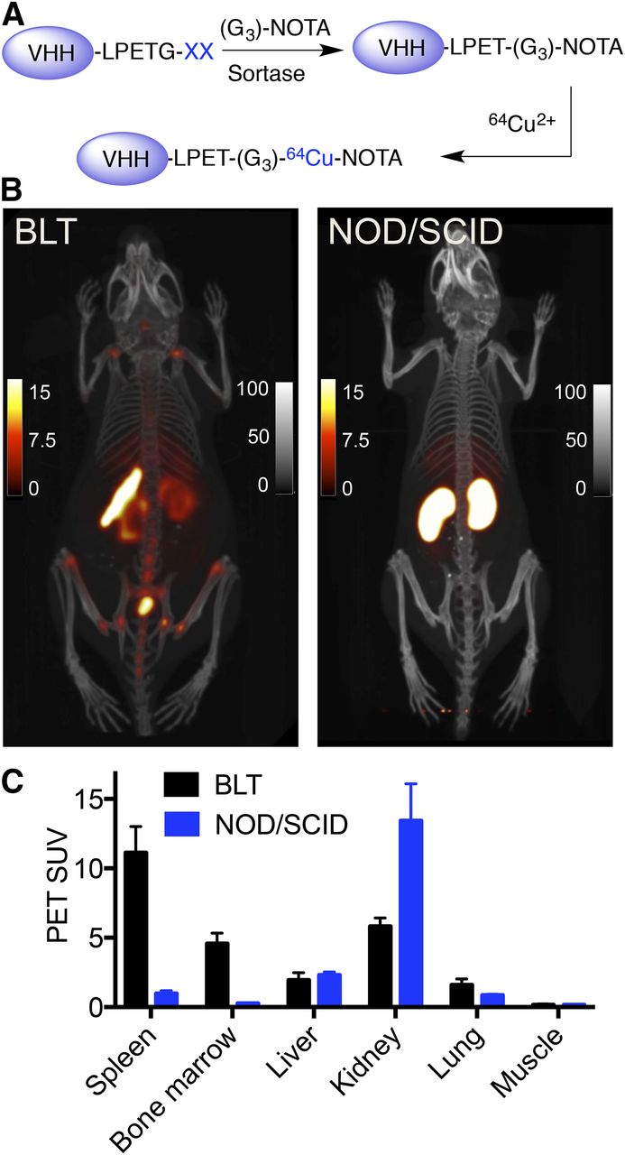
- FIGURE 3.
PET/CT imaging of human reconstitution in BLT mice using radiolabeled VHH4. (A) Schematic representation of radiolabeling of VHHs using sortase. VHH4 was first labeled using sortase with a chelator molecule, 1,4,7-triazacyclononane-triacetic acid, and then was radiolabeled using 64Cu. (B) PET/CT images of BLT mouse (left) and NOD/SCID mouse (right) imaged at 2 h after injection of 64Cu-VHH4. For BLT mouse, PET images show increased signal in spleen and bone marrow. Images are representative data of 4 different BLT mice. For NOD/SCID images, accumulation of signal in kidneys is nonspecific. Images are representative of 2 NOD/SCID mice. (C) PET SUVs of BLT and NOD/SCID mice shown in B. PET scale bars represent SUVs; CT scale bars have arbitrary units. Supplemental Videos 1 and 2 provide better 3-dimensional visualization. Images are all window-leveled to same intensity.
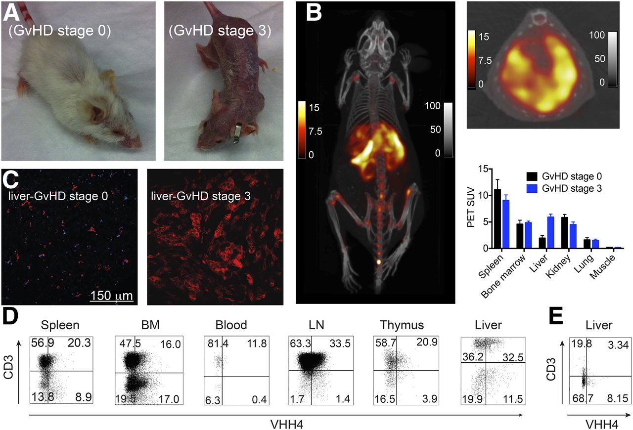
- FIGURE 4.
Imaging of GvHD in hu-BLT mice using VHH4. Mice with and without clinical symptoms of GvHD were injected with Texas Red–labeled or 64Cu-labeled-VHH4. Mice were either euthanized 2 h after injection to collect organs or imaged by PET/CT. (A) Phenotype of BLT mice with GvHD stage 0 or stage 3. (B) PET/CT images of BLT mouse with stage 3 GvHD (n = 2). PET/CT images show intense PET signal in spleen, marrow, and liver. Supplemental Video 3 provides better 3-dimensional visualization. On top right is shown transverse PET/CT image of liver of same mouse for better visualization of signal. PET scale bars represent SUVs; CT scale bars have arbitrary units. Below transverse image is PET SUVs for BLT mice with stage 0 GvHD (n = 2) and stage 3 GvHD (n = 2) imaged with 64Cu-labeled VHH4. (C) Two-photon imaging of the liver of a BLT mouse without clinical sings of GvHD (stage 0) or with stage 3 GvHD. (D and E) Single-cell suspensions of different organs were prepared for mice with GvHD stage 3 (D) or stage 0 (E). By flow cytometry, cells were gated on human CD45 and analyzed for expression of human class II MHC and T cell lineage markers. BM = bone marrow; LN = lymph node.

Additional Files
Supplemental Data
Files in this Data Supplement:


Jump to section
Related Articles
Cited By...
- Molecular Imaging of Acute Graft-Versus-Host Disease
- Single-Domain Antibody Theranostics on the Horizon
- A class II MHC-targeted vaccine elicits immunity against SARS-CoV-2 and its variants
- Molecular Imaging of Chimeric Antigen Receptor T Cells by ICOS-ImmunoPET
- Visualization of Activated T Cells by OX40-ImmunoPET as a Strategy for Diagnosis of Acute Graft-versus-Host Disease
- The Immunoimaging Toolbox
- Predicting the response to CTLA-4 blockade by longitudinal noninvasive monitoring of CD8 T cells