
Main menu

  • Home
  • Content
    • Current
    • Ahead of print
    • Past Issues
    • JNM Supplement
    • SNMMI Annual Meeting Abstracts
    • Continuing Education
    • JNM Podcasts
  • Subscriptions
    • Subscribers
    • Institutional and Non-member
    • Rates
    • Journal Claims
    • Corporate & Special Sales
  • Authors
    • Submit to JNM
    • Information for Authors
    • Assignment of Copyright
    • AQARA requirements
  • Info
    • Reviewers
    • Permissions
    • Advertisers
  • About
    • About Us
    • Editorial Board
    • Contact Information
  • More
    • Alerts
    • Feedback
    • Help
    • SNMMI Journals
  • SNMMI
    • JNM
    • JNMT
    • SNMMI Journals
    • SNMMI

User menu

  • Subscribe
  • My alerts
  • Log in
  • My Cart

Search

  • Advanced search
Journal of Nuclear Medicine
  • SNMMI
    • JNM
    • JNMT
    • SNMMI Journals
    • SNMMI
  • Subscribe
  • My alerts
  • Log in
  • My Cart
Journal of Nuclear Medicine

Advanced Search

  • Home
  • Content
    • Current
    • Ahead of print
    • Past Issues
    • JNM Supplement
    • SNMMI Annual Meeting Abstracts
    • Continuing Education
    • JNM Podcasts
  • Subscriptions
    • Subscribers
    • Institutional and Non-member
    • Rates
    • Journal Claims
    • Corporate & Special Sales
  • Authors
    • Submit to JNM
    • Information for Authors
    • Assignment of Copyright
    • AQARA requirements
  • Info
    • Reviewers
    • Permissions
    • Advertisers
  • About
    • About Us
    • Editorial Board
    • Contact Information
  • More
    • Alerts
    • Feedback
    • Help
    • SNMMI Journals
  • View or Listen to JNM Podcast
  • Visit JNM on Facebook
  • Join JNM on LinkedIn
  • Follow JNM on Twitter
  • Subscribe to our RSS feeds
Research ArticleOncology

Fluorescence-Based Molecular Imaging of Porcine Urinary Bladder Sentinel Lymph Nodes

Hak J. Lee, Christopher V. Barback, Carl K. Hoh, Zhengtao Qin, Kareem Kader, David J. Hall, David R. Vera and Christopher J. Kane
Journal of Nuclear Medicine April 2017, 58 (4) 547-553; DOI: https://doi.org/10.2967/jnumed.116.178582
Hak J. Lee
1Department of Urology, University of California, San Diego, La Jolla, California
  • Find this author on Google Scholar
  • Find this author on PubMed
  • Search for this author on this site
Christopher V. Barback
2Department of Radiology, University of California, San Diego, La Jolla, California; and
3UCSD Molecular Imaging Program, University of California, San Diego, La Jolla, California
  • Find this author on Google Scholar
  • Find this author on PubMed
  • Search for this author on this site
Carl K. Hoh
2Department of Radiology, University of California, San Diego, La Jolla, California; and
3UCSD Molecular Imaging Program, University of California, San Diego, La Jolla, California
  • Find this author on Google Scholar
  • Find this author on PubMed
  • Search for this author on this site
Zhengtao Qin
2Department of Radiology, University of California, San Diego, La Jolla, California; and
3UCSD Molecular Imaging Program, University of California, San Diego, La Jolla, California
  • Find this author on Google Scholar
  • Find this author on PubMed
  • Search for this author on this site
Kareem Kader
1Department of Urology, University of California, San Diego, La Jolla, California
  • Find this author on Google Scholar
  • Find this author on PubMed
  • Search for this author on this site
David J. Hall
2Department of Radiology, University of California, San Diego, La Jolla, California; and
3UCSD Molecular Imaging Program, University of California, San Diego, La Jolla, California
  • Find this author on Google Scholar
  • Find this author on PubMed
  • Search for this author on this site
David R. Vera
2Department of Radiology, University of California, San Diego, La Jolla, California; and
3UCSD Molecular Imaging Program, University of California, San Diego, La Jolla, California
  • Find this author on Google Scholar
  • Find this author on PubMed
  • Search for this author on this site
Christopher J. Kane
1Department of Urology, University of California, San Diego, La Jolla, California
  • Find this author on Google Scholar
  • Find this author on PubMed
  • Search for this author on this site
  • Article
  • Figures & Data
  • Info & Metrics
  • PDF
Loading

Article Figures & Data

Figures

  • Tables
  • FIGURE 1.
    • Download figure
    • Open in new tab
    • Download powerpoint
    FIGURE 1.

    Cystoscopic injection (needle tip at arrow) into pig bladder of molecular imaging agent IRDye800CW-tilmanocept radiolabeled with 68Ga and 99mTc.
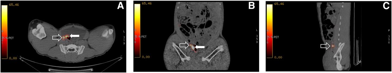
  • FIGURE 2.
    • Download figure
    • Open in new tab
    • Download powerpoint
    FIGURE 2.

    Fused PET/CT transaxial (A), coronal (B), and sagittal (C) cross-sections of right perivesicular LN (open arrow) adjacent to injection site in bladder wall (solid arrow). Injected dose of 68Ga was 4.4 MBq (120 μCi); at time of PET/CT image this SLN contained 0.085 MBq (2.3 μCi) of radioactivity and had an SUV of 65.2.
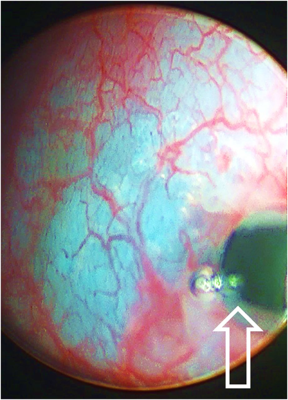
  • FIGURE 3.
    • Download figure
    • Open in new tab
    • Download powerpoint
    FIGURE 3.

    Intraoperative FireFly robotic surgical camera view looking into pelvis with a white light (A) and same location with near-infrared view (B). Fluorescent SLN (open arrow) was visualized within minutes of starting mapping procedure. This SLN contained 88 pmol of fluorescent dye.

Tables

  • Figures
    • View popup
    TABLE 1

    Primary Objective: Dose Parameters and Number of Lymph Nodes Excised and Detected

    Molecular imaging agent
    Study no.Tilmanocept (nmol)68Ga (MBq)99mTc (MBq)IRDye800CW dye (nmol)Time between administration and SLN mapping (h)No. of SLNs visualized by the endoscopic camera (n)Lymph nodes excised (n)No. of SLNs detected ex vivo by γ-counting* (n)
    One1.55.58.12.25382122
    Two1.54.47.42.25361121
    Three1.52.98.92.25322122
    • ↵* Using 10% rule based on 99mTc radioactivity in cpm.

    • View popup
    TABLE 2

    Secondary Objectives: SLN and Injection Site Accumulation During PET/CT and Visualization During Robotic Surgery

    Study oneStudy twoStudy three
    PET/CTγ-counterPET/CTγ-counterPET/CTγ-counter
    Lymph node/injection siteSUVSatisfy 10% rule? (yes/no)FireFly in vivo fluorecence? (yes/no)Percentage injected dose (%)Dye content (pmol)SUVSatisfy 10% rule? (yes/no)FireFly in vivo fluorescence? (yes/no)Percentage injected dose (%)Dye content (pmol)SUVSatisfy 10% rule? (yes/no)FireFly in vivo fluorescence? (yes/no)Percentage injected dose (%)Dye content (pmol)
    L. external iliacNoNo
    R. external iliacNoNoNVYes0.388.50
    PresacralNoNoNo
    Para aorticNoNoNo
    L. common iliacNoNoNo
    R. common iliac12.4YesYes2.7232.3No139YesYes1.3329.8
    L. perivesicularNoNoNo
    R. perivesicular59.0YesYes1.4461.065.2YesYes3.9188.0No
    L. pelvicNoNoNo
    R. pelvicNoNoNo
    L. obturatorNoNoNo
    R. obturatorNoNoNo
    Injection site2,911Yes29.26581,769Yes16.537120,322Yes64.11,441
    • L. = left; R. = right; NV = not visualized by PET/CT.

    • If PET/CT or γ-counter did not satisfy the 10% rule, all entries for LN are blank.

PreviousNext
Back to top

In this issue

Journal of Nuclear Medicine: 58 (4)
Journal of Nuclear Medicine
Vol. 58, Issue 4
April 1, 2017
  • Table of Contents
  • Table of Contents (PDF)
  • About the Cover
  • Index by author
Print
Download PDF
Article Alerts
Sign In to Email Alerts with your Email Address
Email Article

Thank you for your interest in spreading the word on Journal of Nuclear Medicine.

NOTE: We only request your email address so that the person you are recommending the page to knows that you wanted them to see it, and that it is not junk mail. We do not capture any email address.

Enter multiple addresses on separate lines or separate them with commas.
Fluorescence-Based Molecular Imaging of Porcine Urinary Bladder Sentinel Lymph Nodes
(Your Name) has sent you a message from Journal of Nuclear Medicine
(Your Name) thought you would like to see the Journal of Nuclear Medicine web site.
Citation Tools
Fluorescence-Based Molecular Imaging of Porcine Urinary Bladder Sentinel Lymph Nodes
Hak J. Lee, Christopher V. Barback, Carl K. Hoh, Zhengtao Qin, Kareem Kader, David J. Hall, David R. Vera, Christopher J. Kane
Journal of Nuclear Medicine Apr 2017, 58 (4) 547-553; DOI: 10.2967/jnumed.116.178582

Citation Manager Formats

  • BibTeX
  • Bookends
  • EasyBib
  • EndNote (tagged)
  • EndNote 8 (xml)
  • Medlars
  • Mendeley
  • Papers
  • RefWorks Tagged
  • Ref Manager
  • RIS
  • Zotero
Share
Fluorescence-Based Molecular Imaging of Porcine Urinary Bladder Sentinel Lymph Nodes
Hak J. Lee, Christopher V. Barback, Carl K. Hoh, Zhengtao Qin, Kareem Kader, David J. Hall, David R. Vera, Christopher J. Kane
Journal of Nuclear Medicine Apr 2017, 58 (4) 547-553; DOI: 10.2967/jnumed.116.178582
Twitter logo Facebook logo LinkedIn logo Mendeley logo
  • Tweet Widget
  • Facebook Like
  • Google Plus One
Bookmark this article

Jump to section

  • Article
    • Abstract
    • MATERIALS AND METHODS
    • RESULTS
    • DISCUSSION
    • CONCLUSION
    • DISCLOSURE
    • Footnotes
    • REFERENCES
  • Figures & Data
  • Info & Metrics
  • PDF

Related Articles

  • This Month in JNM
  • PubMed
  • Google Scholar

Cited By...

  • Imaging Synaptic Density: A Different Look at Neurologic Diseases
  • Google Scholar

More in this TOC Section

Oncology

  • The Evolving Role of FDG-PET/CT in Assessing Primary Cutaneous Lymphoma
  • Molecular imaging of Renal clear cell carcinoma subtypes as metabolic diseases
  • Role of PET/CT in the management of multiple myeloma
Show more Oncology

Basic

  • Dopamine D1 Receptor Agonist PET Tracer Development: Assessment in Nonhuman Primates
  • Optical Navigation of the Drop-In γ-Probe as a Means to Strengthen the Connection Between Robot-Assisted and Radioguided Surgery
  • Synthesis and Preclinical Evaluation of a 68Ga-Labeled Adnectin, 68Ga-BMS-986192, as a PET Agent for Imaging PD-L1 Expression
Show more Basic

Similar Articles

Keywords

  • fluorescence
  • Molecular imaging
  • cystectomy
  • sentinel lymph node
  • pelvic lymph node dissection
  • robotic surgery
  • Lymphoseek
  • tilmanocept
  • PET/CT
SNMMI

© 2025 SNMMI

Powered by HighWire