


Abstract
2698
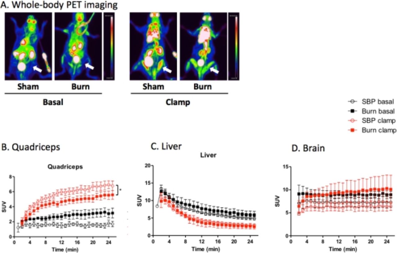
Objectives Investigation of the tissue-specific insulin sensitivity is necessary for better understanding of the pathophysiology of burn-induced insulin resistance and hyperglycemia. A combination of hyperinsulinemic isoglycemic clamp (HIC) and PET can be very useful for obtaining such data. HIC ensures basal (fasting) levels of blood glucose under insulin stimulation, whereas PET provides time-dependent tissue uptake quantification. As such, the aim of the present study was to quantify with the aid of the above techniques the impact of burn injury on tissue-specific FDG kinetics in a mouse model.
Methods Burned (30% total body surface area third-degree burn in 80°C water) and sham (room-temperature water) male C57BL/6 mice underwent PET/CT imaging during either fasting (basal) or HIC conditions (n=5/group). Imaging was performed using a microPET Focus 220/CereTom NL 3000 scanner combination on the 3rd day after the treatment. FDG time-activity (SUV) curves over 25 min were determined in the brain, liver, and skeletal muscles. Biochemical and metabolic tests (glycogen content in liver and muscle, immunoblotting for Akt, phospho-Akt and IRS1 and indirect calorimetry) in separate groups of animals were performed to validate the imaging data.
Results In skeletal muscles, FDG uptake during the basal period was comparable in burned and sham mice, whereas under insulin stimulation (HIC) FDG utilization was significantly decreased in burned mice. In the liver, HIC did not cause changes in FDG uptake in either groups. The brain represented an insulin insensitive tissue and its FDG uptake, as expected, remained unaffected.
Conclusions PET data supported by several biochemical tests strongly suggests that burn injury suppresses insulin sensitivity in skeletal muscles. This phenomenon supposedly plays a key role in the development of systemic burn-induced insulin resistance.

In this issue


Jump to section
Related Articles
Cited By...
- No citing articles found.