


Abstract
116
Objectives Inability to determine the distribution and activity of adoptively transferred T cells in the body presents a great barrier to advancement of adoptive T cell therapy in cancer. Accurate T cell imaging can be used to predict either success or failure in T cell therapy, and to avoid systemic toxicity and fatality by a timely intervention. For specific and sensitive imaging of chimeric antigen receptor (CAR) T cells in the setting of adoptive T cell therapy against solid cancers, we have developed a lentiviral system to transduce T cells with CAR specific to tumor antigen intercellular adhesion molecule (ICAM)-1 and somatostatin receptor 2 (SSTR2) as a genetic reporter. Imaging of SSTR2 by 68Ga-DOTATOC provides a lowest background in the upper body compared to other tested surface markers (e.g., NIS, PSMA), ideal for detecting T cells distributed in the brain, thyroid, lungs, stomach, and liver. Using mouse models of cervical and thyroid cancers, we demonstrate a feasibility of detecting T cell localization and expansion, closely predicting T cell activity against tumors.
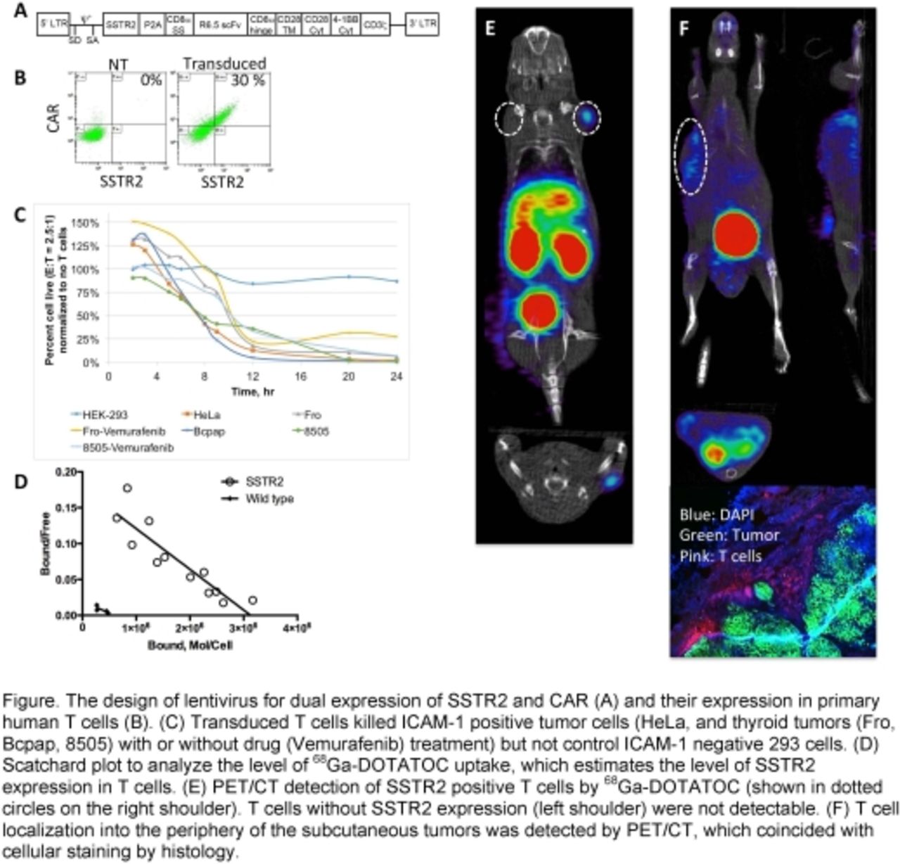
Methods Dual expression of CAR and SSTR2 was constructed by connecting SSTR2 to CAR via ‘ribosome skipping’ P2A sequence (Fig-a). CAR is comprised of ICAM-1-specific scFv (derived from mAb R6.5) and the transmembrane and cytoplasmic domains of CD28, CD137, and CD3ζwithin the lentiviral CAR construct. Lentivirus is produced by transfecting 293T cells with a mixture of packaging (pCMV-dR8.2; Addgene), envelope (pCMV-VSVG; Addgene), and transfer vectors. Mice were xenografted with HeLa and anaplastic thyroid cancer cells (8505) by either subcutaneous, intravenous, or orthotopic injection. PET/CT Images were acquired on a micro-PET/CT scanner (Inveon, Siemens) within 30 min of 68Ga-DOTATOC (1-10 MBq) injection via a tail vein.
Results Lentiviral system has typically produced 30~70% transduction efficiency in T cells for expression of CAR and SSTR2 (Fig-b). Specific target killing of R6.5-CAR T cells has been validated from potent cell killing of ICAM-1 positive thyroid/cervical tumors but not ICAM-1 negative cells (293 T) with the induction of Th1 cytokines specific to CAR and antigen interaction (Fig-c). By titration of DOTATOC, we determined that ~ 1 million molecules of SSTR2 could be expressed in a single T cell (Fig-d). Prior to imaging T cells in xenografted mice, we first confirmed PET/CT imaging of DOTATOC was specific to T cells transduced with SSTR2 (Fig-e). Our imaging studies successfully detected localization of intravenously introduced T cells into the periphery of the tumors, which coincided with T cell mapping by histology (Fig-f).
Conclusions We demonstrated that our lentiviral system for simultaneous expression of CAR and SSTR2 in T cells conferred on T cells the ability to kill ICAM-1 positive tumors and to internalize SSTR2-specific radioligands. SSTR2 imaging by PET/CT was specific and sensitive to T cells localized to the tumors and to other normal organs. Our ongoing studies include PET/CT imaging of T cells over the course of T cell therapy to quantitatively correlate DOTATOC signals with T cell killing of tumors and to predict potential toxicity of adoptive T cell therapy.

In this issue


Jump to section
Related Articles
Cited By...
- No citing articles found.