
Main menu

  • Home
  • Content
    • Current
    • Ahead of print
    • Past Issues
    • JNM Supplement
    • SNMMI Annual Meeting Abstracts
    • Continuing Education
    • JNM Podcasts
  • Subscriptions
    • Subscribers
    • Institutional and Non-member
    • Rates
    • Journal Claims
    • Corporate & Special Sales
  • Authors
    • Submit to JNM
    • Information for Authors
    • Assignment of Copyright
    • AQARA requirements
  • Info
    • Reviewers
    • Permissions
    • Advertisers
  • About
    • About Us
    • Editorial Board
    • Contact Information
  • More
    • Alerts
    • Feedback
    • Help
    • SNMMI Journals
  • SNMMI
    • JNM
    • JNMT
    • SNMMI Journals
    • SNMMI

User menu

  • Subscribe
  • My alerts
  • Log in
  • Log out
  • My Cart

Search

  • Advanced search
Journal of Nuclear Medicine
  • SNMMI
    • JNM
    • JNMT
    • SNMMI Journals
    • SNMMI
  • Subscribe
  • My alerts
  • Log in
  • Log out
  • My Cart
Journal of Nuclear Medicine

Advanced Search

  • Home
  • Content
    • Current
    • Ahead of print
    • Past Issues
    • JNM Supplement
    • SNMMI Annual Meeting Abstracts
    • Continuing Education
    • JNM Podcasts
  • Subscriptions
    • Subscribers
    • Institutional and Non-member
    • Rates
    • Journal Claims
    • Corporate & Special Sales
  • Authors
    • Submit to JNM
    • Information for Authors
    • Assignment of Copyright
    • AQARA requirements
  • Info
    • Reviewers
    • Permissions
    • Advertisers
  • About
    • About Us
    • Editorial Board
    • Contact Information
  • More
    • Alerts
    • Feedback
    • Help
    • SNMMI Journals
  • View or Listen to JNM Podcast
  • Visit JNM on Facebook
  • Join JNM on LinkedIn
  • Follow JNM on Twitter
  • Subscribe to our RSS feeds
Research ArticleClinical Investigations

Multi-Atlas–Based Attenuation Correction for Brain 18F-FDG PET Imaging Using a Time-of-Flight PET/MR Scanner: Comparison with Clinical Single-Atlas– and CT-Based Attenuation Correction

Tetsuro Sekine, Ninon Burgos, Geoffrey Warnock, Martin Huellner, Alfred Buck, Edwin E.G.W. ter Voert, M. Jorge Cardoso, Brian F. Hutton, Sebastien Ourselin, Patrick Veit-Haibach and Gaspar Delso
Journal of Nuclear Medicine August 2016, 57 (8) 1258-1264; DOI: https://doi.org/10.2967/jnumed.115.169045
Tetsuro Sekine
1Department of Nuclear Medicine, University Hospital Zurich, Zurich, Switzerland
2Department of Radiology, Nippon Medical School, Tokyo, Japan
  • Find this author on Google Scholar
  • Find this author on PubMed
  • Search for this author on this site
Ninon Burgos
3Translational Imaging Group, Centre for Medical Image Computing, University College London, NW1 2HE, London, United Kingdom
  • Find this author on Google Scholar
  • Find this author on PubMed
  • Search for this author on this site
Geoffrey Warnock
4Institute of Pharmacology & Toxicology, University of Zurich, Zurich, Switzerland
5PMOD Technologies Ltd., Zurich, Switzerland
  • Find this author on Google Scholar
  • Find this author on PubMed
  • Search for this author on this site
Martin Huellner
1Department of Nuclear Medicine, University Hospital Zurich, Zurich, Switzerland
6Department of Neuroradiology, University Hospital Zurich, Zurich, Switzerland
  • Find this author on Google Scholar
  • Find this author on PubMed
  • Search for this author on this site
Alfred Buck
1Department of Nuclear Medicine, University Hospital Zurich, Zurich, Switzerland
  • Find this author on Google Scholar
  • Find this author on PubMed
  • Search for this author on this site
Edwin E.G.W. ter Voert
1Department of Nuclear Medicine, University Hospital Zurich, Zurich, Switzerland
  • Find this author on Google Scholar
  • Find this author on PubMed
  • Search for this author on this site
M. Jorge Cardoso
3Translational Imaging Group, Centre for Medical Image Computing, University College London, NW1 2HE, London, United Kingdom
7Dementia Research Centre, Institute of Neurology, University College London, WC1N 3AR London, United Kingdom
  • Find this author on Google Scholar
  • Find this author on PubMed
  • Search for this author on this site
Brian F. Hutton
8Institute of Nuclear Medicine, University College London, NW1 2BU London, United Kingdom
9Centre for Medical Radiation Physics, University of Wollongong, NSW 2522 Wollongong, Australia
  • Find this author on Google Scholar
  • Find this author on PubMed
  • Search for this author on this site
Sebastien Ourselin
3Translational Imaging Group, Centre for Medical Image Computing, University College London, NW1 2HE, London, United Kingdom
7Dementia Research Centre, Institute of Neurology, University College London, WC1N 3AR London, United Kingdom
  • Find this author on Google Scholar
  • Find this author on PubMed
  • Search for this author on this site
Patrick Veit-Haibach
1Department of Nuclear Medicine, University Hospital Zurich, Zurich, Switzerland
10Division of Diagnostic and Interventional Radiology, Department of Medical Radiology, University Hospital Zurich, Zurich, Switzerland; and
  • Find this author on Google Scholar
  • Find this author on PubMed
  • Search for this author on this site
Gaspar Delso
11GE Healthcare, Waukesha, Wisconsin
  • Find this author on Google Scholar
  • Find this author on PubMed
  • Search for this author on this site
  • Article
  • Figures & Data
  • Info & Metrics
  • PDF
Loading

Article Figures & Data

Figures

  • Tables
  • FIGURE 1.
    • Download figure
    • Open in new tab
    • Download powerpoint
    FIGURE 1.

    Flowchart illustrating generation of the 3 AC maps. More detailed descriptions are provided in previously published studies (6,10,12).
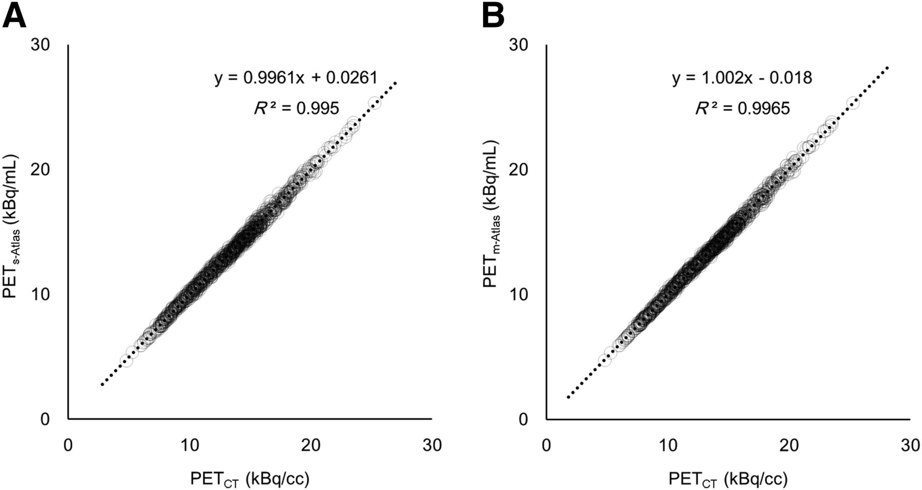
  • FIGURE 2.
    • Download figure
    • Open in new tab
    • Download powerpoint
    FIGURE 2.

    Regression plots between CT-AC and s-Atlas (A) and CT-AC and m-Atlas (B) for 67 VOIs × 15 patients.
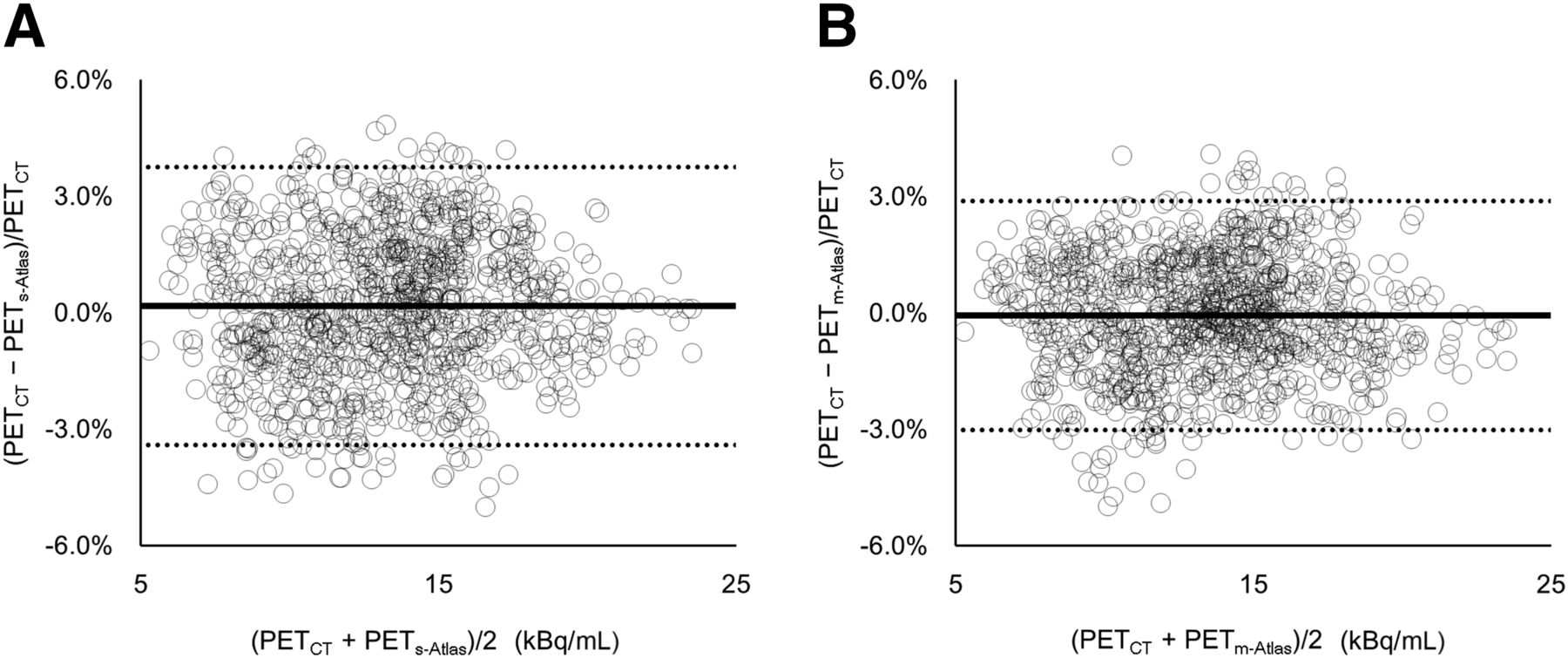
  • FIGURE 3.
    • Download figure
    • Open in new tab
    • Download powerpoint
    FIGURE 3.

    Bland–Altman plots of CT-AC and s-Atlas (A) and CT-AC and m-Atlas (B) for 67 VOIs × 15 patients. Average and SD of %diff are given in Table 1.
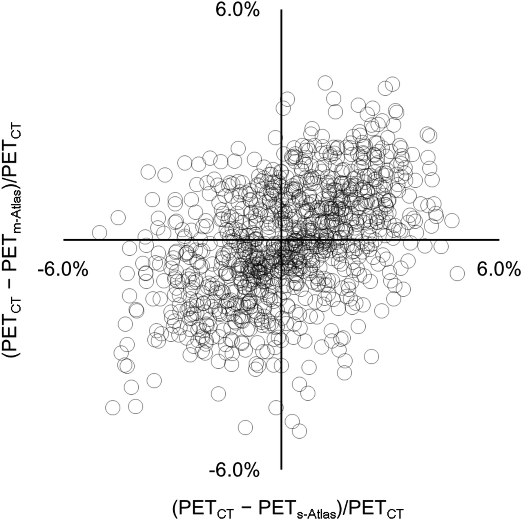
  • FIGURE 4.
    • Download figure
    • Open in new tab
    • Download powerpoint
    FIGURE 4.

    Scatterplot of %diff on s-Atlas and on m-Atlas.
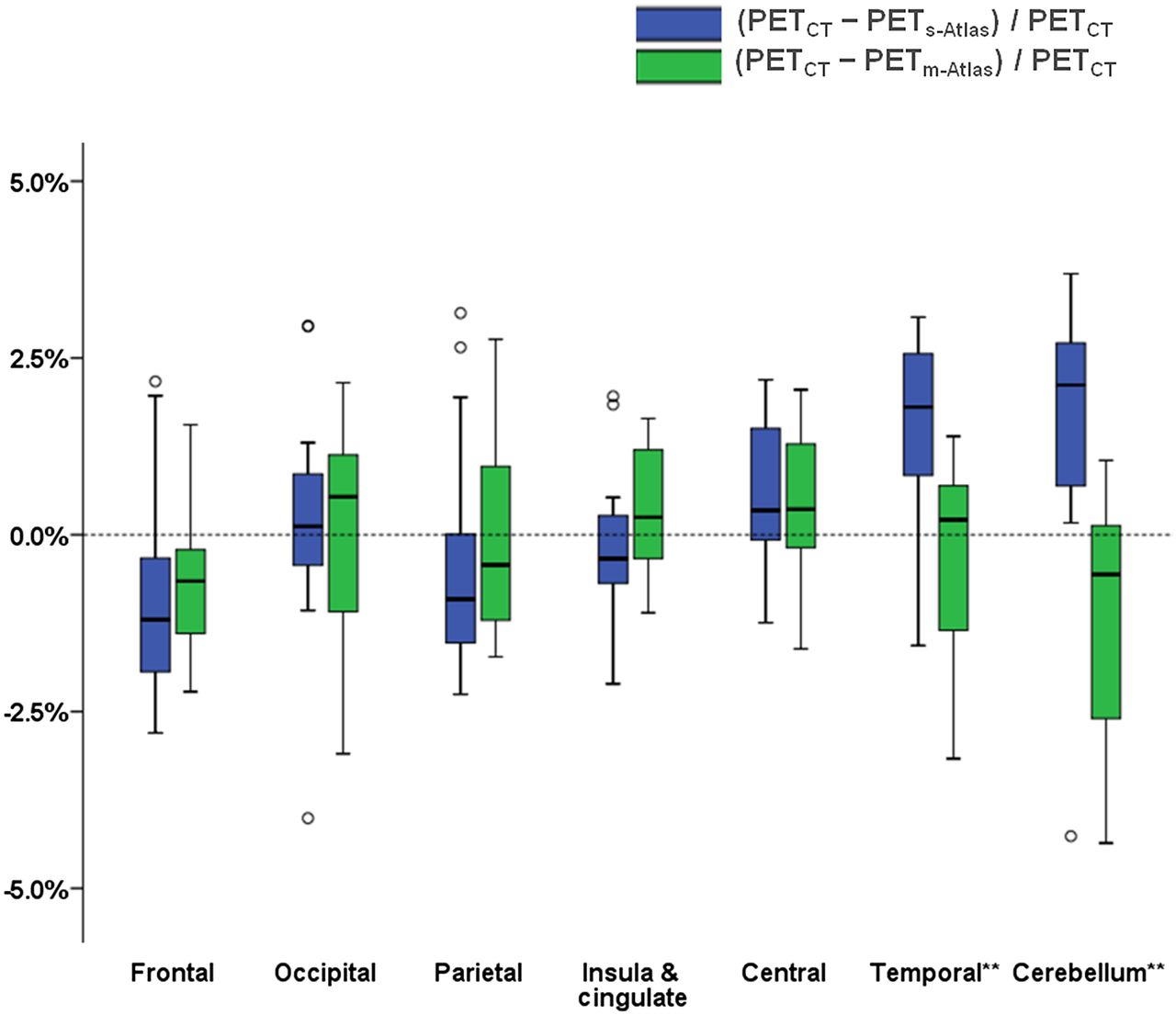
  • FIGURE 5.
    • Download figure
    • Open in new tab
    • Download powerpoint
    FIGURE 5.

    Box plot of each generalized VOI of s-Atlas (blue box) and m-Atlas (green box). In temporal lobe and cerebellum, %diff on m-Atlas is significantly smaller than that on s-Atlas. **P < 0.01 using paired t test with Bonferroni adjustment.
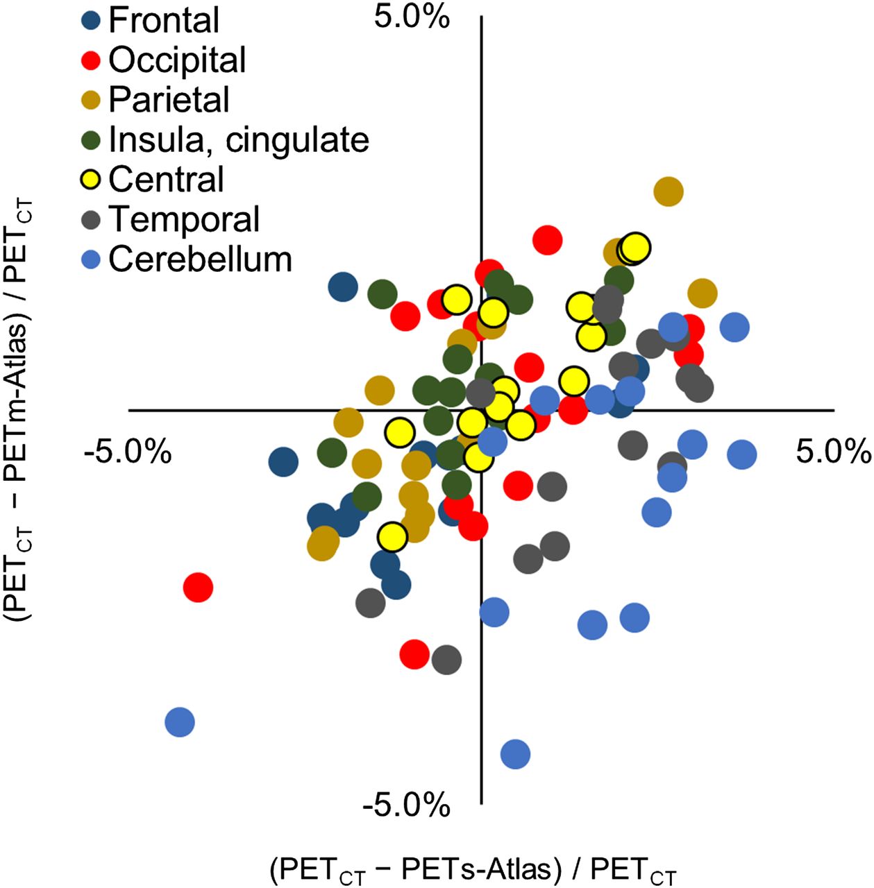
  • FIGURE 6.
    • Download figure
    • Open in new tab
    • Download powerpoint
    FIGURE 6.

    Scatterplot of each generalized VOI of %diff on s-Atlas and on m-Atlas. Dot color corresponds to color of Supplemental Figure 3. R2 and slope of best fit line of each region are given in Table 3.

  • FIGURE 7.
    • Download figure
    • Open in new tab
    • Download powerpoint
    FIGURE 7.

    %diff on s-Atlas and on m-Atlas in 3 representative cases, all of which were normalized to a brain template (SPM5). In pt_01, %diff on m-Atlas is much smaller than on m-Atlas, especially in cerebellar and temporal regions. In pt_02, trend of %diff is somewhat different. In cerebellum, s-Atlas underestimated 18F-FDG uptake, whereas m-Atlas overestimated it. Absolute %diff on m-Atlas is smaller than on s-Atlas. Of all 15 patients, pt_03 is only one in whom %diff on m-Atlas was apparently larger than on s-Atlas, especially in cerebellum. Bottom row is respective high-resolution T1w image for %diff map.

Tables

  • Figures
    • View popup
    TABLE 1

    %diff and |%diff| in 18F-FDG Uptake (kBq/mL) Between CT-AC and Each s-/m-Atlas in All Regions (67 × 15 = 1,005 VOIs)

    ParameterWith TOFWithout TOF
    %diff on s-Atlas*0.17% ± 1.82%0.06% ± 2.13%
    %diff on m-Atlas*−0.06% ± 1.50%0.03% ± 1.92%
    |%diff| on s-Atlas†1.49% ± 1.06%1.71% ± 1.28%
    |%diff| on m-Atlas†1.21% ± 0.89%1.51% ± 1.19%
    • ↵* s-Atlas vs. m-Atlas, P < 0.01.

    • ↵† With TOF vs. without TOF, P < 0.01.

    • %diff is CT-AC minus s-/m-AC, divided by CT-AC. |%diff| is absolute value of %diff.

    • View popup
    TABLE 2

    %diff and |%diff| in 18F-FDG Uptake (kBq/mL) Between CT-AC and Each s-/m-Atlas in Each Merged Region

    ParameterFrontal lobeOccipital lobeParietal lobeInsula and cingulateCentral structureTemporal lobe*Cerebellum*
    %diff on s-Atlas−0.87% ± 1.47%0.21% ± 1.67%−0.37% ± 1.70%−0.20% ± 1.12%0.58% ± 1.09%1.49% ± 1.37%1.55% ± 1.97%
    %diff on m-Atlas−0.64% ± 1.13%0.04% ± 1.53%−0.06% ± 1.41%0.35% ± 0.92%0.53% ± 1.04%−0.37% ± 1.41%−1.15% ± 1.72%
    |%diff| on s-Atlas1.46% ± 0.83%1.15% ± 1.19%1.42% ± 0.92%0.87% ± 0.71%0.97% ± 0.74%1.77% ± 0.95%2.11% ± 1.28%
    |%diff| on m-Atlas1.12% ± 0.61%1.25% ± 0.82%1.18% ± 0.71%0.80% ± 0.55%0.91% ± 0.69%1.15% ± 0.85%1.49% ± 1.40%
    • ↵* s-Atlas vs. m-Atlas, P < 0.01.

    • %diff is CT-AC minus s-/m-AC divided by CT-AC. |%diff| is absolute value of %diff.

    • View popup
    TABLE 3

    Correlation Between %diff on s-Atlas and on m-Atlas

    ParameterAll regions (1,005 VOIs)Frontal lobeOccipital lobeParietal lobeInsula and cingulateCentral structureTemporal lobeCerebellum
    R20.2210.1880.2350.7200.3290.5880.5000.385
    Slope of the best fit line0.3880.3340.4450.7050.4730.7270.7310.541
PreviousNext
Back to top

In this issue

Journal of Nuclear Medicine: 57 (8)
Journal of Nuclear Medicine
Vol. 57, Issue 8
August 1, 2016
  • Table of Contents
  • Table of Contents (PDF)
  • About the Cover
  • Index by author
Print
Download PDF
Article Alerts
Sign In to Email Alerts with your Email Address
Email Article

Thank you for your interest in spreading the word on Journal of Nuclear Medicine.

NOTE: We only request your email address so that the person you are recommending the page to knows that you wanted them to see it, and that it is not junk mail. We do not capture any email address.

Enter multiple addresses on separate lines or separate them with commas.
Multi-Atlas–Based Attenuation Correction for Brain 18F-FDG PET Imaging Using a Time-of-Flight PET/MR Scanner: Comparison with Clinical Single-Atlas– and CT-Based Attenuation Correction
(Your Name) has sent you a message from Journal of Nuclear Medicine
(Your Name) thought you would like to see the Journal of Nuclear Medicine web site.
Citation Tools
Multi-Atlas–Based Attenuation Correction for Brain 18F-FDG PET Imaging Using a Time-of-Flight PET/MR Scanner: Comparison with Clinical Single-Atlas– and CT-Based Attenuation Correction
Tetsuro Sekine, Ninon Burgos, Geoffrey Warnock, Martin Huellner, Alfred Buck, Edwin E.G.W. ter Voert, M. Jorge Cardoso, Brian F. Hutton, Sebastien Ourselin, Patrick Veit-Haibach, Gaspar Delso
Journal of Nuclear Medicine Aug 2016, 57 (8) 1258-1264; DOI: 10.2967/jnumed.115.169045

Citation Manager Formats

  • BibTeX
  • Bookends
  • EasyBib
  • EndNote (tagged)
  • EndNote 8 (xml)
  • Medlars
  • Mendeley
  • Papers
  • RefWorks Tagged
  • Ref Manager
  • RIS
  • Zotero
Share
Multi-Atlas–Based Attenuation Correction for Brain 18F-FDG PET Imaging Using a Time-of-Flight PET/MR Scanner: Comparison with Clinical Single-Atlas– and CT-Based Attenuation Correction
Tetsuro Sekine, Ninon Burgos, Geoffrey Warnock, Martin Huellner, Alfred Buck, Edwin E.G.W. ter Voert, M. Jorge Cardoso, Brian F. Hutton, Sebastien Ourselin, Patrick Veit-Haibach, Gaspar Delso
Journal of Nuclear Medicine Aug 2016, 57 (8) 1258-1264; DOI: 10.2967/jnumed.115.169045
Twitter logo Facebook logo LinkedIn logo Mendeley logo
  • Tweet Widget
  • Facebook Like
  • Google Plus One
Bookmark this article

Jump to section

  • Article
    • Abstract
    • MATERIALS AND METHODS
    • RESULTS
    • DISCUSSION
    • CONCLUSION
    • DISCLOSURE
    • Footnotes
    • REFERENCES
  • Figures & Data
  • Info & Metrics
  • PDF

Related Articles

  • This Month in JNM
  • PubMed
  • Google Scholar

Cited By...

  • No citing articles found.
  • Google Scholar

More in this TOC Section

  • Feasibility of Ultra-Low-Activity 18F-FDG PET/CT Imaging Using a Long–Axial-Field-of-View PET/CT System
  • Cardiac Presynaptic Sympathetic Nervous Function Evaluated by Cardiac PET in Patients with Chronotropic Incompetence Without Heart Failure
  • Validation and Evaluation of a Vendor-Provided Head Motion Correction Algorithm on the uMI Panorama PET/CT System
Show more Clinical Investigations

Similar Articles

Keywords

  • PET/MR
  • attenuation correction
  • atlas-based
  • brain
  • 18F-FDG
SNMMI

© 2025 SNMMI

Powered by HighWire