


Article Figures & Data
Figures
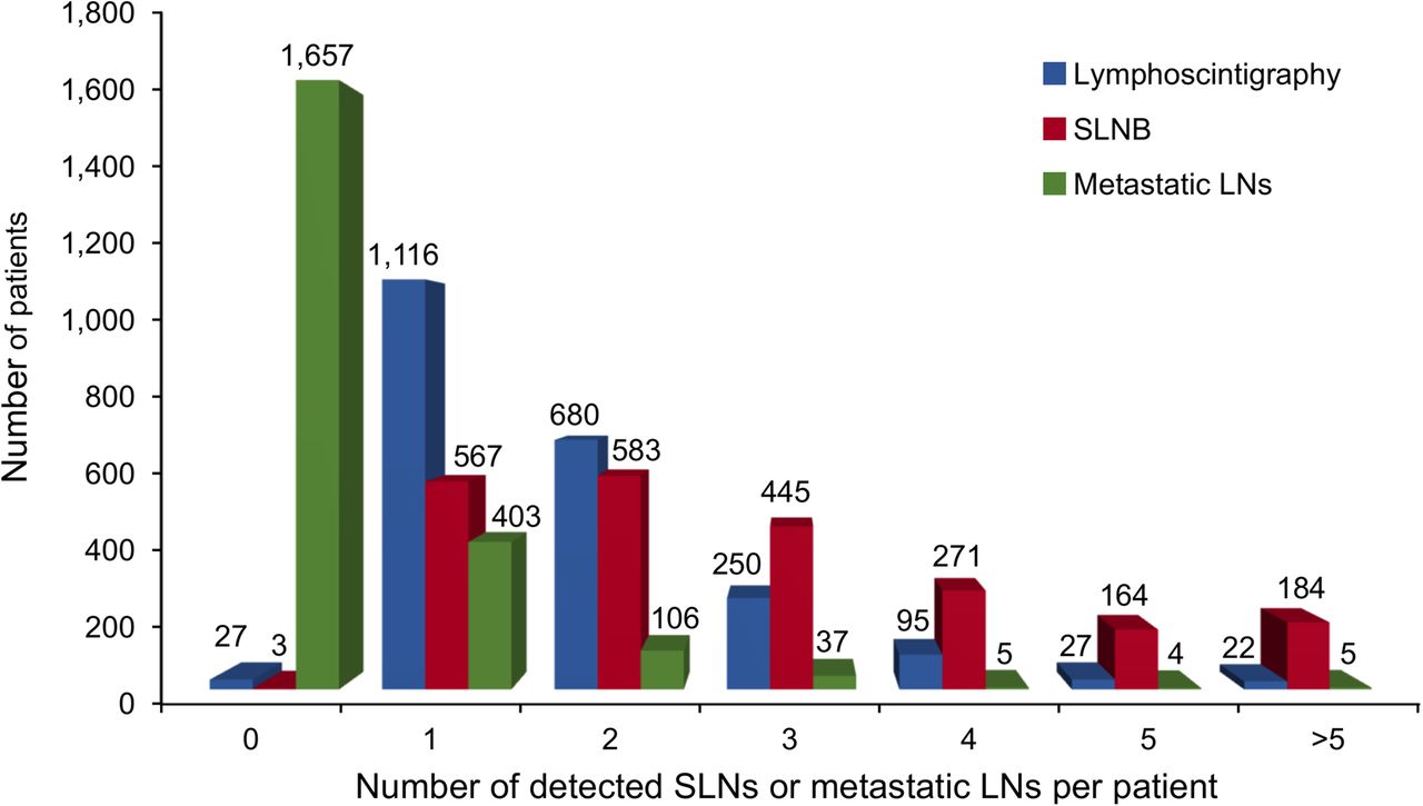
- FIGURE 1.
SLN imaging of breast cancer patients after injection of 99mTc-rituximab (black arrow, point source on patient body surface to mark sternal notch, contralateral sternal margin, and metasternum; red arrow, SLNs; hollow arrow, injection site). (A) One SLN in axilla and 1 SLN in intramammary. (B) Two SLNs in axilla.
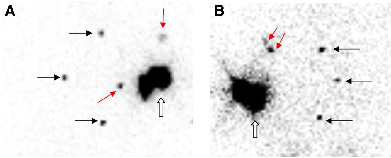
- FIGURE 2.
Lymphoscintigraphy and SLNB frequency distribution results in these 2,217 breast cancer patients.

Tables
Parameter QC specification QC result Appearance Clear, colorless Pass Volume 0.5–1.0 mL 0.5 mL Injection dose 18.5–37 MBq 37 MBq pH 4.0–8.0 7.0 Radio–thin-layer chromatography >95% >99% Radio–high-performance liquid chromatography >95% >99% Ethanol <5% 0 Endotoxins <15 EU/mL Pass Sterility Sterile Pass Specific activity 111 GBq/μmol Pass Characteristic No. of patients Sex Female 100 Age (y) ≤50 49 >50 51 Left or right side breast cancer Left 45 Right 55 Imaging time after injection of tracer 2–4 h 28 16–18 h 72 Primary tumor location Upper inner quadrant 26 Lower inner quadrant 7 Upper outer quadrant 49 Lower outer quadrant 16 Central portion 2 Histopathologic type Invasive ductal carcinoma 88 Invasive lobular carcinoma 4 Other 8 Clinical T stage Tis 1 T1 25 T2 65 T3 9 ER/PR/HER2 +/+/+ 23 −/−/+ 11 +/−/− or −/+/− or +/+/− 49 +/−/+ or −/+/+ 11 −/−/− 6 ER = estrogen receptor; PR = progesterone receptor; HER2 = human epidermal growth factor receptor-2.
Group Metastatic SLN Nonmetastatic SLN Total Metastatic LNs in ALND 26 2 28 Nonmetastatic LNs in ALND 49 23 72 Total 75 25 100 Characteristic Patient Percentage Sex Female 27 100% Age ≤50 10 37.04% >50 17 62.96% Left or right cancer Left 17 62.96% Right 10 37.04% Imaging time 2–4 h after injection 13 48.15% 16–18 h after injection 14 51.85% Primary tumor site Upper inner quadrant 26 96.30% Lower inner quadrant 7 25.93% Upper outer quadrant 49 181.48% Lower outer quadrant 16 59.26% Central portion 2 7.41% Histopathologic type Ductal 24 88.89% Lobular 1 3.70% Other 2 7.41% Clinical T stage T1 10 37.04% T2 16 59.26% T3 1 3.70% ER/PR/HER2 +/+/+ 5 18.52% −/−/+ 4 14.81% +/−/− or −/+/− or +/+/− 14 51.85% +/−/+ or −/+/+ 3 11.11% −/−/− 1 3.70% Pathology of SLN Metastatic SLN 19 70.37% Nonmetastatic SLN 8 29.63% ER = estrogen receptor; PR = progesterone receptor; HER2 = human epidermal growth factor receptor-2.
Lymphoscintigraphy Intraoperative SLN detection SLN pathology Characteristic No. of patients SLNs (  ± s)
± s)Significance SLNs ( ± s)Significance Patients with metastatic SLNS Significance Total 2,217 1.76 ± 1.08 2.85 ± 1.86 Sex P = 0.377 P = 0.613 P = 0.411 Female 2,215 1.76 ± 1.08 2.50 ± 0.71 560 Male 2 2.50 ± 0.71 2.85 ± 1.87 0 Age P = 0.000 P = 0.002 P = 0.355 ≤50 1,182 1.85 ± 1.13 2.96 ± 1.89 308 50 1,035 1.65 ± 1.00 2.72 ± 1.83 252 Left or right cancer P = 0.651 P = 0.719 P = 0.371 Bilateral 4 2 ± 0.82 3.00 ± 1.16 2 Left 1,140 1.74 ± 1.02 2.82 ± 1.84 279 Right 1,073 1.78 ± 1.13 2.88 ± 1.89 279 Imaging time P = 0.275 P = 0.897 P = 0.834 2–4 h after injection 922 1.73 ± 1.04 2.85 ± 1.80 235 16–18 h after injection 1,295 1.78 ± 1.10 2.84 ± 1.91 325 Primary tumor site P = 0.080 P = 0.316 P = 0.166 Upper inner quadrant 302 1.85 ± 0.98 2.66 ± 1.70 66 Lower inner quadrant 254 1.67 ± 0.89 2.83 ± 1.90 54 Upper outer quadrant 882 1.79 ± 1.19 2.89 ± 1.94 243 Lower outer quadrant 438 1.75 ± 1.13 2.94 ± 1.76 110 Central portion 341 1.65 ± 0.89 2.81 ± 1.90 87 Histopathologic subtype P = 0.178 P = 0.398 P = 0.000 Ductal 1,821 1.74 ± 1.06 2.83 ± 1.84 484 Lobular 179 1.90 ± 1.22 2.85 ± 1.85 49 Other 217 1.76 ± 1.09 3.01 ± 2.07 27 Clinical T stage P = 0.098 P = 0.191 P = 0.002 Tis 58 1.90 ± 1.07 3.10 ± 2.26 4 T1 1,179 1.73 ± 1.05 2.79 ± 1.84 294 T2 907 1.77 ± 1.09 2.88 ± 1.85 249 T3 73 2.01 ± 1.23 3.16 ± 2.10 13 ER/PR/HER2 P = 0.311 P = 0.973 P = 0.000 +/+/+ 634 1.78 ± 1.12 2.81 ± 1.86 187 −/−/+ 329 1.83 ± 1.04 2.87 ± 1.56 68 +/−/− or −/+/− or +/+/− 902 1.70 ± 1.04 2.85 ± 1.95 243 +/−/+ or −/+/+ 150 1.86 ± 1.09 2.86 ± 1.89 31 −/−/− 202 1.79 ± 1.22 2.92 ± 1.93 31


Supplemental Data
Files in this Data Supplement:


