


Article Figures & Data
Figures
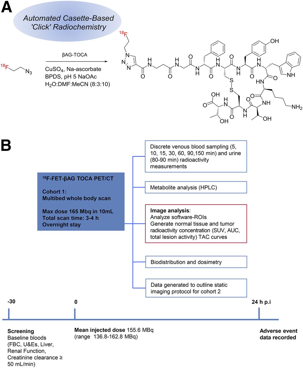
- FIGURE 1.
Chemical structure of 18F-FET-βAG-TOCA and study design. (A) Schematic diagram of chemical structure of 18F-FET-βAG-TOCA. pH 5, NaOAc H2O; sodium acetate buffer (pH5), water; MeCN (8:3:10). (B) PET/CT study timeline. AUC = area under the curve; BPDS = bathophenanthrolinedisulfonate; CuSO4 = copper (II) sulphate; DMF = dimethyl formamide; FBC = full blood count; Max = maximum; Na-ascorbate = sodium ascorbate; p.i. = after injection; ROIs = regions of interest; TAC = time–activity curves; U&Es = urea and electrolytes.
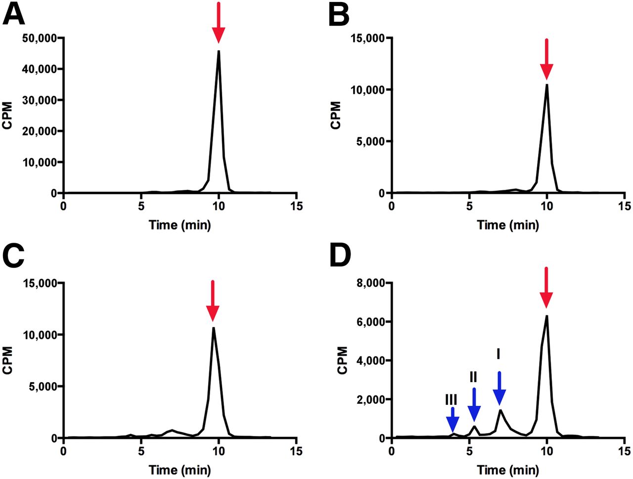
- FIGURE 2.
Metabolite analysis of 18F-FET-βAG-TOCA in patient 1. Typical high-performance liquid chromatogram of 18F-FET-βAG-TOCA in plasma at 5-, 30-, 60-, and 90-min time points (A, B, C and D, respectively). Red arrows indicate parent/unmetabolized 18F-FET-βAG-TOCA. Scaling of B, C, and D adjusted to allow for visualization of metabolite peaks I, II, and III (blue arrow). CPM = counts per minute.
- FIGURE 3.
18F-FET-βAG-TOCA PET/CT images and corresponding maximum-intensity-projection images in patient 1 (small bowel NET with widespread metastases in liver and bone). Sagittal images (A and B) and axial slices (C and D) showing widespread liver and bone metastases.
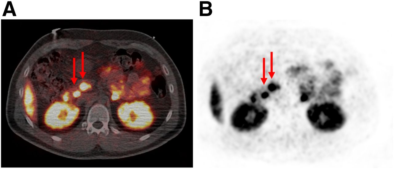
- FIGURE 4.
18F-FET-βAG-TOCA PET/CT images and corresponding maximum-intensity-projection images in patient with MEN1 syndrome, with pancreatic NETs. (A and B) Axial slices showing multiple lesions within pancreas (red arrows).
- FIGURE 5.
Time course biodistribution of 18F-FET-βAG-TOCA in male and female patients. Maximum-intensity-projection images of 18F-FET-βAG-TOCA in female patient with liver metastases (A) and male patient with lung NET (B). p.i. = after injection.
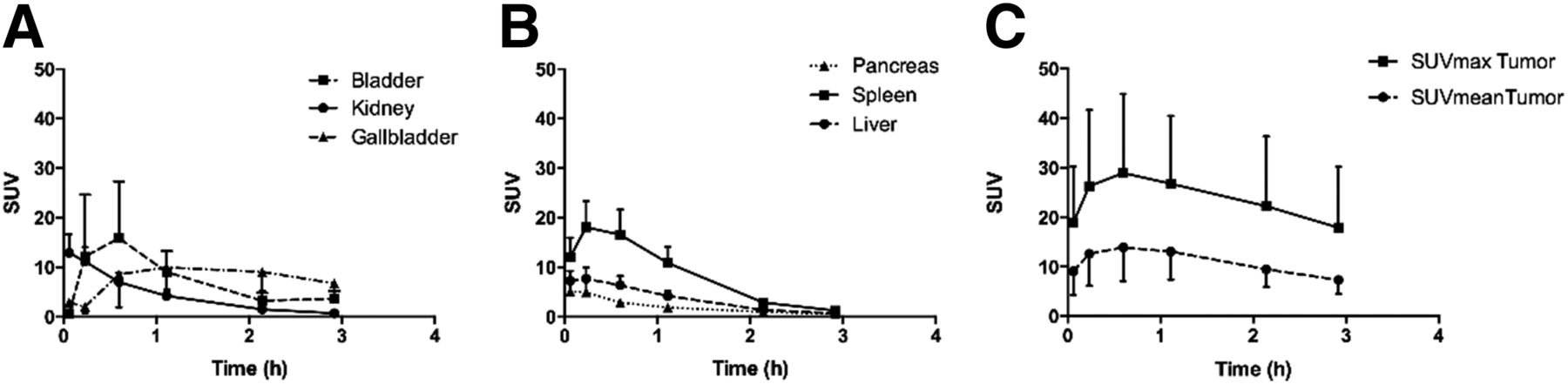
- FIGURE 6.
Mean decay-corrected time–activity curves for source organs and tumors. (A and B) SUVmean for bladder, kidneys, gallbladder, pancreas, spleen, and liver. (C) SUVmean and SUVmax for tumors (maximum of 3 lesions chosen per patient).
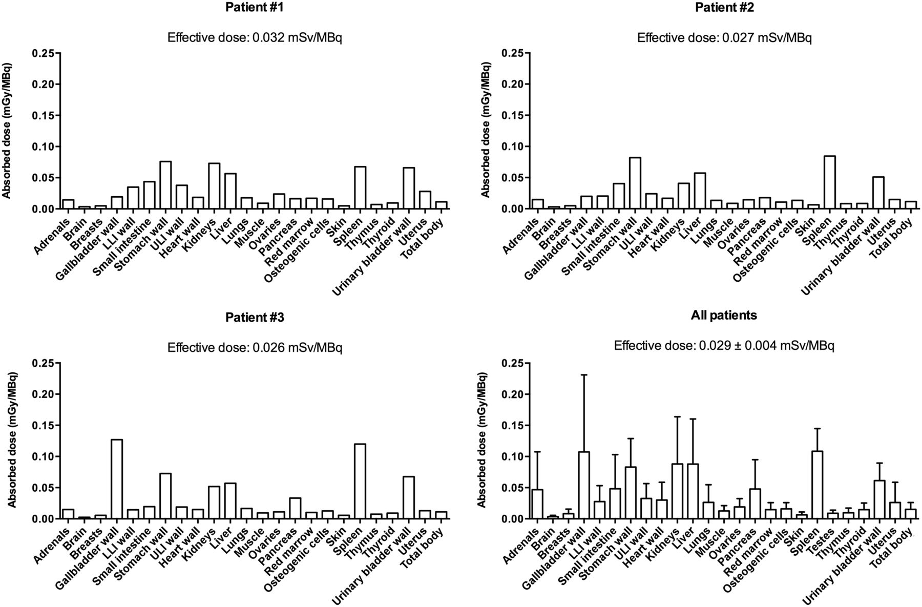
- FIGURE 7.
Biodistribution and dosimetry of 18F-FET-βAG-TOCA. Multibed whole-body PET scanning over 192 min was used to determine absorbed doses per unit administered activity (mGy/MBq) of major organs and tissues for each patient (first 3 patients are shown). Organ absorbed doses in all patients are shown with mean effective dose ± SD. LLI = lower large intestine; ULI = upper large intestine.


Tables
Patient no. Primary site Age (y) Sex Grade Metastatic Ki-67 (%) (primary tumor histology) Biomarker (pmol/L) Previous treatment CgA CgB 1 Small bowel 53 F 2 Liver, bone >10 1,567 170 Surgery/RFA/177Lu*/octreotide 2 Small bowel 73 F 1 Paraaortic lymph nodes, left SCF <1 962 558 Surgery 3 Lung 56 F 2 Bone <5 507 1,328 Octreotide 4 Small bowel 60 F 1 Liver <2 143 73 Surgery/RFA/177Lu 5 Lung 68 F 2 Mediastinal lymph nodes, bone 17 47 58 Surgery/RFA/177Lu 6 Pancreas 70 M 2 None 5 70 174 Surgery 7 Pancreas 53 F 2 Liver, bone 9 95 243 Surgery/chemotherapy 8 Lung 41 M 2 Lung, liver 12 24 126 Surgery/RFA/177Lu 9 Pancreas† 35 M 2 None <5 49 124 Surgery - TABLE 2
Mean Residence Times (MBq⋅h/MBq) of 18F-FET-βAG-TOCA for Different Organs in Male (n = 3) and Female (n = 6) Subjects
Men Women Organ Mean SD Mean SD Adrenals* 0.002 0.0002 0.003 0.0008 Brain 0.006 0.001 0.007 0.001 Breasts 0.004 0.001 Cortical bone 0.049 0.002 0.040 0.016 Gallbladder† 0.110 0.072 0.019 Heart contents 0.029 0.014 0.019 0.005 Heart wall 0.020 0.010 0.013 0.004 Kidneys 0.088 0.007 0.089 0.026 Liver 0.478 0.073 0.089 0.084 Lungs 0.063 0.009 0.067 0.025 Lower large intestine 0.012 0.002 0.019 0.011 Muscle 0.709 0.154 0.600 0.154 Ovaries‡ 0.0005 0.00004 Pancreas 0.017 0.006 0.010 0.0035 Red marrow 0.028 0.004 0.050 0.032 Small intestine 0.094 0.065 0.121 0.055 Stomach 0.005 0.029 0.066 0.222 Spleen 0.122 0.019 0.097 0.026 Testes 0.001 0.0002 Thyroid 0.001 0.0003 0.0008 0.0003 Upper large intestine 0.026 0.004 0.028 0.014 Urinary bladder¶ 0.075 0.019 0.101 0.028 Uterus 0.006 0.001 Remainder 0.523 0.250 0.628 0.025 ↵* Adrenal glands could not be visualized in 3 subjects.
↵† Gallbladder surgically removed in 5 subjects (1 male subject had gallbladder in situ).
↵‡ Ovaries could not be visualized in 1 subject because of postmenopausal atrophy, and 2 subjects had previous hysterectomy and bilateral salpingo-oophorectomy.
↵¶ Urinary bladder mean residence time is for 2-h voiding model.
Supplemental Data
Files in this Data Supplement:


Jump to section
Related Articles
Cited By...
- Somatostatin Receptor Imaging with [18F]FET-{beta}AG-TOCA PET/CT and [68Ga]Ga-DOTA-Peptide PET/CT in Patients with Neuroendocrine Tumors: A Prospective, Phase 2 Comparative Study
- Study protocol of LANTana: a phase Ib study to investigate epigenetic modification of somatostatin receptor-2 with ASTX727 to improve therapeutic outcome with [177Lu]Lu-DOTA-TATE in patients with metastatic neuroendocrine tumours, UK
- 18F-Labeled Somatostatin Analogs as PET Tracers for the Somatostatin Receptor: Ready for Clinical Use
- 18F-Labeled Somatostatin Analogs as PET Tracers for the Somatostatin Receptor: Ready for Clinical Use