
Main menu

  • Home
  • Content
    • Current
    • Ahead of print
    • Past Issues
    • JNM Supplement
    • SNMMI Annual Meeting Abstracts
    • Continuing Education
    • JNM Podcasts
  • Subscriptions
    • Subscribers
    • Institutional and Non-member
    • Rates
    • Journal Claims
    • Corporate & Special Sales
  • Authors
    • Submit to JNM
    • Information for Authors
    • Assignment of Copyright
    • AQARA requirements
  • Info
    • Reviewers
    • Permissions
    • Advertisers
  • About
    • About Us
    • Editorial Board
    • Contact Information
  • More
    • Alerts
    • Feedback
    • Help
    • SNMMI Journals
  • SNMMI
    • JNM
    • JNMT
    • SNMMI Journals
    • SNMMI

User menu

  • Subscribe
  • My alerts
  • Log in
  • Log out
  • My Cart

Search

  • Advanced search
Journal of Nuclear Medicine
  • SNMMI
    • JNM
    • JNMT
    • SNMMI Journals
    • SNMMI
  • Subscribe
  • My alerts
  • Log in
  • Log out
  • My Cart
Journal of Nuclear Medicine

Advanced Search

  • Home
  • Content
    • Current
    • Ahead of print
    • Past Issues
    • JNM Supplement
    • SNMMI Annual Meeting Abstracts
    • Continuing Education
    • JNM Podcasts
  • Subscriptions
    • Subscribers
    • Institutional and Non-member
    • Rates
    • Journal Claims
    • Corporate & Special Sales
  • Authors
    • Submit to JNM
    • Information for Authors
    • Assignment of Copyright
    • AQARA requirements
  • Info
    • Reviewers
    • Permissions
    • Advertisers
  • About
    • About Us
    • Editorial Board
    • Contact Information
  • More
    • Alerts
    • Feedback
    • Help
    • SNMMI Journals
  • View or Listen to JNM Podcast
  • Visit JNM on Facebook
  • Join JNM on LinkedIn
  • Follow JNM on Twitter
  • Subscribe to our RSS feeds
Research ArticleClinical Investigations

Reproducibility of MRI Dixon-Based Attenuation Correction in Combined PET/MR with Applications for Lean Body Mass Estimation

Ivo Rausch, Petra Rust, Matthew D. DiFranco, Martin Lassen, Andreas Stadlbauer, Marius E. Mayerhoefer, Markus Hartenbach, Marcus Hacker and Thomas Beyer
Journal of Nuclear Medicine July 2016, 57 (7) 1096-1101; DOI: https://doi.org/10.2967/jnumed.115.168294
Ivo Rausch
1Center for Medical Physics and Biomedical Engineering, Medical University of Vienna, Vienna, Austria
  • Find this author on Google Scholar
  • Find this author on PubMed
  • Search for this author on this site
Petra Rust
2Department of Nutritional Sciences, University of Vienna, Vienna, Austria
  • Find this author on Google Scholar
  • Find this author on PubMed
  • Search for this author on this site
Matthew D. DiFranco
1Center for Medical Physics and Biomedical Engineering, Medical University of Vienna, Vienna, Austria
  • Find this author on Google Scholar
  • Find this author on PubMed
  • Search for this author on this site
Martin Lassen
1Center for Medical Physics and Biomedical Engineering, Medical University of Vienna, Vienna, Austria
  • Find this author on Google Scholar
  • Find this author on PubMed
  • Search for this author on this site
Andreas Stadlbauer
3Division of General and Pediatric Radiology, Department of Biomedical Imaging and Image-Guided Therapy, Medical University of Vienna, Vienna, Austria; and
  • Find this author on Google Scholar
  • Find this author on PubMed
  • Search for this author on this site
Marius E. Mayerhoefer
3Division of General and Pediatric Radiology, Department of Biomedical Imaging and Image-Guided Therapy, Medical University of Vienna, Vienna, Austria; and
  • Find this author on Google Scholar
  • Find this author on PubMed
  • Search for this author on this site
Markus Hartenbach
4Division of Nuclear Medicine, Department of Biomedical Imaging and Image-guided Therapy, Medical University of Vienna, Vienna, Austria
  • Find this author on Google Scholar
  • Find this author on PubMed
  • Search for this author on this site
Marcus Hacker
4Division of Nuclear Medicine, Department of Biomedical Imaging and Image-guided Therapy, Medical University of Vienna, Vienna, Austria
  • Find this author on Google Scholar
  • Find this author on PubMed
  • Search for this author on this site
Thomas Beyer
1Center for Medical Physics and Biomedical Engineering, Medical University of Vienna, Vienna, Austria
  • Find this author on Google Scholar
  • Find this author on PubMed
  • Search for this author on this site
  • Article
  • Figures & Data
  • Info & Metrics
  • PDF
Loading

Article Figures & Data

Figures

  • Tables
  • FIGURE 1.
    • Download figure
    • Open in new tab
    • Download powerpoint
    FIGURE 1.

    Examples of typical image artifacts in Dixon-based attenuation maps: missing tissue artifact (A), air cavities (B), truncation artifacts (C), and tissue swap artifact (D) with inverted ST and fat distributions in central bed position.

  • FIGURE 2.
    • Download figure
    • Open in new tab
    • Download powerpoint
    FIGURE 2.

    CV of total-body volume (TB). CVs are given for all MR-AC maps and for MR-AC maps excluding missing tissue artifacts, before and after exclusion of lung compartment.
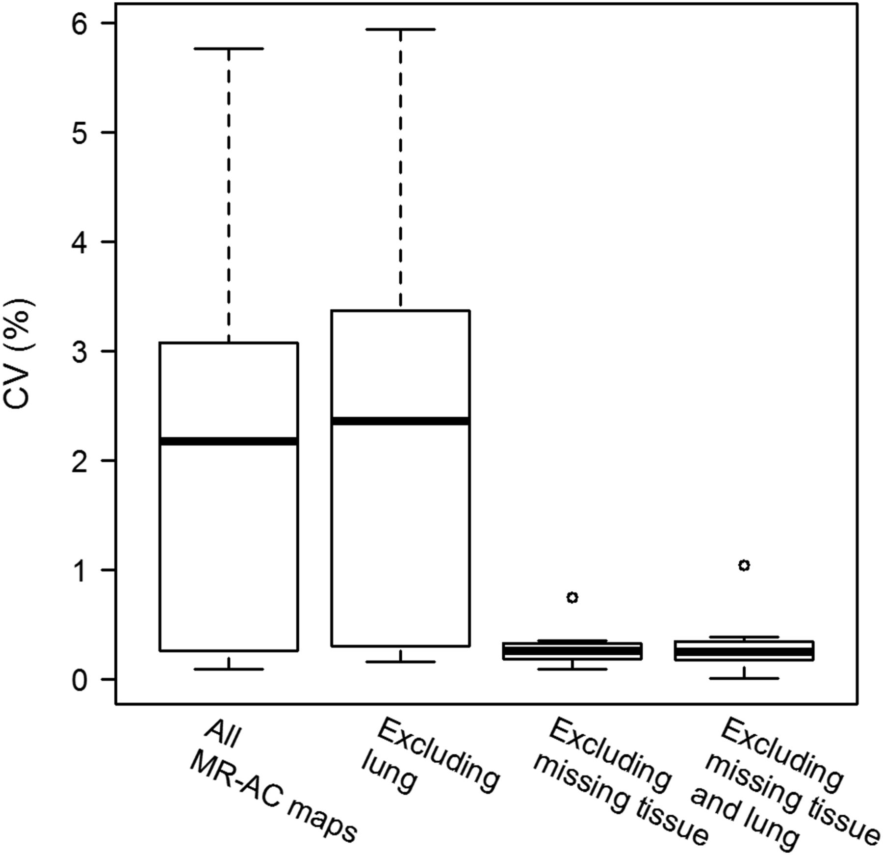
  • FIGURE 3.
    • Download figure
    • Open in new tab
    • Download powerpoint
    FIGURE 3.

    CV of subcompartment volumes. CVs are shown for all MR-AC maps and for MR-AC maps excluding tissue swap and missing tissue artifacts (i.e., artifact-free).
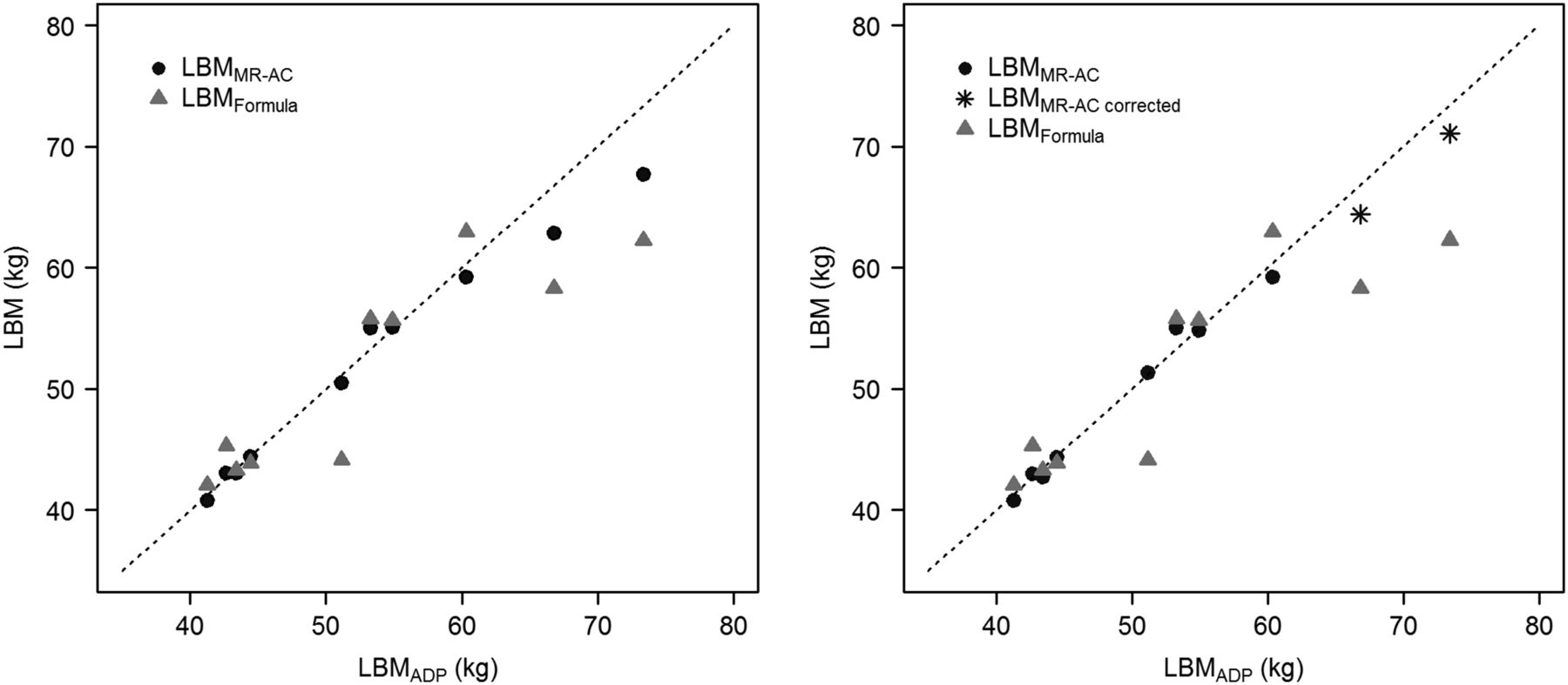
  • FIGURE 4.
    • Download figure
    • Open in new tab
    • Download powerpoint
    FIGURE 4.

    Comparison of scaled LBMMR-AC and LBMFormula with LBMADP: using all MR-AC acquisitions (left) and excluding tissue swap and missing tissue artifacts (right). LBMMR-AC corrected values (*) are LBMMR-AC for the 2 volunteers with tissue swaps in all acquisitions for a single acquisition in which tissue swaps have been manually corrected.

Tables

  • Figures
    • View popup
    TABLE 1

    Summary of Results for All Subjects

    SubjectSexHeight (cm)Body weight (kg)FatADP (kg)LBMADP (kg)LBMFormula (kg)LBMMR-AC (all MR-AC maps/excluding artifacts) (kg)Scaled LBMMR-AC (kg)
    1F16956.411.944.543.942.9/42.845.4
    2M17782.522.260.363.055.9/55.959.2
    3M17867.414.253.355.851.9/51.955.0
    4M16971.416.554.955.752.0/51.854.9
    5F17059.016.442.745.340.6/40.543.0
    6F16756.012.643.443.340.6/40.342.7
    7F16056.815.541.342.138.5/38.540.8
    8M18079.05.673.462.363.9/— (67.1*)— (71.1*)
    9M17971.75.066.858.359.3/— (60.7*)— (64.4*)
    10F16956.95.851.244.147.6/48.451.3
    • ↵* For these subjects, tissue swaps occurred in all repeated scans. Values in parentheses are extracted from 1 scan each for which tissue swaps were corrected manually. This was done by inverting ST and fat values in MR-AC maps in respective regions. This is predictive for LBM with no tissue swaps but is not supposed to result in same LBM gained from artifact-free MR-AC image.

    • LBMMR-AC column shows values of LBM gained for all acquisitions with and without tissue swap and missing tissue artifacts. Scaled LBMMR-AC column shows values scaled by scaling factor gained from linear regression.

    • View popup
    TABLE 2

    Artifacts in Repeated Scans

    ParameterMissing tissueAir cavitiesTruncationTissue swaps
    Subjects4 of 1010 of 1010 of 104 of 10
    Scans6 of 3028 of 3030 of 308 of 30
    • Subjects row gives number of subjects in whom respective artifacts were observed. Scans row gives number of scans in total in which respective artifacts were observed.

PreviousNext
Back to top

In this issue

Journal of Nuclear Medicine: 57 (7)
Journal of Nuclear Medicine
Vol. 57, Issue 7
July 1, 2016
  • Table of Contents
  • Table of Contents (PDF)
  • About the Cover
  • Index by author
Print
Download PDF
Article Alerts
Sign In to Email Alerts with your Email Address
Email Article

Thank you for your interest in spreading the word on Journal of Nuclear Medicine.

NOTE: We only request your email address so that the person you are recommending the page to knows that you wanted them to see it, and that it is not junk mail. We do not capture any email address.

Enter multiple addresses on separate lines or separate them with commas.
Reproducibility of MRI Dixon-Based Attenuation Correction in Combined PET/MR with Applications for Lean Body Mass Estimation
(Your Name) has sent you a message from Journal of Nuclear Medicine
(Your Name) thought you would like to see the Journal of Nuclear Medicine web site.
Citation Tools
Reproducibility of MRI Dixon-Based Attenuation Correction in Combined PET/MR with Applications for Lean Body Mass Estimation
Ivo Rausch, Petra Rust, Matthew D. DiFranco, Martin Lassen, Andreas Stadlbauer, Marius E. Mayerhoefer, Markus Hartenbach, Marcus Hacker, Thomas Beyer
Journal of Nuclear Medicine Jul 2016, 57 (7) 1096-1101; DOI: 10.2967/jnumed.115.168294

Citation Manager Formats

  • BibTeX
  • Bookends
  • EasyBib
  • EndNote (tagged)
  • EndNote 8 (xml)
  • Medlars
  • Mendeley
  • Papers
  • RefWorks Tagged
  • Ref Manager
  • RIS
  • Zotero
Share
Reproducibility of MRI Dixon-Based Attenuation Correction in Combined PET/MR with Applications for Lean Body Mass Estimation
Ivo Rausch, Petra Rust, Matthew D. DiFranco, Martin Lassen, Andreas Stadlbauer, Marius E. Mayerhoefer, Markus Hartenbach, Marcus Hacker, Thomas Beyer
Journal of Nuclear Medicine Jul 2016, 57 (7) 1096-1101; DOI: 10.2967/jnumed.115.168294
Twitter logo Facebook logo LinkedIn logo Mendeley logo
  • Tweet Widget
  • Facebook Like
  • Google Plus One
Bookmark this article

Jump to section

  • Article
    • Abstract
    • MATERIALS AND METHODS
    • RESULTS
    • DISCUSSION
    • CONCLUSION
    • DISCLOSURE
    • Acknowledgments
    • Footnotes
    • REFERENCES
  • Figures & Data
  • Info & Metrics
  • PDF

Related Articles

  • This Month in JNM
  • PubMed
  • Google Scholar

Cited By...

  • Impact of ComBat Harmonization on PET Radiomics-Based Tissue Classification: A Dual-Center PET/MRI and PET/CT Study
  • Reproducibility of MR-Based Attenuation Maps in PET/MRI and the Impact on PET Quantification in Lung Cancer
  • PET/MRI for Oncologic Brain Imaging: A Comparison of Standard MR-Based Attenuation Corrections with a Model-Based Approach for the Siemens mMR PET/MR System
  • Google Scholar

More in this TOC Section

  • Feasibility of Ultra-Low-Activity 18F-FDG PET/CT Imaging Using a Long–Axial-Field-of-View PET/CT System
  • Cardiac Presynaptic Sympathetic Nervous Function Evaluated by Cardiac PET in Patients with Chronotropic Incompetence Without Heart Failure
  • Validation and Evaluation of a Vendor-Provided Head Motion Correction Algorithm on the uMI Panorama PET/CT System
Show more Clinical Investigations

Similar Articles

Keywords

  • PET/MR
  • Dixon-based MR-AC reproducibility
  • Lean Body Mass estimation
  • Body composition measurement
SNMMI

© 2025 SNMMI

Powered by HighWire