


Article Figures & Data
Figures
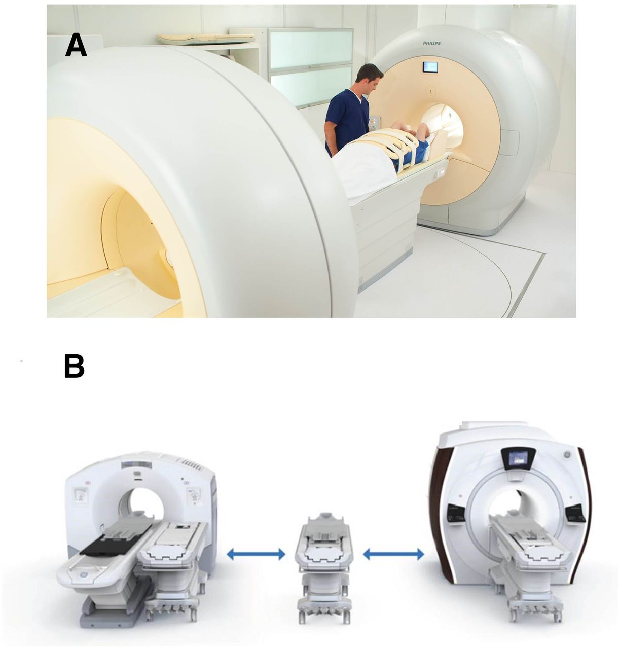
- FIGURE 1.
Sequential PET/MRI systems: Ingenuity TF (Philips) (A) and PET/CT+MR trimodality setup (GE Healthcare) (B). Both are connected by scanner bed shuttle system and allow sequential PET and MRI data acquisition at 3-T field strengths. (Courtesy of Philips and GE Healthcare.)
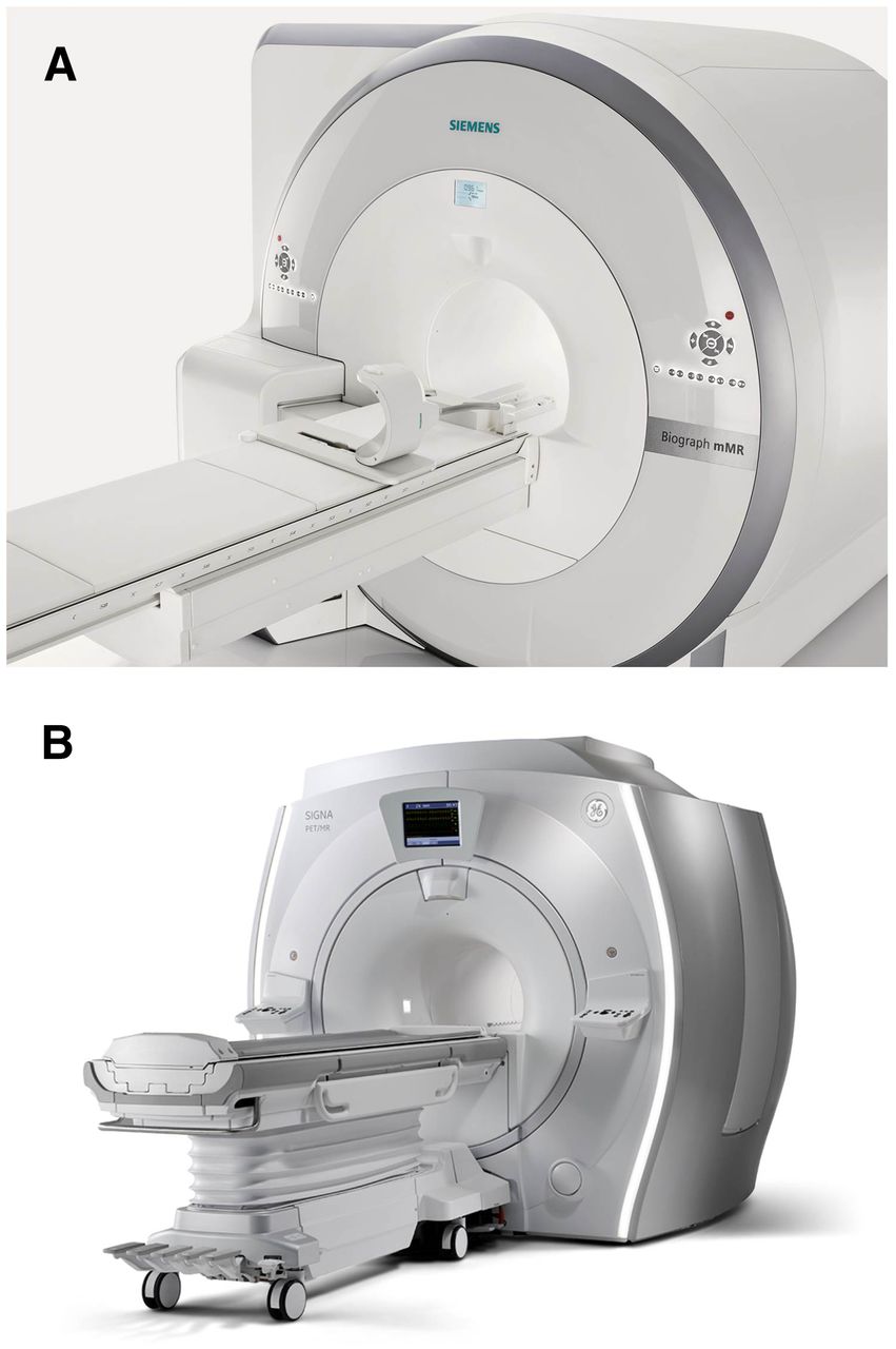
- FIGURE 2.
Integrated PET/MRI systems: Biograph mMR (Siemens) (A) and Signa PET/MR (GE Healthcare) (B). Both allow for simultaneous PET and MRI data acquisitions at 3-T field strengths. (Courtesy of Siemens and GE Healthcare.)
Tables
Study Design Patients (n) PET/MRI Indication T-staging N-staging M-staging Superiority Kuhn (28) Prospective 150 Sequential Staging, restaging — NS — ND Queiroz (29) Prospective 87 Sequential Restaging NS NS — ND Kubiessa (25) Prospective 17 Simultaneous Staging, restaging — — — ND Partovi (26) Prospective 14 Sequential Staging, restaging — — — ND Varoquaux (27) Prospective 32 Sequential Staging, restaging — — — ND Covello (30) Retrospective 44 Sequential Staging, restaging NS — — ND Schaarschmidt (31) Retrospective 25 Simultaneous Staging, restaging NS NS — ND Total 369 NS = nonsignificant; ND = no difference; — = not reported.
Study Design Patients (n) PET/MRI Indication T-staging N-staging M-staging Superiority Schwenzer (36) Not stated 10 Simultaneous Staging — — — ND Fraioli (37) Prospective 50 Simultaneous Staging — — — ND Heusch (38) Prospective 22 Simultaneous Staging NS NS — ND Stolzmann (39) Prospective 40 Sequential Lung nodules — — — CT superior for 18F-FDG–negative lesions Rauscher (40) Prospective 40 Simultaneous Lung nodules — — — CT for lesions < 1 cm Chandarana (41) Prospective 32 Simultaneous Lung nodules — — — ND Total 194 NS = nonsignificant; ND = no difference; — = not reported.
Study Design Patients (n) PET/MRI Cancer type Indication T-staging N-staging M-staging Superiority Lee (44) Prospective 15 Sequential Esophageal Staging — NS — ND Paspulati (47) Prospective 12 Sequential Colorectal Staging, restaging — — — ND (no enhanced CT) Brendle (48) Retrospective 15 Simultaneous Colorectal Staging, restaging — NS NS ND Reiner (49) Prospective 55 Sequential Liver lesions Staging, restaging — — — ND Beiderwellen (50) Prospective 70 Simultaneous Liver lesions Staging, restaging — — — ND Gaertner (76) Prospective 24 Simultaneous NET Staging, restaging — — — ND Hope (77) Prospective 10 Simultaneous NET Staging, restaging — — — MRI superior for liver lesions; no validation Total 201 NS = nonsignificant; ND = no difference; — = not reported.
Study Design Patients (n) PET/MRI Cancer type Indication T-staging N-staging M-staging Superiority Beiderwellen (53) Prospective 19 Simultaneous Gynecologic Restaging — NS NS ND Queiroz (54) Prospective 26 Sequential Gynecologic Staging, restaging NS NS NS ND Grueneisen (55) Retrospective 24 Simultaneous Gynecologic Restaging NS NS NS ND Pace (57) Prospective 36 Simultaneous Breast Staging, restaging — — — ND Grueneisen (58) Prospective 49 Simultaneous Breast Staging P < 0.05 NS — PET/MRI superior for T-staging Total 154 NS = nonsignificant; ND = no difference; — = not reported.
Study Design Patients (n) PET/MRI PET ligand Indication T-staging N-staging M-staging Superiority Wetter (64) Not stated 36 Simultaneous 18F-choline Staging, restaging — — — ND Afshar-Oromieh (62) Not stated 20 Simultaneous 68Ga-PSMA Restaging — — — ND Souvatzoglou (63) Prospective 32 Simultaneous 11C-choline Staging, restaging NS NS NS PET/MRI superior for prostatic and bone lesions Total 88 NS = nonsignificant; ND = no difference; — = not reported.
Study Design Patients (n) PET/MRI Cancer type Indication TNM-staging Superiority Heacock (69) Prospective 28 Simultaneous Lymphoma Staging — ND Eiber (79) Retrospective 119 Simultaneous Malignant bone disease Staging, restaging — PET/MRI superior for allocation of bone lesions Beiderwellen (80) Prospective 67 Simultaneous Malignant bone disease Staging, restaging — ND Catalano (81) Prospective 109 Simultaneous Malignant bone disease Staging, restaging — PET/MRI superior for bone lesion detection Afshar-Oromieh (84) Prospective 15 Simultaneous Meningioma Staging — ND Total 338 NS = nonsignificant; ND = no difference; — = not reported.
Study Design Patients (n) PET/MRI Indication T-staging N-staging M-staging Superiority Drzezga (85) Prospective 32 Simultaneous Staging, restaging NS NS NS ND Quick (86) Prospective 80 Simultaneous Staging, restaging — — — ND Al-Nabhani (87) Prospective 50 Simultaneous Staging — — — NS Catalano (88) Retrospective 134 Simultaneous Staging, restaging — — — PET/MRI superior for patient management (P < 0.001) Wiesmuller (89) Prospective 46 Simultaneous Staging, restaging — — — ND Appenzeller (90) Prospective 63 Sequential Staging, restaging — — — ND Jeong (91) Not stated 12 Simultaneous Staging, restaging — — — ND Huellner (92) Prospective 106 Sequential Staging, restaging NS NS NS ND (more incidental findings by PET/CT) Schäfer (83) Prospective 18 Simultaneous Staging, restaging — — — ND Iagaru (10) Prospective 36 Simultaneous Staging, restaging — — — ND Tian (93) Retrospective 285 Simultaneous Staging, restaging — — — ND Heusch (94) Retrospective 73 Simultaneous Staging NS NS ND ND Schaarschmidt (95) Retrospective 61 Simultaneous Staging NS NS NS ND Total 996 NS = nonsignificant; ND = no difference; — = not reported.


Jump to section
Related Articles
Cited By...
- Lung imaging methods: indications, strengths and limitations
- Optimized Whole-Body PET MRI Sequence Workflow in Pediatric Hodgkin Lymphoma Patients
- Development of 89Zr-anti-CD103 PET imaging for non-invasive assessment of cancer reactive T cell infiltration
- PET/MRI Versus PET/CT for Whole-Body Staging: Results from a Single-Center Observational Study on 1,003 Sequential Examinations
- The Contribution of Multiparametric Pelvic and Whole-Body MRI to Interpretation of 18F-Fluoromethylcholine or 68Ga-HBED-CC PSMA-11 PET/CT in Patients with Biochemical Failure After Radical Prostatectomy
- Additional Clinical Value for PET/MRI in Oncology: Moving Beyond Simple Diagnosis
- New horizons in multimodality molecular imaging and novel radiotracers
- Variations in PET/MRI Operations: Results from an International Survey Among 39 Active Sites
- Value of 68Ga-PSMA HBED-CC PET for the Assessment of Lymph Node Metastases in Prostate Cancer Patients with Biochemical Recurrence: Comparison with Histopathology After Salvage Lymphadenectomy
- PET and MRI: Is the Whole Greater than the Sum of Its Parts?
- Evaluation of Prostate Cancer with PET/MRI